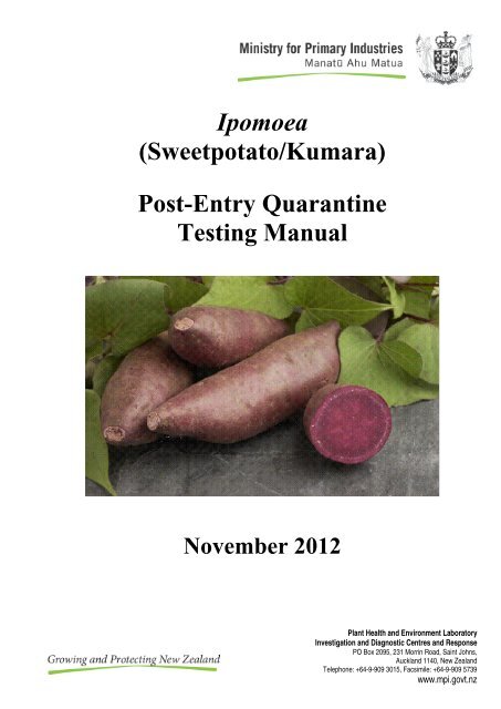
Ipomoea (Sweetpotato/Kumara) Post-Entry Quarantine Testing Manual
Ipomoea (Sweetpotato/Kumara) Post-Entry Quarantine Testing Manual
Ipomoea (Sweetpotato/Kumara) Post-Entry Quarantine Testing Manual

You also want an ePaper? Increase the reach of your titles
YUMPU automatically turns print PDFs into web optimized ePapers that Google loves.
<strong>Ipomoea</strong><br />
(<strong>Sweetpotato</strong>/<strong>Kumara</strong>)<br />
<strong>Post</strong>-<strong>Entry</strong> <strong>Quarantine</strong><br />
<strong>Testing</strong> <strong>Manual</strong><br />
November 2012<br />
Plant Health and Environment Laboratory<br />
Investigation and Diagnostic Centres and Response<br />
PO Box 2095, 231 Morrin Road, Saint Johns,<br />
Auckland 1140, New Zealand<br />
Telephone: +64-9-909 3015, Facsimile: +64-9-909 5739<br />
www.mpi.govt.nz
Contents<br />
<strong>Ipomoea</strong> <strong>Post</strong>-<strong>Entry</strong> <strong>Quarantine</strong> <strong>Testing</strong> <strong>Manual</strong><br />
1. SCOPE ....................................................................................................................................................... 1<br />
2. INTRODUCTION ..................................................................................................................................... 1<br />
3. IMPORT REQUIREMENTS................................................................................................................... 3<br />
4. PESTS ........................................................................................................................................................ 3<br />
4.1 Regulated pests for which generic measures are required ........................................................... 3<br />
4.2 Regulated pests for which specific tests are required ................................................................... 4<br />
5. PROPAGATION, CARE AND MAINTENANCE IN POST-ENTRY QUARANTINE .................... 4<br />
5.1 Whole plants..................................................................................................................................... 4<br />
5.2 Plants in tissue culture .................................................................................................................... 5<br />
5.3 Pollen ................................................................................................................................................ 5<br />
6. INSPECTION ............................................................................................................................................ 5<br />
7. TESTING ................................................................................................................................................... 6<br />
7.1 Specific tests for nursery stock ....................................................................................................... 7<br />
7.1.1 Graft inoculation ......................................................................................................................... 8<br />
7.1.2 Herbaceous indexing................................................................................................................. 10<br />
7.1.3 Serological and molecular assays ............................................................................................ 11<br />
7.1.3.1 Enzyme-linked immunosorbent assay (ELISA) .......................................................... 11<br />
7.1.3.2 Polymerase chain reaction (PCR)................................................................................. 12<br />
7.1.3.2.1 Virus reverse transcription-PCR .............................................................................. 14<br />
7.1.3.2.1.1 Sweet potato chlorotic stunt virus ........................................................................ 18<br />
7.1.3.2.1.2 <strong>Sweetpotato</strong> leaf curl virus ................................................................................... 18<br />
7.1.3.2.1.3 <strong>Sweetpotato</strong> mild speckling virus ......................................................................... 18<br />
7.1.3.2.1.4 <strong>Sweetpotato</strong> vein mosaic virus .............................................................................. 18<br />
7.1.3.2.1.5 Tobacco streak virus ............................................................................................. 18<br />
7.1.3.2.2 Phytoplasma PCR ....................................................................................................... 19<br />
7.1.3.2.2.2 <strong>Sweetpotato</strong> little leaf phytoplasma ................................................................... 21<br />
7.1.3.2.3 Bacteria PCR .............................................................................................................. 21<br />
7.1.3.2.3.1 Dickeya chrysanthemi .......................................................................................... 21<br />
7.1.4 Bacterial isolation on media ..................................................................................................... 22<br />
7.1.4.1 Dickeya chrysanthemi (basonym. Erwinia chrysanthemi) ........................................... 22<br />
7.1.5 Microscopic inspection for mites ............................................................................................. 23<br />
7.1.5.1 Tetranychus evansi ......................................................................................................... 23<br />
8. CONTACT POINT ................................................................................................................................. 24<br />
9. ACKNOWLEDGEMENTS .................................................................................................................... 24<br />
10. REFERENCES ........................................................................................................................................ 24<br />
Appendix 1. Symptoms of significant regulated pests of <strong>Ipomoea</strong> batatas .................................................. 27<br />
1.1 Meliodogyne incognita ................................................................................................................... 27<br />
1.2 Rotylenchulus reniformis ............................................................................................................... 27<br />
1.3 Tetranychus evansi ......................................................................................................................... 27<br />
1.4 Plant damage caused by mites ...................................................................................................... 27<br />
1.5 Streptomyces ipomoea .................................................................................................................... 28<br />
1.6 Elsinoë batatas ................................................................................................................................ 28<br />
<strong>Ipomoea</strong> <strong>Post</strong>-<strong>Entry</strong> <strong>Quarantine</strong> <strong>Testing</strong> <strong>Manual</strong> · November 2012<br />
ii
1.7 Dickeya chrysanthemi .................................................................................................................... 28<br />
1.8 <strong>Ipomoea</strong> batatas infected with a mixture of viruses .................................................................... 29<br />
1.9 <strong>Sweetpotato</strong> chlorotic stunt virus ................................................................................................... 29<br />
1.10 <strong>Sweetpotato</strong> leaf curl virus ............................................................................................................. 29<br />
1.11 <strong>Sweetpotato</strong> little leaf phytoplasma ............................................................................................. 29<br />
Appendix 2. Virus symptoms on graft inoculated <strong>Ipomoea</strong> setosa ............................................................... 30<br />
2.1 <strong>Sweetpotato</strong> chlorotic stunt virus + <strong>Sweetpotato</strong> feathery mottle virus ......................................... 30<br />
2.2 <strong>Sweetpotato</strong> virus 2 ......................................................................................................................... 30<br />
2.3 <strong>Sweetpotato</strong> virus C6....................................................................................................................... 30<br />
2.4 <strong>Sweetpotato</strong> leaf curl virus ............................................................................................................. 31<br />
2.5 <strong>Sweetpotato</strong> leaf curl virus + <strong>Sweetpotato</strong> virus 2 ......................................................................... 31<br />
2.6 <strong>Sweetpotato</strong> leaf curl virus + <strong>Sweetpotato</strong> feathery mottle virus ................................................... 31<br />
Appendix 3. Protocols referenced in manual ................................................................................................. 32<br />
3.1 Silica-milk RNA extraction protocol ............................................................................................ 32<br />
3.2 Phytoplasma DNA enrichment CTAB extraction protocol ........................................................ 32<br />
©Ministry for Primary Industries, November, 2012<br />
<strong>Ipomoea</strong> <strong>Post</strong>-<strong>Entry</strong> <strong>Quarantine</strong> <strong>Testing</strong> <strong>Manual</strong> · November 2012<br />
iii
1. SCOPE<br />
The scope of this manual is limited to <strong>Ipomoea</strong> batatas and <strong>Ipomoea</strong> setosa nursery stock<br />
(whole plants and plants in tissue culture), seed for sowing and pollen of <strong>Ipomoea</strong> species<br />
permitted entry into New Zealand as listed in the Ministry for Primary Industries (MPI)<br />
Plants Biosecurity Index (http://www.maf.govt.nz/cgi-bin/bioindex/bioindex.pl). At the date<br />
of publication of this manual, these species were as follows:<br />
<strong>Ipomoea</strong> alba<br />
<strong>Ipomoea</strong> aquatica<br />
<strong>Ipomoea</strong> arborescens<br />
<strong>Ipomoea</strong> batatas<br />
<strong>Ipomoea</strong> brasiliensis<br />
<strong>Ipomoea</strong> cairica<br />
<strong>Ipomoea</strong> carnea<br />
<strong>Ipomoea</strong> horsfalliae<br />
<strong>Ipomoea</strong> imperialis<br />
<strong>Ipomoea</strong> lobata<br />
<strong>Ipomoea</strong> minuta<br />
<strong>Ipomoea</strong> nil<br />
<strong>Ipomoea</strong> <strong>Post</strong>-<strong>Entry</strong> <strong>Quarantine</strong> <strong>Testing</strong> <strong>Manual</strong> · November 2012<br />
<strong>Ipomoea</strong> noctiflora<br />
<strong>Ipomoea</strong> palmata (syn. <strong>Ipomoea</strong> cairica)<br />
<strong>Ipomoea</strong> pes-caprae<br />
<strong>Ipomoea</strong> platensis<br />
<strong>Ipomoea</strong> purpurea<br />
<strong>Ipomoea</strong> quamoclit<br />
<strong>Ipomoea</strong> sepacuitensis<br />
<strong>Ipomoea</strong> setosa<br />
<strong>Ipomoea</strong> sloteri<br />
<strong>Ipomoea</strong> tricolor<br />
<strong>Ipomoea</strong> tuberosa (syn. Merremia<br />
tuberosa)<br />
Note: The importation of <strong>Ipomoea</strong> caerulea, <strong>Ipomoea</strong> hederacea, <strong>Ipomoea</strong> indica,<br />
<strong>Ipomoea</strong> learii (syn. <strong>Ipomoea</strong> indica), <strong>Ipomoea</strong> plebeia and <strong>Ipomoea</strong> triloba is prohibited.<br />
This manual describes the testing requirements specified in the import health standards for<br />
<strong>Ipomoea</strong>. The manual also provides an introduction to the crop and guidance on the<br />
establishment and maintenance of healthy plants in quarantine.<br />
2. INTRODUCTION<br />
<strong>Sweetpotato</strong> (<strong>Ipomoea</strong> batatas (L.) Lam.), a member of the family Convolvulaceae, probably<br />
originated in Central or South America, where it has been a food source for over 55,000<br />
years. <strong>Sweetpotato</strong> was taken to Spain and early Spanish explorers are believed to have taken<br />
it to the Philippines and East Indies; from there it was soon carried to India, China, and<br />
Malaysia by Portuguese voyagers. It is not fully known how sweetpotato arrived in<br />
Polynesia, but it has been used on many of the islands in the South Pacific Ocean for at least<br />
2000 years (Clark & Moyer, 1988).<br />
<strong>Sweetpotato</strong> is grown in a wide range of environments under a range of farming systems,<br />
from the humid tropics to mild temperate zones, and from sea level to 2700 m altitude.<br />
Annual global production of sweetpotato currently exceeds 124 million tonnes. More than<br />
95% of the global sweetpotato crop is grown in developing countries. China is the world's<br />
largest producer, accounting for more than 90%. Vietnam, Indonesia, and Uganda all grow<br />
more than two million tonnes per year. India and Rwanda each harvest more than a million<br />
tonnes annually. Of the 82 developing countries where sweetpotatoes grow, 36 are in Africa,<br />
22 in Asia, and 24 in Latin America. Around 40 countries count sweetpotato among the five<br />
most important food crops produced on an annual basis.<br />
1
The per capita income provided by sweetpotato is one of the lowest among the major food<br />
crops. Its potential benefit to poor farm households and urban consumers is only now being<br />
considered. <strong>Sweetpotato</strong> actually produces more edible energy per hectare per day than any<br />
other major food crop.<br />
<strong>Sweetpotato</strong> is a perennial plant cultivated as an annual crop and propagated vegetatively.<br />
The sweetpotato plant is a prostrate vine system that expands horizontally and develops a<br />
shallow canopy. The sweetpotato plant produces several different types of thick and thin<br />
roots. Thick roots can differentiate into either ‘pencil roots’ or ‘storage roots’, the latter<br />
being used for human consumption. <strong>Sweetpotato</strong>es are not tubers as they are initiated at the<br />
first stem node below the soil line, to which they are attached by a stalk of thinner root (Clark<br />
& Moyer, 1988).<br />
Although known for its tolerance to drought and its sensitivity to saturated soil condition,<br />
sweetpotato requires sufficient water and nutrients to produce good yield. Non-rooted stem<br />
cuttings (20-40 cm) with 5-8 nodes are harvested from storage roots laid out in nursery beds,<br />
and transplanted into the field. <strong>Sweetpotato</strong> can be cultivated continuously throughout the<br />
year in tropical regions, but in temperate regions the crop is planted in spring when the risk of<br />
frost is reduced. The crop requires a minimum frost-free period of 120-150 days and average<br />
daily temperatures of 22-24ºC along with good rainfall and good drainage (Clark & Moyer,<br />
1988).<br />
The flesh of sweetpotato can be white, purple, orange or yellow. Yellow and orange-fleshed<br />
varieties are valuable for their carotene (provitamin A) content. Skin colour ranges from<br />
nearly white through shades of buff to brown, or through pink to copper, even magenta and<br />
purple.<br />
In New Zealand, sweetpotato (known as kumara) is a crop of cultural importance and an<br />
important food source. <strong>Kumara</strong> was introduced by Maori when they settled in New Zealand<br />
from Polynesia. Cultivation of this crop was undertaken on a large scale because of its<br />
importance as a food source. With the arrival of European settlers, other carbohydrate crops<br />
such as potato, wheat, and corn displaced sweetpotato in dietary importance but not the<br />
crop’s place in Maori culture. Since the 1950s, after efforts to select improved clones,<br />
production has steadily increased along with consumption as people rediscover kumara.<br />
The local cultivar Owairaka Red, released in 1954, comprises 80% of the crop. Other<br />
cultivars include Toka Toka Gold, selected in 1972 (14%) and Beauregard (introduced in<br />
1993). A range of other local varieties are also grown, usually in garden plots.<br />
The area around Dargaville in the Kaipara district produces 85% of the national crop.<br />
Smaller plantings of approximately 5% are found around Auckland and Bay of Plenty. The<br />
area planted annually is approximately 1,100 hectares producing about 26,500 tonnes, with<br />
yields averaging 20 t/ha. Plantings range in area from garden plots to 30 ha, averaging 10 ha.<br />
Most of New Zealand’s production is for local fresh consumption although increasing<br />
amounts are processed and/or exported. A thorough review of this crop and its place in New<br />
Zealand agriculture is presented by Lewthwaite (1997).<br />
<strong>Ipomoea</strong> <strong>Post</strong>-<strong>Entry</strong> <strong>Quarantine</strong> <strong>Testing</strong> <strong>Manual</strong> · November 2012<br />
2
3. IMPORT REQUIREMENTS<br />
The import requirements for I. batatas and I. setosa nursery stock (whole plant and plants in<br />
tissue culture) are set out in MPI’s import health standard “Importation of Nursery Stock”<br />
(http://www.biosecurity.govt.nz/files/ihs/155-02-06.pdf). Imported nursery stock must meet<br />
the general requirements (sections 1-2) and the specific requirements detailed in the<br />
“<strong>Ipomoea</strong> batatas” schedule. On arrival in New Zealand, the nursery stock must be grown<br />
for a minimum period of 3 months in a Level 3 post-entry quarantine facility where it will be<br />
inspected, treated and/or tested for regulated pests.<br />
The import requirements for <strong>Ipomoea</strong> seed for sowing are set out in MPI’s import health<br />
standard “Importation of Seed for Sowing” (http://www.biosecurity.govt.nz/files/ihs/155-02-<br />
05.pdf). Imported seed is only required to meet the general requirements (sections 1-2) and<br />
there are no specific requirements for the genus. An import permit is not required and seed<br />
meeting the import requirements is given biosecurity clearance at the border without the need<br />
for post-entry quarantine.<br />
The import requirements for pollen are stated in section 2.2.3 in MPI’s import health standard<br />
“Importation of Nursery Stock” (http://www.biosecurity.govt.nz/files/ihs/155-02-06.pdf ) and<br />
further details can be found in section 5.3 of this manual.<br />
4. PESTS<br />
The following section lists regulated pests of I. batatas and I. setosa nursery stock that<br />
require generic or specific measures.<br />
4.1 Regulated pests for which generic measures are required<br />
Insects:<br />
Cylas formicarius<br />
Cylas puncticollis<br />
Euscepes postfasciatus<br />
Nematodes:<br />
Meliodogyne incognita [Fig. 1.1]<br />
Rotylenchulus reniformis [Fig. 1.2]<br />
Pratylenchus coffeae<br />
Pratylenchus brachyurus<br />
Fungi:<br />
Elsinoë batatas [Fig. 1.6]<br />
Helicobasidium mompa<br />
Bacteria:<br />
Pseudomonas batatas<br />
Streptomyces ipomoea [Fig. 1.5]<br />
Xanthomonas batatae<br />
Xylella fastidiosa<br />
<strong>Ipomoea</strong> <strong>Post</strong>-<strong>Entry</strong> <strong>Quarantine</strong> <strong>Testing</strong> <strong>Manual</strong> · November 2012<br />
3
Viruses:<br />
<strong>Sweetpotato</strong> chlorotic fleck virus<br />
<strong>Sweetpotato</strong> latent virus<br />
<strong>Sweetpotato</strong> ringspot virus<br />
<strong>Sweetpotato</strong> virus C6<br />
[Fig. 1.8]<br />
<strong>Ipomoea</strong> <strong>Post</strong>-<strong>Entry</strong> <strong>Quarantine</strong> <strong>Testing</strong> <strong>Manual</strong> · November 2012<br />
[Fig. 1.8, 2.3]<br />
4.2 Regulated pests for which specific tests are required<br />
Mites:<br />
Tetranychus evansi [Fig. 1.3, 1.4]<br />
Bacteria:<br />
Dickeya chrysanthemi [Fig. 1.7]<br />
Phytoplasma:<br />
<strong>Sweetpotato</strong> little leaf phytoplasma [Fig. 1.11]<br />
Viruses:<br />
<strong>Sweetpotato</strong> caulimo-like virus<br />
<strong>Sweetpotato</strong> chlorotic stunt virus [Fig. 1.9, 2.1]<br />
<strong>Sweetpotato</strong> leaf curl virus [Fig. 1.10, 2.4, 2.5, 2.6]<br />
<strong>Sweetpotato</strong> leaf speckling virus<br />
<strong>Sweetpotato</strong> mild speckling virus<br />
<strong>Sweetpotato</strong> vein mosaic virus<br />
<strong>Sweetpotato</strong> yellow dwarf virus<br />
Tobacco streak virus<br />
5. PROPAGATION, CARE AND MAINTENANCE IN POST-ENTRY<br />
QUARANTINE<br />
5.1 Whole plants<br />
<strong>Sweetpotato</strong> plants can be grown in the glasshouse all year round as long as a day-time<br />
temperature between 18-26ºC is maintained. Night-time temperatures should not fall below<br />
12ºC to avoid chilling injury. Supplementary lighting may be required in winter.<br />
<strong>Sweetpotato</strong>es require free-draining planting which is initially only moistened to avoid<br />
development of rots in quiescent storage roots. The plants can be watered more freely when<br />
the canopy has established and the plants are actively growing. The sweetpotato is a<br />
perennial plant and harvesting takes place when storage roots reach the desired size.<br />
Harvesting may be plant destructive, but if larger storage roots are removed with minimal<br />
plant disturbance, the remaining storage roots will continue to grow and new plants will form.<br />
Harvested roots should be stored in the dark at 13ºC. Storage temperatures should not fall<br />
below 12ºC which can cause chilling injury. Relative humidity during storage should be<br />
maintained at 80 to 90%. Roots stored in multi-walled paper bags can respire and maintain<br />
their own humid environment. Hessian or net bags should be avoided as they can cause<br />
abrasive injury and allow moisture and pathogen entry.<br />
4
Whole plants should be planted into sufficiently sized pots (eg 3 L minimum) containing<br />
50:50 (v/v) pasteurised peat:pumice planting media and a few grams of slow-release fertiliser<br />
with trace elements (e.g. Osmocote ® ). Nodal cuttings should be taken from growing vines to<br />
maintain the clone and facilitate quarantine examination and testing. Cuttings with one or<br />
two leaves and at least two nodes can be rooted directly in pasteurised pumice sand or perlite<br />
before transferring to planting media. It would be worth preserving clonal material in tissue<br />
culture as a back-up resource, and to preserve any established virus-free status.<br />
Plants in tissue culture<br />
Tissue culture plantlets can be sub-cultured after arrival by cutting into nodal sections and<br />
placing into new tissue culture vessels with fresh nutrient media (e.g. Murashige and Skoog<br />
media).<br />
Plantlets to be tested are carefully excised from the tissue culture vessel and washed to<br />
remove any remaining agar and planted into pots of planting media containing 50:50 (v/v)<br />
pasturised peat:perlite or 50:50 (v/v) peat:vermiculite. The plantlets must be protected from<br />
desiccation for approximately three weeks by covering initially with a vented plastic tub or<br />
bag. Alternatively, the plants can be misted regularly to keep the planting media moist, and<br />
to maintain a high relative humidity. Pots should be placed in bright light, but not direct<br />
sunlight during the three weeks. After this period, any coverings should be removed and the<br />
plants moved to higher light intensity.<br />
5.3 Pollen<br />
Anthers can be collected from mature but unopened flowers and dried in warm, light<br />
conditions. Following this drying period, pollen should be collected into a centrifuge vial or<br />
into gel capsules and stored at 4°C in a sealed container in the presence of a strong desiccant<br />
such as calcium chloride.<br />
6. INSPECTION<br />
The inspection requirements for the operator of the facility are set out in the “MPI<br />
Biosecurity Authority Standard PBC-NZ-TRA-PQCON”<br />
(http://www.biosecurity.govt.nz/files/regs/stds/pbc-nz-tra-pqcon.pdf )<br />
Photographs of symptoms caused by significant regulated diseases can be found in Appendix<br />
1. However, please note that pot-grown sweetpotato plants can be prone to nutrient<br />
deficiencies if not adequately fertilised and nutrient deficiencies can resemble virus infection,<br />
e.g. chlorosis and necrosis. Symptoms related to nutrient deficiencies can be found in<br />
Appendix 2. Further information on nutrient deficiencies is described in Clark & Moyer<br />
(1988).<br />
<strong>Ipomoea</strong> <strong>Post</strong>-<strong>Entry</strong> <strong>Quarantine</strong> <strong>Testing</strong> <strong>Manual</strong> · November 2012<br />
5
7. TESTING<br />
Each of the specific tests required in the import health standard (as described in section 4 and<br />
summarised in Table 1) must be done irrespective of whether plants exhibit symptoms. This<br />
testing is required to detect latent infections.<br />
Samples should be tested as soon as possible after removal from the plant. If samples have to<br />
be stored before testing, the plant material must be kept whole, all surface water must be<br />
removed, and the material stored in a plastic bag at 4 o C. Samples that become partially<br />
decayed or mouldy must not be tested, and further samples should be collected.<br />
Inspection for mites<br />
Inspection for mites is performed once whole plants or tissue culture plants have established<br />
successfully in planting media and have produced stems with at least 10-15 nodes.<br />
Inspection should take place before samples are taken for other testing methods. Using a<br />
hand lens, the underside of all leaves must be inspected for mite eggs, nymphs, adults and<br />
symptoms of mite presence. Following this, the 3 youngest leaves of each plant, plus any<br />
suspect leaves showing the presence of mites must be collected for further examination under<br />
a binocular microscope. See section 7.1.5 for further details.<br />
Indexing tests<br />
Graft inoculation: Grafting can begin when the whole plants or tissue culture plants have<br />
established successfully in planting media and have produced stems with at least 10-15<br />
nodes. Grafting should take place before leaf samples are collected for other testing methods.<br />
See section 7.1.1 for further details. Each plant in the glasshouse must be tested individually<br />
by graft indexing<br />
Herbaceous indexing: Virus testing should be done in spring (or under spring-like<br />
conditions) when new growth has occurred. At least two fully expanded leaves must be<br />
sampled from each of two different branches of the main stem, one a younger leaf and one an<br />
older leaf from a mid-way position. Each plant in the glasshouse must be tested individually<br />
by herbaceous indexing. See section 7.1.2 for further details<br />
PCR and ELISA testing<br />
Viruses: For virus-testing of I. batatas by PCR and ELISA, it is recommended to test the<br />
graft-inoculated indicator plants rather than the original test plants. However, original I.<br />
setosa plants can be tested directly. Virus testing should be done in spring (or under springlike<br />
conditions) when new growth has occurred. At least two fully expanded leaves must be<br />
sampled from each of two different branches of the main stem, one a younger leaf and one an<br />
older leaf from a mid-way position. The sampled leaves from each plant must be bulked<br />
together and tested as soon as possible after removal from the host. See section 7.1.3 for<br />
further details.<br />
Bacteria and phytoplasma: Bacteria and phytoplasma testing must be carried out using the<br />
original I. batatas and I. setosa plants and should be done in summer (or under summer-like<br />
conditions). For each plant, at least two fully expanded leaves must be sampled from each of<br />
two different branches of the main stem, one a younger leaf and one an older leaf from a midway<br />
position. Detection of both bacteria and phytoplasma requires testing of leaf petioles<br />
and mid-veins. The sampled leaves from each plant must be bulked together and tested as<br />
soon as possible after removal from the host. See section 7.1.3 for further details<br />
<strong>Ipomoea</strong> <strong>Post</strong>-<strong>Entry</strong> <strong>Quarantine</strong> <strong>Testing</strong> <strong>Manual</strong> · November 2012<br />
6
Bacterial isolation on media<br />
Isolation of regulated bacteria testing must be carried out using the original I. batatas and I.<br />
setosa plants and should be done in summer (or under summer-like conditions). For each<br />
plant, at least two fully expanded leaves must be sampled from each of two different branches<br />
of the main stem, one a younger leaf and one an older leaf from a mid-way position.<br />
Detection of bacteria requires plating vascular tissue from petioles mid-veins. Each plant in<br />
the glasshouse must be tested individually. See section 7.1.4 for further details.<br />
Table 1: Summary of the regulated pests for I. batatas and I. setosa indicating the<br />
specific tests that are required (■), alternative (□) or optional ()<br />
Organism Type Graft<br />
Inoculation 1<br />
Herbaceous<br />
Indexing<br />
<strong>Ipomoea</strong> <strong>Post</strong>-<strong>Entry</strong> <strong>Quarantine</strong> <strong>Testing</strong> <strong>Manual</strong> · November 2012<br />
ELISA PCR<br />
Isolation<br />
on media<br />
Inspection<br />
Mites<br />
Tetranychus evansi ■<br />
Bacterium<br />
Dickeya chrysanthemi □ □<br />
Phytoplasma<br />
<strong>Sweetpotato</strong> little leaf<br />
phytoplasma<br />
Viruses<br />
■<br />
<strong>Sweetpotato</strong> caulimo-like virus ■<br />
<strong>Sweetpotato</strong> chlorotic stunt<br />
virus<br />
■ ■<br />
<strong>Sweetpotato</strong> leaf curl virus 2 ■ ■<br />
<strong>Sweetpotato</strong> leaf speckling<br />
virus<br />
■<br />
<strong>Sweetpotato</strong> mild speckling ■ <br />
virus<br />
<strong>Sweetpotato</strong> vein mosaic virus ■ □ □<br />
<strong>Sweetpotato</strong> yellow dwarf virus ■ ■<br />
Tobacco streak virus ■ □ □<br />
1 2<br />
Not required for I. setosa; ssDNA Geminivirus<br />
7.1 Specific tests for nursery stock<br />
Each plant must be tested separately with the following exceptions, samples from up to 5<br />
plants may be bulked for testing provided that either:<br />
(a) the plants are derived from a single imported plant or plant established from a storage<br />
root from which separate cuttings have been taken upon arrival in New Zealand, in<br />
the presence of a MPI inspector; or<br />
(b) in the case of tissue culture where plants are clonal, and this is confirmed by evidence<br />
from the national plant protection organisation in the exporting country.<br />
7
7.1.1 Graft inoculation<br />
Each I. batatas plant must be tested by graft inoculation using a minimum of 3 replicate<br />
indicator plants of either I. setosa or I. nil ‘Scarlet O’ Hara’. Indicator plants must be<br />
maintained in a healthy, vigorous state, as symptoms associated with abiotic stresses, such as<br />
water and nutrient deficiencies, may mask and interfere with observations of disease<br />
symptoms. The indicator plants can be grown from seed or from young cuttings. If using<br />
seed, sow 3-4 weeks before grafting.<br />
<strong>Sweetpotato</strong> seed requires scarification prior to germination. Indicator seeds are soaked in<br />
concentrated sulphuric acid (98%) for 20 minutes (I. nil) or 60 minutes (I. batatas). Seeds<br />
are then rinsed in running tap water 3-4 times prior to planting in moist planting media.<br />
The method for propagating sweetpotato plants from seed is described in full by Saladaga et<br />
al. (1991).<br />
The indicator plants are ready for grafting when they have two or more fully expanded<br />
leaves.<br />
To avoid cross-contamination of plants during the grafting process, use a sterile scalpel for<br />
each sweetpotato plant to be tested.<br />
Recommended method<br />
1. Begin grafting by cutting indicator plants back to 2 true leaves.<br />
2. <strong>Sweetpotato</strong> plants are tested by wedge-grafting. Each sweetpotato plant that is to be<br />
used for indexing should be established with a minimum of five nodes. Remove a branch<br />
from the sweetpotato plant to be indexed and cut the branch into 5 sections, each<br />
containing a node with a fully expanded leaf attached.<br />
3. Wedge-graft each node section onto a separate indicator plant.<br />
4. To prevent desiccation, wrap the graft with parafilm, or similar.<br />
5. Cover the whole plant with a plastic bag to reduce airflow around the graft.<br />
6. Remove the plastic bag 5-7 days after grafting.<br />
7. Fertilise the indicator plant with a slow-release fertiliser (e.g. Osmocote ® ) and insert a<br />
bamboo-stake into the pot to support the growth of the plant.<br />
8. Grow the indicator plants to at least 10-15 nodes; this will take approximately 3-5 weeks.<br />
During the growth period, monitor the indicator plants daily for virus symptoms which<br />
may only show for a short period of time.<br />
9. Some sweetpotato grafts may grow faster than the indicator plant, cut any sweetpotato<br />
growth back to ensure the indicator plant grows well.<br />
10. At the end of the 3-5 week growth period, cut the indicator plants back to 1-2 buds and<br />
re-grow for 3-5 weeks. Re-growth should again be closely monitored daily for virus<br />
symptoms.<br />
11. A positive control must be included with each batch of inoculations. For the positive<br />
control, graft a sweetpotato plant known to be infected with a non-regulated virus, e.g.<br />
<strong>Sweetpotato</strong> feathery mottle virus (SPFMV).<br />
12. It is recommended to include a negative control with each batch of inoculations. For the<br />
negative indicator, cut back to 2 true leaves, as for grafted plants, but do not graft.<br />
<strong>Ipomoea</strong> <strong>Post</strong>-<strong>Entry</strong> <strong>Quarantine</strong> <strong>Testing</strong> <strong>Manual</strong> · November 2012<br />
8
Note: <strong>Sweetpotato</strong> plants are sensitive to some pesticides and spray damage can induce<br />
mosaic-like symptoms. In addition, plants suffering from nutrient deficiencies can show<br />
leaf chlorosis and necrosis.<br />
Interpretation of results<br />
Symptoms on I. setosa usually appear within 2-4 weeks, and on I. nil around one week.<br />
However, the severity of virus symptoms and length of time before they appear on the<br />
indicator plants depends upon the virus and the amount of virus inoculum present in the<br />
scion. The graft inoculation results will only be considered valid if:<br />
(a) no symptoms are produced on the negative control (non-grafted) indicator plant; and<br />
(b) the expected symptoms are produced on the indicator hosts with the positive control<br />
(non-regulated virus). If SPFMV was used as the positive control, the following<br />
symptoms will be produced on the indicator plants:<br />
• I. setosa – vein clearing followed by remission.<br />
• I. nil – systemic vein clearing, vein banding, ringspots.<br />
The symptoms produced by each of the regulated viruses on the indicator species I. setosa<br />
and I. nil are described below.<br />
<strong>Sweetpotato</strong> caulimo-like virus:<br />
• I. setosa – chlorotic flecks along the secondary veins and interveinal chlorotic spots on<br />
leaves.<br />
<strong>Sweetpotato</strong> chlorotic stunt virus:<br />
• I. setosa – stunting, yellowing and leaf deformation, although symptoms maybe mild<br />
depending on isolate.<br />
• I. nil – stunting, yellowing and leaf deformation, although symptoms maybe mild<br />
depending on isolate.<br />
<strong>Sweetpotato</strong> leaf curl virus:<br />
• I. setosa – curling of young leaves.<br />
• I. nil – curling of young leaves.<br />
<strong>Sweetpotato</strong> leaf speckling virus:<br />
• I. setosa – chlorotic and necrotic spotting, dwarfing and leaf curling.<br />
• I. nil – chlorotic and necrotic spotting, dwarfing and leaf curling.<br />
<strong>Sweetpotato</strong> mild speckling virus:<br />
• I. setosa – mild mosaic sometimes observed in first two true leaves.<br />
<strong>Sweetpotato</strong> vein mosaic virus:<br />
• I. setosa – systemic vein-clearing and mosaic.<br />
• I. nil – systemic vein-clearing and mosaic.<br />
<strong>Sweetpotato</strong> yellow dwarf virus:<br />
• I. setosa – chlorotic leaf mottling.<br />
<strong>Ipomoea</strong> <strong>Post</strong>-<strong>Entry</strong> <strong>Quarantine</strong> <strong>Testing</strong> <strong>Manual</strong> · November 2012<br />
9
7.1.2 Herbaceous indexing<br />
Each I. batatas and I. setosa plant must be tested for mechanically-transmitted regulated<br />
viruses using herbaceous indicators, this is in addition to graft inoculation. Sap must be<br />
inoculated onto two plants of each herbaceous species as follows: Chenopodium quinoa,<br />
Nicotiana benthamiana, N. clevelandii and N. tabacum.<br />
It is important that the pre- and post-inoculation growing conditions of the herbaceous<br />
indicator plants promote their susceptibility. Plants must be grown at 18-25 o C. The stage of<br />
development to ideally inoculate the indicator plants is 4-6 fully expanded true leaves for<br />
Chenopodium spp., and 4 fully expanded leaves for Nicotiana spp.<br />
Recommended method<br />
1. Place indicator plants in dark for 16-24 hours prior to inoculation to increase<br />
susceptibility.<br />
2. Grind leaf tissue (approximately 1/4; w/v) in 0.1 M sodium phosphate buffer (pH 7.5),<br />
containing 5% (w/v) polyvinylpyrrolidone (PVP-40) and 0.12% (w/v) sodium sulphite<br />
(Na2SO3). A negative (inoculation buffer only) and a positive control must be included in<br />
each batch of inoculations. The positive control is a non-regulated virus which is<br />
moderately transmissible and produces clear symptoms on the herbaceous indicators, (e.g.<br />
Arabis mosaic virus). The plants must be inoculated in the following order:<br />
(a) inoculation buffer only; then<br />
(b) imported plants to be tested; then<br />
(c) positive control (non-regulated virus).<br />
3. Select two young fully expanded leaves preferably opposite leaves, to be inoculated on<br />
each plant and mark them by piercing holes with a pipette tip.<br />
4. Lightly dust the leaves with Celite or carborundum powder. Alternatively, a small<br />
amount of Celite or carborundum powder may be mixed with the sap extract.<br />
5. Using a gloved finger gently apply the sap to the marked leaves of the indicator plants,<br />
stroking from the petiole towards the leaf tip while supporting the leaf below with the<br />
other hand.<br />
6. After 3-5 minutes rinse inoculated leaves with water.<br />
7. Grow inoculated plants for a minimum of 4 weeks. Inspect and record plants twice per<br />
week for symptoms of virus infection.<br />
The Arabis mosaic virus positive control may be obtained from:<br />
1. ATCC Cat. No. PV-192, PV-589, PV-590 (http://www.atcc.org).<br />
2. DSMZ Cat. No. PV-0045, PV-0046, PV-0215, PV-0216, PV-0217, PV-0230, PV-0232<br />
(http://www.dsmz.de).<br />
3. The MPI (see the Contact Point, section 8) (available as freeze-dried leaf material or<br />
nucleic acid). A charge may be imposed to recover costs.<br />
Interpretation of results<br />
The herbaceous indexing results will only be considered valid if:<br />
(a) no symptoms are produced on the indicator hosts with the negative control<br />
(inoculation buffer only); and<br />
(b) the correct symptoms are produced on the indicator hosts with the positive control<br />
(non-regulated virus). If Arabis mosaic virus was used as the positive control, the<br />
following symptoms will be produced on the herbaceous indicators:<br />
• C. quinoa – local lesions, and systemic chlorotic mottling.<br />
<strong>Ipomoea</strong> <strong>Post</strong>-<strong>Entry</strong> <strong>Quarantine</strong> <strong>Testing</strong> <strong>Manual</strong> · November 2012<br />
10
• N. benthamiana – not susceptible.<br />
• N. clevelandii – local lesions, systemic chlorotic spots, rings and lines.<br />
• N. tabacum – local lesions, systemic chlorotic spots, rings and lines.<br />
The virus symptoms produced on herbaceous indicators are described below.<br />
<strong>Sweetpotato</strong> yellow dwarf virus:<br />
• C. quinoa – susceptible, but no information is available on symptoms.<br />
Tobacco streak virus:<br />
• N. tabacum – systemic vein clearing, then downward curling of the leaf and its margins.<br />
7.1.3 Serological and molecular assays<br />
ELISA OR PCR MUST be carried out for the following viruses:<br />
• <strong>Sweetpotato</strong> vein mosaic virus<br />
• Tobacco streak virus<br />
ELISA OR PCR is OPTIONAL for the following virus:<br />
• <strong>Sweetpotato</strong> mild speckling virus<br />
PCR MUST be carried out for the following organisms:<br />
• <strong>Sweetpotato</strong> chlorotic stunt virus<br />
• <strong>Sweetpotato</strong> leaf curl virus<br />
• <strong>Sweetpotato</strong> little leaf phytoplasma<br />
PCR OR selective media MUST be carried out for the following bacterium:<br />
• Dickeya chrysanthemi<br />
7.1.3.1 Enzyme-linked immunosorbent assay (ELISA)<br />
Recommended method<br />
1. Perform the ELISA according to the manufacturer’s instructions. The following controls<br />
must be included on each ELISA plate:<br />
(a) positive control: infected leaf tissue or equivalent (Table 2); and<br />
(b) negative control: sweetpotato tissue that is known to be healthy; and<br />
(c) buffer control: extraction buffer only.<br />
2. Add each of the samples and controls to the ELISA plate as duplicate wells. It is not<br />
recommended to perform ELISA with plant samples or sap that has been frozen.<br />
3. Measure the optical density 60 minutes after addition of the substrate (or as per<br />
manufacturer’s instructions).<br />
<strong>Ipomoea</strong> <strong>Post</strong>-<strong>Entry</strong> <strong>Quarantine</strong> <strong>Testing</strong> <strong>Manual</strong> · November 2012<br />
11
Table 2: Source of antisera and positive controls for ELISA<br />
Pathogen Antisera Positive/negative<br />
<strong>Sweetpotato</strong> vein mosaic virus<br />
and<br />
<strong>Sweetpotato</strong> mild speckling<br />
virus<br />
<strong>Ipomoea</strong> <strong>Post</strong>-<strong>Entry</strong> <strong>Quarantine</strong> <strong>Testing</strong> <strong>Manual</strong> · November 2012<br />
Agdia Cat No. PSA27200 (Potyvirus<br />
group: Pathoscreen kit) 1,2<br />
Tobacco streak virus Agdia Cat No. PSA25500<br />
(Pathoscreen kit) 1,2<br />
control 2<br />
Agdia Cat No.<br />
LNC 27200<br />
Agdia Cat No.<br />
LNP 27200<br />
Agdia Cat No.<br />
LPC25500<br />
1<br />
Catalogue numbers for the complete reagent sets are given, the antisera and reagents can<br />
also be purchased separately.<br />
2<br />
The positive control is included if the Pathoscreen set is purchased.<br />
Further information about the kits and the supplier listed in Table 2 can be found at the<br />
following website:<br />
• Agdia Incorporated, USA (http://www.agdia.com).<br />
Interpretation of results<br />
A result is considered positive if the mean absorbance of the two replicate wells is greater<br />
than 2 times the mean absorbance of the negative control. The test will only be considered<br />
valid if:<br />
(a) the absorbance for the positive and negative controls are within the acceptable range<br />
specified by the manufacturer; and<br />
(b) the coefficient of variation (standard deviation / mean × 100), between the duplicate<br />
wells is less than 20%.<br />
If the test is invalid, it must be repeated with freshly-extracted sample. Samples that are close<br />
to the cut-off must be retested or tested using an alternative method recommended in the<br />
import health standard (see Table 1).<br />
7.1.3.2 Polymerase chain reaction (PCR)<br />
The following section describes the molecular tests required for regulated pests listed on the<br />
import health standard for I. batatas and I. setosa. The recommended published PCR primers<br />
for these tests are listed in Table 3 along with plant internal control primers for RNA and<br />
DNA. The inclusion of an internal control assay is recommended to eliminate the possibility<br />
of PCR false negatives due to extraction failure, nucleic acid degradation or the presence of<br />
PCR inhibitors.<br />
It is strongly recommended to extract nucleic acid from indicator plants (I. setosa or I. nil),<br />
3 to 5 weeks after grafting rather than extracting directly from I. batatas test plants. Viruses<br />
present in I. batatas can be unevenly distributed in the plant and virus titre can fluctuate over<br />
time. Virus levels in grafted indicator plants have been found to be higher in comparison<br />
with I. batatas (Kokkinos & Clark, 2006).<br />
The PCR reagents listed for the methods described in this section have been tested by the<br />
Plant Health & Environment Laboratory, MPI. Alternative reagents may give similar results<br />
but will require validation.<br />
12
Table 3: PCR primers used for the detection of regulated pests of I. batatas and I. setosa,<br />
and plant internal controls<br />
Target<br />
organism<br />
Bacterium<br />
Dickeya chrysanthemi<br />
(Use both assays to<br />
detect all pathovars)<br />
Phytoplasma<br />
<strong>Sweetpotato</strong> little leaf<br />
phytoplasma<br />
Viruses<br />
<strong>Sweetpotato</strong> chlorotic<br />
stunt virus<br />
(Use both assays to<br />
detect East & West<br />
African strains)<br />
<strong>Sweetpotato</strong> leaf curl<br />
virus 1<br />
<strong>Sweetpotato</strong> mild<br />
speckling virus and<br />
<strong>Sweetpotato</strong> vein<br />
mosaic virus<br />
Primer<br />
name<br />
ADE1<br />
ADE2<br />
recAF<br />
recAR<br />
P1<br />
P7<br />
R16F2<br />
R16R2<br />
Phyto-F<br />
Phyto-R<br />
Phyto-P 2<br />
SPCSV-F<br />
SPCSV-R<br />
SPCSV-P 2<br />
EASPCSV-38F<br />
EASPCSV-126R<br />
EASPCSV-67P 2<br />
SPG1<br />
SPG2<br />
SPLCV-F<br />
SPLCV-R<br />
SPLCV-P 2<br />
Oligo1n<br />
Oligo2n<br />
Tobacco streak virus IlarlF5<br />
IlarlR7<br />
Internal Control<br />
Plant DNA control Gd1<br />
Berg54<br />
Plant RNA control Nad5-s<br />
Nad5-as<br />
Plant NA control COX-F<br />
COX-R<br />
COX- P 2<br />
<strong>Ipomoea</strong> <strong>Post</strong>-<strong>Entry</strong> <strong>Quarantine</strong> <strong>Testing</strong> <strong>Manual</strong> · November 2012<br />
Sequence (5´-3´) TM<br />
(ºC)<br />
GATCAGAAAGCCCGCAGCCAGAT<br />
CTGTGGCCGATCAGGATGGTTTTGT<br />
CGTGC<br />
GGTAAAGGGTCTATCATGCG<br />
CCTTCACCATACATAATTTGGA<br />
AAGAGTTTGATCCTGGCTCAGGATT<br />
CGTCCTTCATCGGCTCTT<br />
ACGACTGCTAAGACTGG<br />
TGACGGGCGGTGTGTACAAACCCCG<br />
CGTACGCAAGTATGAAACTTAAAG<br />
GA<br />
TCTTCGAATTAAACAACATGATCCA<br />
FAM-TGACGGGACTCCGCACAAGCG<br />
-NFQ 3<br />
CGAATCAACGGATCGGAATT<br />
CCACCGACTATTACATCACCACTCT<br />
(MGB)FAM-ATCCCAACGTGTTTATCT<br />
A-NFQ 3<br />
GGAGTTTATTCCCACCTGTYTATCT<br />
GTAATTGCGAAGAATCYAAAACCT<br />
FAM-CGGCTACAGGCGACGTGGTTG<br />
TTG-NFQ 3<br />
ATCCVAAYWTYCAGGGAGCTAA<br />
CCCCKGTGCGWRAATCCAT<br />
GGCGCCTAAGTATGGCTGAA<br />
AACCGTATAAAGTATCTGGGAGT<br />
GGT<br />
(MGB)FAM-GTGGGACCCTTTGC-<br />
NFQ 3<br />
ATGGTHTGGTGYATHGARAAYGG<br />
TGCTGCKGCYTTCATYTG<br />
GCNGGWTGYGGDAARWCNAC<br />
AMDGGWAYYTGYTYNGTRTCACC<br />
ACGGAGAGTTTGATCCTG<br />
AAAGGAGGTGATCCAGCCGCACCTT<br />
C<br />
GATGCTTCTTGGGGCTTCTTGTT<br />
CTCCAGTCACCAACATTGGCATAA<br />
CGTCGCATTCCAGATTATCCA<br />
CAACTACGGATATATAAGAGCCAA<br />
AACTG<br />
FAM-TGCTTACGCTGGATGGAATG<br />
CCCT- NFQ 3<br />
Band<br />
(bp)<br />
Reference<br />
72 420 Nassar et al.,<br />
1996<br />
47 760 Waleron et al.,<br />
2002<br />
53 1800 Deng & Hiruki,<br />
1991;<br />
Schneider et al.,<br />
1995<br />
50 1248 Lee et al., 1993<br />
60 75 Christensen et<br />
al., 2004<br />
60 71 Kokkinos &<br />
Clark, 2006<br />
60 90 N. Boonham<br />
(Unpublished)<br />
58 934 Li et al., 2004<br />
66 60 Kokkinos &<br />
Clark, 2006<br />
50 327 Marie-Jeanne et<br />
al., 2000<br />
48 300 Untiveros et al.,<br />
2010<br />
50-62 1500 Andersen et al.,<br />
1998<br />
50-60 180 Menzel et al.,<br />
2002<br />
60 74 Weller et al.,<br />
2000<br />
1 Single stranded DNA virus; 2 Real-time probe; 3 NFQ = Non-fluorescent quencher.<br />
13
7.1.3.2.1 Virus reverse transcription-PCR<br />
Recommended method for RNA viruses: conventional RT-PCR<br />
1. Extract total RNA from leaf tissue according to a standard protocol. Successful RT-PCR<br />
amplification can be achieved using the following RNA extraction procedures:<br />
(a) Qiagen RNeasy ® Plant Mini Kit (Qiagen Cat. No. 74904); or<br />
(b) a silica-based method as described by Menzel et al. (2002); or<br />
(c) InviMag® Plant Mini Kit (Invitek Cat. No. 243711300) used in a Kingfisher mL<br />
workstation.<br />
Commercial kits are used as described by the manufacturer. See Appendix 3 for details<br />
of other extraction methods. Alternative methods may also be used after validation.<br />
2. Optional: Perform a one-step RT-PCR on the RNA with the Nad5 internal control<br />
primers (Table 3) using the components and concentrations listed in Table 4 and cycle<br />
under the conditions listed in Table 6. The Nad5 primers amplify mRNA from plant<br />
mitochondria.<br />
3. Perform a one-step RT-PCR on the RNA with the pathogen-specific primers (Table 3)<br />
using the components and concentrations listed in Table 4 and cycle under the conditions<br />
listed in Table 6. The following controls must be included for each set of RT-PCR<br />
reactions:<br />
(a) positive control: RNA extracted from virus-infected leaf tissue or equivalent; and<br />
(b) no template control: water is added instead of RNA template.<br />
When setting up the test initially, it is advised that a negative control (RNA extracted<br />
from healthy <strong>Ipomoea</strong> leaf tissue) is included. Please note that the Nad5 internal control<br />
primers do not reliably amplify a product from RNA extracted from freeze-dried material.<br />
We therefore recommend mixing fresh healthy <strong>Ipomoea</strong> leaf material with freeze-dried<br />
positive control material (3:1 w/w) prior to carrying out the extraction.<br />
4. Analyse the PCR products by agarose gel electrophoresis.<br />
Recommended method for DNA viruses: conventional PCR<br />
1. Extract total DNA from leaf tissue according to a standard protocol. Successful PCR<br />
amplification can be achieved using<br />
(a) Qiagen DNeasy ® Plant Mini Kit (Qiagen Cat. No. 69104); or<br />
(b) InviMag® Plant Mini Kit (Invitek Cat. No. 243711300) used in a Kingfisher mL<br />
workstation.<br />
Commercial kits are used as described by the manufacturer. Alternative methods may<br />
also be used after validation.<br />
2. Optional: Perform a PCR on the DNA with the Gd1/Berg54 internal control primers<br />
(Table 3) using the components and concentrations listed in Table 5 and cycle under the<br />
conditions listed in Table 6. The Gd1/Berg54 primers amplify the 16S rRNA gene from<br />
most prokaryotes as well as from chloroplasts.<br />
3. Perform a PCR on the DNA with the pathogen-specific primers (Table 3) using the<br />
components and concentrations listed in Table 5 and cycle under the conditions listed in<br />
Table 6. The following controls must be included for each set of PCR reactions:<br />
(a) positive control: DNA extracted from virus-infected leaf tissue or equivalent; and<br />
(b) no template control: water is added instead of DNA template.<br />
When setting up the test initially, it is advised that a negative control (DNA extracted<br />
from healthy <strong>Ipomoea</strong> leaf tissue) is included.<br />
4. Analyse the PCR products by agarose gel electrophoresis.<br />
<strong>Ipomoea</strong> <strong>Post</strong>-<strong>Entry</strong> <strong>Quarantine</strong> <strong>Testing</strong> <strong>Manual</strong> · November 2012<br />
14
Interpretation of results for conventional (RT) PCR<br />
The RT-PCR or PCR test will only be considered valid if:<br />
(a) the positive control produces the correct size product as indicated in Table 3; and<br />
(b) no bands are produced in the negative control (if used) and the no template control.<br />
If the Nad5 or Gd1/Berg54 internal control primers are also used, then the negative control (if<br />
used), positive control and each of the test samples must produce a 181 bp (Nad5) or 1500 bp<br />
(Gd1/Berg54) band. Failure of the samples to amplify with the internal control primers<br />
suggests that the nucleic acid extraction has failed or compounds inhibitory to PCR are<br />
present in the nucleic acid extract, or the nucleic acid has degraded.<br />
Table 4: RT-PCR reaction components for RNA templates using Invitrogen<br />
SuperScript ® III One-step RT-PCR System with Platinum ® Taq DNA polymerase<br />
Reagent Volume per reaction (µl)<br />
Nuclease-free water 4.2<br />
10 × Reaction mix (Invitrogen 12574-026) 10.0<br />
5 µM Forward primer (250 nM) 1.0<br />
5 µM Reverse primer (250 nM) 1.0<br />
SuperScript ® III/ RT/ Platinum ® Taq Mix 0.8<br />
10 mg/ml Bovine Serum Albumin (BSA) (Sigma A7888) 1.0<br />
RNA template 2.0<br />
Total volume 20.0<br />
Table 5: PCR reaction components for DNA templates using<br />
Promega GoTaq ® Green Master Mix<br />
Reagent Volume per reaction (µl)<br />
Nuclease-free water 4.0<br />
GoTaq ® Green Master Mix (Promega M7122) 10.0<br />
50 mM MgSO4 (4 mM final)* 1.0*<br />
5 µM Forward primer (250 nM) 1.0<br />
5 µM Reverse primer (250 nM) 1.0<br />
10 mg/ml Bovine Serum Albumin (BSA) (Sigma A7888) 1.0<br />
DNA template 2.0<br />
Total volume 20.0<br />
*Li et al. (2004) PCR only, for all other primers, adjust water volume accordingly<br />
Table 6: Generic PCR cycling conditions<br />
Step Temperature Time No. of cycles<br />
RT step only 50 o C 30 min 1<br />
Initial denaturation 94 o C 2 min 1<br />
Denaturation 94 o C 30 sec<br />
Annealing See Table 3 30 sec<br />
Elongation 72<br />
40<br />
o C<br />
30 to 45 sec (virus/bacteria)<br />
1 min (phytoplasma)<br />
Final elongation 72 o C 7 min 1<br />
<strong>Ipomoea</strong> <strong>Post</strong>-<strong>Entry</strong> <strong>Quarantine</strong> <strong>Testing</strong> <strong>Manual</strong> · November 2012<br />
15
Recommended method for RNA viruses: real-time RT-PCR<br />
1. Extract total RNA from leaf tissue according to a standard protocol (as described above).<br />
2. Set-up a one-step RT-PCR using pathogen-specific primers (Table 3) and the components<br />
and concentrations listed in Table 7 and cycle under the conditions listed in Table 8.<br />
Please note that reaction and cycling conditions can be changed depending on the realtime<br />
machine used, but this would require validation.<br />
3. Optional: Perform a one-step RT-PCR on the nucleic acid using the COX internal control<br />
primers (Table 3) and the components and concentrations listed in Table 7 and cycle<br />
under the conditions listed in Table 8. The COX primers amplify the constitutive<br />
cytochrome oxidase 1 gene found in plant mitochondria (note: this assay is not RNA<br />
specific).<br />
4. The following controls must be included for each set of reactions:<br />
(a) positive control: RNA extracted from virus-infected leaf tissue or equivalent; and<br />
(b) no template control: water is added instead of RNA template.<br />
5. When setting up the test initially, it is advised that a negative control (RNA extracted<br />
from healthy <strong>Ipomoea</strong> leaf tissue) is included.<br />
6. Analyse real-time amplification data according to the manufacturer’s instructions<br />
accompanying the real-time PCR machine.<br />
Recommended method for DNA viruses: real-time PCR<br />
1. Extract total DNA from leaf tissue according to a standard protocol (as described above).<br />
2. Set-up the PCR using pathogen-specific primers (Table 3) and the components and<br />
concentrations listed in Table 9 and cycle under the conditions listed in Table 10. Please<br />
note that reaction and cycling conditions can be changed depending on the real-time<br />
machine used, but this would require validation.<br />
3. Optional: Perform PCR on the nucleic acid using the COX internal control primers<br />
(Table 3), and using the components and concentrations listed in Table 9 and cycle under<br />
the conditions listed in Table 10.<br />
4. The following controls must be included for each set of reactions:<br />
(a) positive control: DNA extracted from virus-infected leaf tissue or equivalent; and<br />
(b) no template control: water is added instead of DNA template<br />
5. When setting up the test initially, it is advised that a negative control (DNA extracted<br />
from healthy <strong>Ipomoea</strong> leaf tissue) is included.<br />
6. Analyse real-time amplifcation data according to the manufacturer’s instructions<br />
accompanying the real-time PCR machine.<br />
Table 7: Real-time RT-PCR reaction components for RNA templates using<br />
Invitrogen Superscript ® III One-step qRT PCR system<br />
Reagent Volume per reaction (µl)<br />
Nuclease-free water 4.3<br />
2 × Reaction Mix (Invitrogen 11730-017) 10.0<br />
10 µg/µl Bovine Serum Albumin (BSA) (Sigma A7888) 0.5<br />
5 µM Forward primer (300 nM) 1.2<br />
5 µM Reverse primer (300 nM) 1.2<br />
5 µM Dual-labelled fluorogenic probe (100 nM) 0.4<br />
Superscript ® III RT/Platinum ® Taq Mix 0.4<br />
RNA 2.0<br />
Total volume 20.0<br />
<strong>Ipomoea</strong> <strong>Post</strong>-<strong>Entry</strong> <strong>Quarantine</strong> <strong>Testing</strong> <strong>Manual</strong> · November 2012<br />
16
Table 8: Generic cycling conditions for RNA real-time RT-PCR<br />
Step Temperature Time No. of cycles<br />
RT-Step 50ºC 30 min 1<br />
Initial denaturation 95 o C 2 min 1<br />
Denaturation 95 o C 10 sec 40<br />
Annealing & elongation See Table 3 40 sec<br />
Table 9: Real-time PCR reaction componnets for DNA templates using<br />
Invitrogen Platinum ® qPCR SuperMix-UDG<br />
Reagent Volume per reaction (µl)<br />
Nuclease-free water 4.6<br />
Platinum ® Quantitative PCR Supermix-UDG (Invitrogen 11730-017) 10.0<br />
10 µg/µl Bovine Serum Albumin (BSA) (Sigma A7888) 0.6<br />
5 µM Forward primer (300 nM) 1.2<br />
5 µM Reverse primer (300 nM) 1.2<br />
5 µM Dual-labelled fluorogenic probe (100 nM) 0.4<br />
DNA 2.0<br />
Total volume 20.0<br />
Table 10: Generic cycling conditions for DNA real-time PCR<br />
Step Temperature Time No. of cycles<br />
UDG incubation hold<br />
(Invitrogen only)<br />
50ºC 2 min 1<br />
Initial denaturation 95ºC 2 min (Invitrogen)<br />
5 min (Roche)<br />
1<br />
Denaturation 95ºC 10 sec 40<br />
Annealing & elongation See Table 3 40 sec<br />
Interpretation of results for real-time PCR<br />
The real-time PCR or RT-PCR test will only be considered valid if:<br />
(a) the positive control produces an amplification curve with the pathogen-specific<br />
primers; and<br />
(b) no amplification curve is seen (i.e. cycle threshold [CT] value is 40) with the negative<br />
control (if used) and the no template control.<br />
If the COX internal control primers are also used, then the negative control (if used), positive<br />
control and each of the test samples must produce an amplification curve. Failure of the<br />
samples to produce an amplification plot with the internal control primers suggests that the<br />
nucleic acid extraction has failed or compounds inhibitory to PCR are present in the nucleic<br />
acid extract, or the nucleic acid has degraded.<br />
Virus positive controls for PCR<br />
Tobacco streak virus positive control controls may be obtained from the following sources:<br />
1. American Type Culture Collection (ATCC; http://www.atcc.org): No. PV-276, PV-31,<br />
PV-352, PV-353, PV-360.<br />
2. DSMZ Culture Collection (http://www.dsmz.de): PV-0309, PV-0612, PV-0738.<br />
3. The commercial source listed in Table 2.<br />
<strong>Ipomoea</strong> <strong>Post</strong>-<strong>Entry</strong> <strong>Quarantine</strong> <strong>Testing</strong> <strong>Manual</strong> · November 2012<br />
17
Positive control material, in the form of nucleic acid, for <strong>Sweetpotato</strong> chlorotic stunt virus<br />
and <strong>Sweetpotato</strong> leaf curl virus and Tobacco streak virus may be obtained from MPI (see the<br />
Contact Point, section 8). Positive control material for <strong>Sweetpotato</strong> vein mosaic virus and<br />
<strong>Sweetpotato</strong> mild speckling virus is currently unobtainable; however, an alternative Potyvirus<br />
may be used for the PCR. Potyvirus positive controls in the form of nucleic acid may also be<br />
obtained from the MPI. A charge may be imposed to recover costs.<br />
7.1.3.2.1.1 Sweet potato chlorotic stunt virus<br />
Plants must be tested for <strong>Sweetpotato</strong> chlorotic stunt virus by real-time PCR using the primer<br />
pairs listed in Table 3. See section 7.1.3.2.1 for details of test methods and interpretation of<br />
results. Please note that SPCSV should be tested with both sets of primers listed in Table 3 in<br />
order to detect both East and West African strains.<br />
7.1.3.2.1.2 <strong>Sweetpotato</strong> leaf curl virus<br />
Plants must be tested for <strong>Sweetpotato</strong> leaf curl virus by PCR or real-time PCR using the<br />
primer pairs listed in Table 3. See section 7.1.3.2.1 for details of test methods and<br />
interpretation of results. Please note the Li et al., (2004) PCR should be cycled as shown in<br />
Table 11<br />
Table 11: Cycling conditions for SPLCV PCR<br />
Step Temperature Time No. of Cycles<br />
Initial denaturation 94 o C 2 min 1<br />
Denaturation 94 o C 30 sec<br />
Annealing 58ºC 30 sec<br />
40<br />
Elongation 68 o C 90 sec<br />
Final elongation 68 o C 3 min 1<br />
7.1.3.2.1.3 <strong>Sweetpotato</strong> mild speckling virus<br />
Plants can be tested for <strong>Sweetpotato</strong> mild speckling virus by RT-PCR using the primer pairs<br />
listed in Table 3. Please note that a suitable positive control is not available for <strong>Sweetpotato</strong><br />
mild speckling virus, however, the PCR has been validated with other potyviruses. See<br />
section 7.1.3.2.1 for details of test methods and interpretation of results.<br />
7.1.3.2.1.4 <strong>Sweetpotato</strong> vein mosaic virus<br />
Plants must be tested for <strong>Sweetpotato</strong> vein mosaic virus by RT-PCR using the primer pairs<br />
listed in Table 3. Please note that a suitable positive control is not available for <strong>Sweetpotato</strong><br />
vein mosaic virus, however, the PCR has been validated with other potyviruses. See section<br />
7.1.3.2.1 for details of test methods and interpretation of results.<br />
7.1.3.2.1.5 Tobacco streak virus<br />
Plants must be tested for Tobacco streak virus by RT-PCR using the primer pair listed in<br />
Table 3. See section 7.1.3.2.1 for details of test methods and interpretation of results.<br />
<strong>Ipomoea</strong> <strong>Post</strong>-<strong>Entry</strong> <strong>Quarantine</strong> <strong>Testing</strong> <strong>Manual</strong> · November 2012<br />
18
7.1.3.2.2 Phytoplasma PCR<br />
Recommended method phytoplasma: conventional PCR<br />
1. Extract total DNA from leaf petioles and mid-veins according to a standard protocol.<br />
Successful PCR amplification can be achieved using the following DNA extraction<br />
procedures:<br />
(a) Qiagen DNeasy ® Plant Mini Kit (Qiagen Cat. No. 69104); or<br />
(b) phytoplasma enrichment procedure as described by Kirkpatrick et al. (1987) and<br />
modified by Ahrens & Seemüller (1992); or<br />
(c) InviMag® Plant Mini Kit (Invitek Cat. No. 243711300) used in a Kingfisher mL<br />
workstation.<br />
Commercial kits are used as described by the manufacturer. See Appendix 3 for details<br />
of other extraction methods. Alternative methods may also be used after validation.<br />
2. Optional: Perform a PCR with the Gd1/Berg54 internal control primers (Table 3) using<br />
the components and concentrations listed in Table 5 (section 7.1.3.2.1) and cycle under<br />
the conditions listed in Table 6 (section 7.1.3.2.1). The Gd1/Berg54 primers amplify the<br />
16S rRNA gene from most prokaryotes as well as from chloroplasts.<br />
3. Perform a nested PCR on the purified DNA using the universal phytoplasma primer pair<br />
P1/P7 (Table 3), for the first-stage PCR, followed by the R16F2/R16R2 primer pair<br />
(Table 3) for the second-stage PCR.<br />
4. Set-up the first-stage and second-stage PCR reactions using the components and<br />
concentrations listed in Table 5 (section 7.1.3.2.1) and cycle under the conditions listed in<br />
Table 6 (section 7.1.3.2.1). The first-stage PCR products are diluted 1:25 (v/v) in water<br />
prior to re-amplification using the second-stage PCR primers.<br />
5. The following controls must be included for each set of PCR reactions:<br />
(a) positive control: total DNA or a cloned fragment from the appropriate organism may<br />
be used. If the internal control primers are not used, then the DNA must be mixed<br />
with healthy <strong>Ipomoea</strong> DNA to rule out the presence of PCR inhibitors; and<br />
(b) no template control: water is added instead of DNA template.<br />
When setting up the test initially, it is advised that a negative control (DNA extracted<br />
from healthy <strong>Ipomoea</strong> leaf tissue) is included.<br />
6. Analyse the PCR products (second-stage PCR products only) by agarose gel<br />
electrophoresis.<br />
Interpretation of results<br />
The pathogen-specific PCR test will only be considered valid if:<br />
(a) the positive control produces the correct size product as indicated in Table 3; and<br />
(b) no bands are produced in the negative control (if used) and the no template control.<br />
If the Gd1/Berg54 internal control primers are also used, then the negative control (if used),<br />
positive control and each of the test samples must produce a 1500 bp band. Failure of the<br />
samples to amplify with the control primers suggests that either the DNA extraction has<br />
failed or compounds inhibitory to PCR are present in the DNA or the DNA has degraded. An<br />
effective method to further purify the DNA is by using MicroSpin S-300 HR columns (GE<br />
Healthcare Cat. No. 27-5130-01).<br />
Recommended method for phytoplasma: Real-time PCR<br />
1. Extract total DNA from leaf petioles and mid-veins according to a standard protocol (as<br />
described above).<br />
2. Set-up the real-time PCR using pathogen-specific primers (Table 3) and the components<br />
and concentrations listed in Table 12 and cycle under the conditions listed in Table 10.<br />
<strong>Ipomoea</strong> <strong>Post</strong>-<strong>Entry</strong> <strong>Quarantine</strong> <strong>Testing</strong> <strong>Manual</strong> · November 2012<br />
19
The reaction and cycling conditions can be changed depending on the real-time reagents<br />
and machine used, but this would require validation.<br />
3. Optional: Perform PCR on the nucleic acid using the COX internal control primers<br />
(Table 3), and using the components and concentrations listed in Table 12 and cycle<br />
under the conditions listed in Table 10.<br />
4. The following controls must be included for each set of reactions:<br />
(a) Positive control: total DNA or a cloned fragment from the appropriate organism<br />
may be used. If the internal control primers are not used, then the DNA must be<br />
mixed with healthy <strong>Ipomoea</strong> DNA to rule out the presence of PCR inhibitors; and<br />
(b) no template control: water is added instead of DNA template<br />
5. When setting up the test initially, it is advised that a negative control (DNA extracted<br />
from healthy <strong>Ipomoea</strong> leaf tissue) is included.<br />
6. Analyse real-time amplification data according to the real-time thermocycler<br />
manufacturer’s instructions.<br />
Table 12: Real-time PCR reaction components for phytoplasma using<br />
Roche LightCycler 480 Probes Mastermix<br />
Reagent Volume per reaction (µl)<br />
Nuclease-free water 4.3<br />
2 × Reaction Mix (Roche 04707494001) 10.0<br />
10 µg/µl Bovine Serum Albumin (BSA) (Sigma A7888) 0.8<br />
5 µM Forward primer (300 nM) 1.2<br />
5 µM Reverse primer (300 nM) 1.2<br />
5 µM Dual-labelled fluorogenic probe (100 nM) 0.5<br />
DNA 2.0<br />
Total volume 20.0<br />
Interpretation of results for real-time PCR<br />
The real-time PCR test will only be considered valid if:<br />
(a) the positive control produces an amplification curve with the pathogen-specific<br />
primers; and<br />
(b) no amplification curve is seen (i.e. cycle threshold [CT] value is 40) with the negative<br />
control (if used) and the no template control.<br />
If the COX internal control primers are also used, then the negative control (if used), positive<br />
control and each of the test samples must produce an amplification curve. Failure of the<br />
samples to produce an amplification plot with the internal control primers suggests that the<br />
DNA extraction has failed or compounds inhibitory to PCR are present in the DNA extract or<br />
the DNA has degraded. The effect of inhibitors may be overcome by adding Bovine Serum<br />
Albumin (BSA) to a final concentration of 0.5µg/µl. Alternatively, DNA may be further<br />
purified using MicroSpin S-300 HR columns (GE Healthcare Cat. No. 27-5130-01).<br />
Phytoplasma positive controls for PCR<br />
Positive control material for <strong>Sweetpotato</strong> little leaf phytoplasma (available as DNA) may be<br />
obtained from MPI (see the Contact Point, section 8). A charge may be imposed to recover<br />
costs.<br />
<strong>Ipomoea</strong> <strong>Post</strong>-<strong>Entry</strong> <strong>Quarantine</strong> <strong>Testing</strong> <strong>Manual</strong> · November 2012<br />
20
7.1.3.2.2.2 <strong>Sweetpotato</strong> little leaf phytoplasma<br />
Plants must be tested for <strong>Sweetpotato</strong> little leaf phytoplasma using the universal primers<br />
listed in Table 3. See section 7.1.3.2.2 for details of test methods and interpretation of<br />
results.<br />
7.1.3.2.3 Bacteria PCR<br />
Recommended method bacteria: conventional PCR<br />
1. Extract total DNA from leaf petioles and mid-veins according to a standard protocol.<br />
Successful PCR amplification can be achieved using the following DNA extraction<br />
procedures:<br />
(a) Qiagen DNeasy ® Plant Mini Kit (Qiagen Cat. No. 69104); or<br />
(b) InviMag® Plant Mini Kit (Invitek Cat. No. 243711300) used in a Kingfisher mL<br />
workstation.<br />
2. Optional: Perform a PCR with the Gd1/Berg54 internal control primers listed in Table 3<br />
using the components and concentrations listed in Table 5 and cycled as shown in table 6.<br />
3. Perform a PCR with bacteria-specific primers on the purified DNA using the components<br />
and concentrations listed in Table 5. See Table 13, section 7.1.3.2.3.1 for details of PCR<br />
cycling conditions. The following controls must be included for each set of PCR<br />
reactions:<br />
(a) positive control: total DNA or a cloned fragment from the appropriate organism may<br />
be used. If the internal control primers are not used, then the DNA must be mixed<br />
with healthy <strong>Ipomoea</strong> DNA to rule out the presence of PCR inhibitors;<br />
(b) no template control: water is added instead of DNA template.<br />
When setting up the test initially, it is advised that a negative control (DNA extracted<br />
from healthy <strong>Ipomoea</strong> tissue) is included.<br />
4. Analyse the PCR products by agarose gel electrophoresis.<br />
Interpretation of results<br />
The pathogen-specific PCR test will only be considered valid if:<br />
(a) the positive control produces the correct size product as indicated in Table 3; and<br />
(b) no bands are produced in the negative control (if used) and the no template control.<br />
If the Gd1/Berg54 internal control primers are also used, then the negative control (if used),<br />
positive control and each of the test samples must produce a 1500 bp band. Failure of the<br />
samples to amplify with the control primers suggests that either the DNA extraction has<br />
failed or compounds inhibitory to PCR are present in the DNA or the DNA has degraded. An<br />
effective method to further purify the DNA is by using MicroSpin S-300 HR columns (GE<br />
Healthcare Cat. No. 27-5130-01).<br />
Bacterial positive controls for PCR<br />
Positive control material for Dickeya chrysanthemi (available as DNA) may be obtained from<br />
MPI (see the Contact Point, section 8). A charge may be imposed to recover costs.<br />
7.1.3.2.3.1 Dickeya chrysanthemi<br />
Plants must be tested for Dickeya chrysanthemi using the primer pairs ADE1/ADE2 and<br />
recAF/recAR (Table 3). See section 7.1.3.2.3 for details of test methods and interpretation of<br />
results. Please note that PCRs are cycled as shown in Tables 13 and 14.<br />
<strong>Ipomoea</strong> <strong>Post</strong>-<strong>Entry</strong> <strong>Quarantine</strong> <strong>Testing</strong> <strong>Manual</strong> · November 2012<br />
21
(a) Primers recAF/recAR (Waleron et al., 2002) detects bacteria at the generic level belonging<br />
to the former Erwinia genus; however, sequencing of the resulting recA PCR product will<br />
provide resolution to the sub-species level.<br />
(b) Primers ADE1/ADE2 (Nassar et al., 1996) will detect pectinolytic strains of Dickeya<br />
spp.<br />
Table 13: Cycling conditions for Waleron et al., 2002 PCR<br />
Step Temperature Time No. of cycles<br />
Initial denaturation 94 o C 3 min 1<br />
Denaturation 94 o C 1 min<br />
Annealing 47ºC 1 min<br />
35<br />
Elongation 72 o C 2 min<br />
Final elongation 72 o C 5 min 1<br />
Table 14: Cycling conditions for Nassar et al., 1996 PCR<br />
Step Temperature Time No. of cycles<br />
Initial denaturation 94 o C 3 min 1<br />
Denaturation 94 o C 1 min<br />
Annealing 72ºC 1 min<br />
25<br />
Elongation 72 o C 2 min<br />
Final elongation 72 o C 5 min 1<br />
7.1.4 Bacterial isolation on media<br />
Isolation of regulated bacteria from plants is a required test on the <strong>Ipomoea</strong> IHS. Plants<br />
should be tested separately. Aseptic techniques should be used throughout the test procedure.<br />
7.1.4.1 Dickeya chrysanthemi (basonym. Erwinia chrysanthemi)<br />
Dickeya chrysanthemi primarily occurs on storage roots but the bacteria can also move into<br />
the vascular tissues of the aerial parts of the plant and become systemic. For testing the aerial<br />
parts of sweetpotato plants, leaf petioles and mid-veins (vascular strands) should be tested in<br />
summer or under summer-like conditions. At least two fully expanded leaves must be<br />
sampled from the indicator plant, one young leaf from the top of the plant and one older leaf<br />
from a mid-way position. Leaves should be tested as soon as possible after removal from the<br />
plant.<br />
Recommended method<br />
Macerate a small amount of tissue in 500 µl of sterile distilled water. Pipette 100 µl of<br />
macerate into 5 ml of PT (pectate tergitol) broth and incubate anaerobically at 27°C for 48 h.<br />
Undiluted broth (100 µl) and a 10-fold serial dilution of broth are spread onto crystal violet<br />
pectate agar plates and incubated at 27°C for 3 days. Suspected pectolytic Dickeya can be<br />
transferred to Potato Dextrose Agar (PDA) and colony morphology examined.<br />
Interpretation of results<br />
D. chrysanthemi bacteria are mottled, gram-negative, non-sporing, straight rods with rounded<br />
ends. The bacteria can occur as single cells or in pairs. The average cell size is 1.8 × 0.6 µm<br />
<strong>Ipomoea</strong> <strong>Post</strong>-<strong>Entry</strong> <strong>Quarantine</strong> <strong>Testing</strong> <strong>Manual</strong> · November 2012<br />
22
and the average number of peritichous flagellae is 8-11. On PDA media, depending on the<br />
moisture content, young colonies can be circular, convex, smooth and entire, or sculptured<br />
with irregular margins. After 4-5 days, both types of colonies resemble a fried egg, with a<br />
pinkish, round, raised centre and a lobed periphery, which later becomes feathery.<br />
7.1.5 Microscopic inspection for mites<br />
Microscopic examination of plants for regulated mites is a required test on the <strong>Ipomoea</strong> IHS.<br />
7.1.5.1 Tetranychus evansi<br />
Recommended method<br />
For each plant, use a hand lens to inspect the underside of all leaves for mite eggs, nymphs,<br />
adults and symptoms of mite presence. Following this, for each plant, the 3 youngest leaves<br />
of each plant plus any suspect leaves showing the presence of mites must be collected for<br />
further examination using a binocular microscope. For species identification, both male and<br />
female mites must be collected. Male mites should be mounted laterally onto a microscope<br />
slide and female mites should be mounted dorsally to expose the diagnostic characters. To<br />
improve transparency, the mites can be cleared in lactic acid under a table lamp prior to<br />
mounting.<br />
Interpretation of results<br />
If mites are present the following symptoms may be observed on the underside of leaves;<br />
webbing, distinct small yellow spots (which get larger over time), leaf browning and in<br />
extreme cases the leaves may shrivel up and die. Overall, plant vigour and growth may be<br />
affected (Fig. 1.4).<br />
Mites of the Tetranychus genus can be green, yellow, orange or red in colour. Adult males<br />
are smaller than the females for all Tetranychus spp. T. evansi female mites are reddish in<br />
colour and the males are straw-coloured with a more pointed abdomen (Fig. 1.3).<br />
Species level identification requires examination of the male aedeagus (i.e. the male<br />
genitalia). For T. evansi, the male adeagus will appear upright at a 90ºC angle.<br />
<strong>Ipomoea</strong> <strong>Post</strong>-<strong>Entry</strong> <strong>Quarantine</strong> <strong>Testing</strong> <strong>Manual</strong> · November 2012<br />
23
8. CONTACT POINT<br />
This manual was developed by:<br />
Dr Lisa Ward<br />
Plant Health & Environment Laboratory,<br />
Investigation and Diagnostic Centres and Response,<br />
Ministry for Primary Industries (MPI),<br />
231 Morrin Road,<br />
St Johns,<br />
PO Box 2095,<br />
Auckland 1140<br />
Tel: +64 9 909 3015<br />
Fax: +64 9 909 5739<br />
Email: peqtesting@mpi.govt.nz<br />
Website: http://www.biosecurity.govt.nz/regs/imports/plants/high-value-crops<br />
9. ACKNOWLEDGEMENTS<br />
We would like to acknowledge the following people who contributed to the preparation of<br />
this manual:<br />
• Mr John Fletcher (The New Zealand Institute for Plant & Food Research Ltd, Lincoln,<br />
New Zealand) for drafting the introduction and propagation sections of the manual, for<br />
valuable discussion and advice on sweetpotato viruses, and for providing photographs of<br />
virus-infected sweetpotato.<br />
• Dr Chris Clark and Ms Mary Hoy (Louisiana State University, USA) for valuable<br />
discussion on sweetpotato viruses, for supplying isolates of SPV2 and SPLCV, and for<br />
supplying several photographs of virus-infected I. batatas and I. setosa.<br />
• Dr Steve Lewthwaite (The New Zealand Institute for Plant & Food Research Ltd,<br />
Pukekohe, New Zealand) for providing the front cover image of the sweetpotato cultivar<br />
'Radical' (the first New Zealand sweetpotato cultivar to receive plant variety rights) and<br />
for providing information on seed propagation.<br />
• Dr Segundo Fuentes (International Potato Centre (CIP), Peru) for valuable discussion on<br />
sweetpotato viruses.<br />
• Ms Susan Sim (Foundation Plant Services, University of California, Davis, USA) for<br />
valuable advice on graft inoculation.<br />
• Dr Karen Gibb (Charles Darwin University, Australia) for supplying phytoplasma DNA<br />
and the photograph of the <strong>Sweetpotato</strong> little leaf phytoplasma.<br />
• The American Phytopathological Society (APS) for permission to use images from the<br />
Diseases of Root and Tuber Crops CD-Rom, 2000, St Paul, MN, USA.<br />
10. REFERENCES<br />
Ahrens, U; Seemüller, E (1992) Detection of DNA of plant pathogenic mycoplasma-like<br />
organisms by a polymerase chain reaction that amplifies a sequence of the 16S rRNA gene.<br />
Phytopathology 82: 828-832.<br />
<strong>Ipomoea</strong> <strong>Post</strong>-<strong>Entry</strong> <strong>Quarantine</strong> <strong>Testing</strong> <strong>Manual</strong> · November 2012<br />
24
Andersen, M T; Beever, R E; Gilman, A C; Liefting, L W; Balmori, E; Beck, D L;<br />
Sutherland, P W; Bryan, G T; Gardner, R C Forster, R L S (1998) Detection of Phormium<br />
yellow leaf phytoplasma in New Zealand flax (Phormium tenax) using nested PCRs. Plant<br />
Pathology 47: 188-196.<br />
Chen, J; Chen, J; Adams, M J (2001) A universal PCR primer to detect members of the<br />
Potyviridae, and its use to examine the taxonomic status of several members of the family.<br />
Archives of Virology 146: 757-766.<br />
Christensen, N M; Nicolaisen, M; Hansen, M; Schulz, A (2004) Distribution of phytoplasmas<br />
in infected plants as revealed by real-time PCR and bioimaging. Molecular Plant Microbe<br />
Interactions 17: 1175-1184.<br />
Clark, C A; Moyer, J W (1988) Compendium of sweetpotato diseases. The American<br />
Phytopathological Society Press.<br />
Deng, S; Hiruki, D (1991) Amplification of 16S rRNA genes from culturable and<br />
nonculturable mollicutes. Journal of Microbiological Methods 14: 53-61.<br />
Fletcher, J D; Lewthwaite, S L; Fletcher, P J; Dannock, J (2000) <strong>Sweetpotato</strong> (<strong>Kumara</strong>) virus<br />
disease surveys in New Zealand. International Workshop on <strong>Sweetpotato</strong> Cultivar Decline<br />
Study, Miyakonojo, Japan.<br />
Kirkpatrick, B C; Stenger, D C; Morris, T J; Purcell, A H (1987) Cloning and detection of<br />
DNA from a nonculturable plant pathogenic mycoplasma-like organism. Science 238: 197-<br />
200.<br />
Kokkinos, C D; Clark, C A (2006) Real-time PCR assays for detection and quantification of<br />
sweetpotato viruses. Plant Disease 90:783-788.<br />
Lee, I M; Hammond, R W; Davis, R E; Gundersen, D E (1993) Universal amplification and<br />
analysis of pathogen 16S rDNA for classification and identification of mycoplasmalike<br />
organisms. Phytopathology 83: 834-842.<br />
Lewthwaite, S L (1997) Commercial sweetpotato production in New Zealand: foundations<br />
for the future. In: Proceedings of the International Workshop on <strong>Sweetpotato</strong> production<br />
System toward the 21st Century, Miyakonojo, Miyazaki, Japan, (eds, D R La Bonte;<br />
Yamashita, M; Mochida, H), Kyushu National Agricultural Experiment Station, Japan. p. 33-<br />
50.<br />
Li, R; Salih, S; Hurtt, S (2004) Detection of geminiviruses in sweetpotato by polymerase<br />
chain reaction. Plant Disease 88: 1347-1351.<br />
Marie-Jeanne, V; Ioos, R; Peyre, J; Alliot, B; Signoret, P (2000) Differentiation of Poaceae<br />
Potyviruses by reverse transcription-polymerase chain reaction and restriction analysis.<br />
Journal of Phytopthaology 148: 141-151.<br />
Menzel, W; Jelkmann, W; Maiss, E (2002) Detection of four apple viruses by multiplex RT-<br />
PCR assays with coamplification of plant mRNA as internal control. Journal of Virological<br />
Methods 99: 81-92.<br />
<strong>Ipomoea</strong> <strong>Post</strong>-<strong>Entry</strong> <strong>Quarantine</strong> <strong>Testing</strong> <strong>Manual</strong> · November 2012<br />
25
Nassar, A; Darrasse, A; Lemattre, M; Kotoujansky, M;. Dervin, A; Vedel, R; Bertheau, Y<br />
(1996) Characterisation of Erwinia chrysanthemi by pectinolytic isozyme polymorphism and<br />
restriction fragment length polymorphism analysis of PCR-amplification fragments of pel<br />
genes. Applied and Environmental Microbiology 62: 2228-2235.<br />
Saladaga, F A; Takagi, H; Cherng, S J; Opena, R T (1991) Handling and selecting improved<br />
clones from true seed populations of sweetpotato. Asian Vegetable Research and<br />
Development Centre International Cooperator’ Guide 91: 384.<br />
Schneider, B; Seemüller, E; Smart, C D; Kirkpatrick, B C (1995) Phylogenetic classification<br />
of plant pathogenic mycoplasma-like organisms or phytoplasmas. In Razin, S & Tully, J G<br />
(eds) Molecular and Diagnostic Procedures in Mycoplasmology, Vol. 1. Academic Press,<br />
San Diego, CA; p. 369-380.<br />
Univertos, M; Perez-Egusquiza, Z; Clover, G R (2010) PCR assays for the detection of<br />
members of the genus Ilarvirus and family Bromoviridae. Journal of Virological Methods<br />
165: 97-104<br />
Waleron, M; Waleron, K; Podhajska, A.J; Kojkowska, E (2002) Genotyping of bacteria<br />
belonging to the former Erwinia genus by PCR-RFLP analysis of a recA gene fragment.<br />
Microbiology 148: 583-595.<br />
Weller, S A; Elphinstone, J G; Smith, N C; Boonham, N; Stead, D E (2000) Detection of<br />
Ralstonia solanacearum strains with a quantitative multiplex real-time, fluorogenic PCR<br />
(TaqMan) assay. Applied and Environmental Microbiology 66: 2853-2858.<br />
<strong>Ipomoea</strong> <strong>Post</strong>-<strong>Entry</strong> <strong>Quarantine</strong> <strong>Testing</strong> <strong>Manual</strong> · November 2012<br />
26
Appendix 1. Symptoms of significant regulated pests of <strong>Ipomoea</strong> batatas<br />
1.1 Meliodogyne incognita<br />
(a) (b)<br />
(a) Galls and egg masses produced by M. incognita on fibrous roots; (b) cracking of fleshy storage roots<br />
associated with injury by M. incognita. (Courtesy W.J. Martin (a) & G.W. Lawrence (b) reproduced with<br />
permission from the Diseases of Root and Tuber Crops CD-ROM, 2002, APS, St Paul, MN, USA).<br />
1.2 Rotylenchulus reniformis<br />
Cracking of fleshy storage roots associated with injury by R. reniformis (Courtesy C.A. Clark reproduced with<br />
permission from the Diseases of Root and Tuber Crops CD-ROM, 2002, APS, St Paul, MN, USA).<br />
1.3 Tetranychus evansi<br />
T. evansi mites, male (left) and female (right).<br />
(Courtesy EcoPort http://www.ecoport.org<br />
: image 13039, E.A. Ueckerman).<br />
<strong>Ipomoea</strong> <strong>Post</strong>-<strong>Entry</strong> <strong>Quarantine</strong> <strong>Testing</strong> <strong>Manual</strong> · November 2012<br />
1.4 Plant damage caused by mites<br />
Plant damage caused by feeding Tetranychus<br />
evansi (Courtesy EcoPort http://www.ecoport.org<br />
: image 13043, ARC-PPRI)<br />
27
.<br />
1.5 Streptomyces ipomoea<br />
(a) (b)<br />
(a) Soil pox lesions caused by S. ipomoea on storage roots of I. batatas clone L4-89 (b) I. batatas rootlet rot on<br />
sweetpotato fibrous roots, caused by S. ipomoea. (Courtesy W.J. Martin (a) & (b) reproduced with permission<br />
from the Diseases of Root and Tuber Crops CD-ROM, 2002, APS, St Paul, MN, USA).<br />
1.6 Elsinoë batatas<br />
Petiole and stem lesions on I. batatas caused by<br />
Elsinoë batatas. (Courtesy R. Gapsin reproduced<br />
with permission from the Diseases of Root and<br />
Tuber Crops CD-ROM, 2002, APS, St Paul, MN,<br />
USA).<br />
<strong>Ipomoea</strong> <strong>Post</strong>-<strong>Entry</strong> <strong>Quarantine</strong> <strong>Testing</strong> <strong>Manual</strong> · November 2012<br />
1.7 Dickeya chrysanthemi<br />
Bacterial rot on I. batatas ‘Jewel’ storage root<br />
caused by Dickeya chrysanthemi. (Courtesy C. A.<br />
Clark reproduced with permission from the<br />
Diseases of Root and Tuber Crops CD-ROM, 2002,<br />
APS, St Paul, MN, USA).<br />
28
1.8 <strong>Ipomoea</strong> batatas infected with a mixture of viruses<br />
Leaf symptoms on I. batatas ‘Toka Toka Gold’ infected with <strong>Sweetpotato</strong> feathery mottle virus, <strong>Sweetpotato</strong><br />
chlorotic fleck virus, and <strong>Sweetpotato</strong> virus C6. (Courtesy J. Fletcher, The New Zealand Institute for Plant &<br />
Food Research Ltd, Lincoln, New Zealand).<br />
1.9 <strong>Sweetpotato</strong> chlorotic stunt virus<br />
Interveinal purpling on Regal sweetpotato infected<br />
with the ‘White Bunch’ or ‘US’ strain of<br />
<strong>Sweetpotato</strong> chlorotic stunt virus (Courtesy C.<br />
Clark, Louisiana State University., USA).<br />
1.11 <strong>Sweetpotato</strong> little leaf phytoplasma<br />
<strong>Ipomoea</strong> <strong>Post</strong>-<strong>Entry</strong> <strong>Quarantine</strong> <strong>Testing</strong> <strong>Manual</strong> · November 2012<br />
1.10 <strong>Sweetpotato</strong> leaf curl virus<br />
<strong>Sweetpotato</strong> leaf curl virus symptoms in plant bed<br />
on sweetpotato line W-359 (Courtesy C. Clark,<br />
Louisiana State University, USA).<br />
<strong>Sweetpotato</strong> little leaf phytoplasma infecting I.<br />
batatas ‘LO323’. (Courtesy K. Gibb, Charles<br />
Darwin University, Australia).<br />
29
Appendix 2. Virus symptoms on graft inoculated <strong>Ipomoea</strong> setosa<br />
2.1 <strong>Sweetpotato</strong> chlorotic stunt virus +<br />
<strong>Sweetpotato</strong> feathery mottle virus<br />
Symptoms on <strong>Ipomoea</strong> setosa inoculated with the<br />
‘White Bunch’ or ‘US’ strain of <strong>Sweetpotato</strong><br />
chlorotic stunt virus and the russet crack strain of<br />
<strong>Sweetpotato</strong> feathery mottle virus (Courtesy C.<br />
Clark, Louisiana State University, USA).<br />
2.3 <strong>Sweetpotato</strong> virus C6<br />
<strong>Ipomoea</strong> <strong>Post</strong>-<strong>Entry</strong> <strong>Quarantine</strong> <strong>Testing</strong> <strong>Manual</strong> · November 2012<br />
2.2 <strong>Sweetpotato</strong> virus 2<br />
Symptoms induced in <strong>Ipomoea</strong> setosa by C6 virus<br />
(Courtesy C. Clark, Louisiana State University, USA).<br />
Initial symptoms of <strong>Sweetpotato</strong> virus 2 in <strong>Ipomoea</strong><br />
setosa (Courtesy C. Clark, Louisiana State<br />
University, USA).<br />
30
2.4 <strong>Sweetpotato</strong> leaf curl virus<br />
<strong>Ipomoea</strong> setosa infected with SWFT-1 showing leaf<br />
curling (Courtesy C. Clark, Louisiana State University,<br />
USA).<br />
<strong>Ipomoea</strong> <strong>Post</strong>-<strong>Entry</strong> <strong>Quarantine</strong> <strong>Testing</strong> <strong>Manual</strong> · November 2012<br />
2.5 <strong>Sweetpotato</strong> leaf curl virus + <strong>Sweetpotato</strong><br />
virus 2<br />
<strong>Ipomoea</strong> setosa infected with SPLCV (SWFT-1) and<br />
SPV-2 (LSU-2) showing prominent leaf curling<br />
(Courtesy C. Clark, Louisiana State University, USA).<br />
2.6 <strong>Sweetpotato</strong> leaf curl virus + <strong>Sweetpotato</strong> feathery mottle virus<br />
<strong>Ipomoea</strong> setosa showing leaf rolling, chlorosis and<br />
interveinal necrosis after being grafted with a sweetpotato, infected<br />
with SPLCV and SPFMV-Russet Crack<br />
(Courtesy C. Clark, Louisiana State University, USA).<br />
31
Appendix 3. Protocols referenced in manual<br />
3.1 Silica-milk RNA extraction protocol (Menzel et al., 2002)<br />
1. Grind 0.2-0.5 g leaf tissue (1/10; w/v) in RNA extraction buffer (6 M guanidine<br />
hydrochloride, 0.2 M sodium acetate, 25 mM EDTA, 2.5% [w/v] PVP-40 adjusted to pH 5<br />
with acetic acid).<br />
2. Transfer 500 µl of the homogenised extract to a micro-centrifuge tube containing 100 µl of<br />
10% (w/v) SDS.<br />
3. Incubate at 70 o C for 10 minutes with intermittent shaking, and then place on ice for 5<br />
minutes.<br />
4. Centrifuge at 13,000 rpm for 10 minutes.<br />
5. Transfer 300 µl supernatant to a new micro-centrifuge tube and add 300 µl high salt buffer<br />
(6 M sodium iodide, 0.15 M sodium sulphite), 150 µl absolute ethanol and 25 µl silica milk<br />
(1 g/ml silicon dioxide, 1-5 µM size particles, suspended in 100 mM glycine, 100 mM NaCl,<br />
100 mM HCl, pH 2).<br />
6. Incubate at room temperature for 10 minutes with intermittent shaking.<br />
7. Centrifuge at 3,000 rpm for 1 minute and discard the supernatant.<br />
8. Resuspend the pellet in 500 µl of wash buffer (10 mM Tris-HCl pH 7.5, 0.05 mM EDTA, 50<br />
mM NaCl, 50% [v/v] absolute ethanol), centrifuge at 3,000 rpm for 1 minute and discard the<br />
supernatant. Repeat this wash step.<br />
9. Centrifuge at 3,000 rpm for 1 minute and remove any remaining wash buffer from the pellet.<br />
10. Resuspend the pellet in TE buffer (10mM Tris-HCl pH 7.5, 0.05 mM EDTA).<br />
11. Incubate at 70 o C for 4 minutes then centrifuge at 13,000 rpm for 5 minutes.<br />
12. Transfer 100 µl of the supernatant to a sterile nuclease-free micro-centrifuge tube, being<br />
careful not to disturb the pellet. Store at -80 o C.<br />
3.2 Phytoplasma DNA enrichment CTAB extraction protocol (Kirkpatrick et al., 1987 and<br />
modified by Ahrens & Seemüller, 1992)<br />
1. Grind approximately 0.3 g tissue (petioles, veins) in 3 ml ice-cold isolation buffer (0.1 M<br />
Na2HPO4, 0.03 M NaH2PO4, 10 mM EDTA (pH 8.0), 10% (w/v) sucrose, 2% (w/v) PVP-40;<br />
Adjust pH to 7.6 and filter sterilise. Just prior to use add 0.15% (w/v) Bovine Serum<br />
Albumin (BSA) and 1 mM ascorbic acid).<br />
2. Transfer crude sap to a cold 2 ml micro-centrifuge tube.<br />
3. Centrifuge at 4ºC for 5 min at 4500 rpm.<br />
4. Transfer supernatant into a clean 2 ml micro-centrifuge tube.<br />
5. Centrifuge at 4ºC for 15 min at 13000 rpm.<br />
6. Discard the supernatant.<br />
7. Resuspend the pellet in 750 µl of hot (55º C) CTAB buffer (2% (w/v) CTAB, 100 mM Tris-<br />
HCl [pH 8.0], 20 mM EDTA [pH 8.0], 1.4 M NaCl, 1% (w/v) PVP-40). The pellet is<br />
easier to resuspend in a smaller volume of CTAB buffer (e.g. 100 µl) then the remaining<br />
volume of CTAB buffer is added (e.g. 650 µl).<br />
8. Incubate tubes at 55 ºC for 30 min with intermittent shaking.<br />
9. Cool the tubes on ice for 30 sec.<br />
10. Add 750 µl chloroform:octanol (24:1 v/v) and vortex thoroughly.<br />
11. Centrifuge at 4ºC or at room temperature for 4 min at 13000 rpm.<br />
12. Carefully remove upper aqueous layer into a clean 1.5 ml micro-centrifuge tube.<br />
13. Add 1 volume ice-cold isopropanol and vortex thoroughly.<br />
14. Incubate on ice for 4 min.<br />
15. Centrifuge at 4ºC or at room temperature for 10 min at 13000 rpm.<br />
16. Discard supernatant.<br />
<strong>Ipomoea</strong> <strong>Post</strong>-<strong>Entry</strong> <strong>Quarantine</strong> <strong>Testing</strong> <strong>Manual</strong> · November 2012<br />
32
17. Wash DNA pellets with 500 µl ice-cold 70% (v/v) ethanol, centrifuge at 4 o C or at room<br />
temperature for 10 min at 13000 rpm.<br />
18. Dry DNA pellets in the DNA concentrator or air-dry.<br />
19. Resuspend in 20 µl sterile distilled water. Incubating the tubes at 55 o C for 10 min can aid<br />
DNA resuspension.<br />
20. Store DNA at -20ºC for short-term storage or -80ºC for long-term storage.<br />
<strong>Ipomoea</strong> <strong>Post</strong>-<strong>Entry</strong> <strong>Quarantine</strong> <strong>Testing</strong> <strong>Manual</strong> · November 2012<br />
33