Article

Phytochemical Constituents, Antimicrobial Properties and Bioactivity of Marine Red Seaweed (Kappaphycus alvarezii) and Seagrass (Cymodocea serrulata)

by
Deep Das
1,
Abimannan Arulkumar
2,
Sadayan Paramasivam
1,*,
Aroa Lopez-Santamarina
3,
Alicia del Carmen Mondragon
3 and
Jose Manuel Miranda Lopez
3
1
Department of Oceanography and Coastal Area Studies, School of Marine Sciences, Alagappa University, Karaikudi 630 003, Tamil Nadu, India
2
Department of Biotechnology, Achariya Arts and Science College (Affiliated to Pondicherry University), Villiabur, Puducherry 605 110, Tamil Nadu, India
3
Laboratorio de Higiene Inspección y Control de Alimentos, Departamento de Química Analítica, Nutrición y Bromatología, Universidad de Santiago de Compostela, 27002 Lugo, Spain
*
Author to whom correspondence should be addressed.
Foods 2023, 12(14), 2811; https://doi.org/10.3390/foods12142811
Submission received: 29 June 2023 / Revised: 17 July 2023 / Accepted: 20 July 2023 / Published: 24 July 2023
(This article belongs to the Special Issue Natural Preservatives for Foods)

Abstract

:
The present work was performed to evaluate the levels of phytochemical constituents and the antioxidant and antibacterial properties of marine red seaweed (Kappaphycus alvarezii) and seagrass (Cymodocea serrulata). Quantitative phytochemical analysis, antioxidant activity and antimicrobial activity against five potential pathogenic bacteria was investigated. In each case, we found the presence of flavonoids, tannins, phenolic compounds, glycosides, steroids, carbohydrates and ashes. Alkaloids were only found in K. alvarezii, though they were not found in C. serrulata. The antimicrobial properties of both K. alvarezii and C. serrulata chloroform extracts were found to be antagonistically effective against the Gram-positive bacteria Bacillus subtilis and the Gram-negative bacteria Vibrio parahaemolyticus, Vibrio alginolyticus, Vibrio harveyi and Klebsiella pneumoniae. GC-MS analysis revealed the presence of 94 bioactive compounds in K. alvarezii and 104 bioactive compounds in C. serrulata, including phenol, decane, dodecane, hexadecane, vanillin, heptadecane, diphenylamine, benzophenone, octadecanoic acid, dotriaconate, benzene, phytol, butanoic acid and 2-hydroxyl-ethyl ether, which all played important roles in antioxidant and antibacterial activities. Thus, in view of the results, both K. alvarezii and C. serrulata could be considered to be sources of ingredients with appreciable nutritional and medicinal value.

1. Introduction

Marine organisms are valuable sources of bioactive compounds used by both the food and pharmaceutical industries. Bioactive compounds can be obtained from a wide range of marine foods. Nowadays, more than 36,000 compounds with potential effects on human health have been isolated from marine organisms [1]. Significantly, such bioactive compounds can minimize chronic non-communicable disease risk by reducing the onset of inflammation and oxidation [2]. In recent years, seaweeds have been reported to be an important source of bioactive compounds [3,4]. Another marine organism that contains a large variety of bioactive compound is seagrass, which, although it is less used than seaweeds, has been used in food and medicine by populations of coastal region [5].
Seaweeds are a group of autotrophic, halophytic and complex communities that live in marine environments and have the potential to be used as renewable resources [6,7]. Biologically, they are classified as either Phaeophyta (brown algae), Rhodophyta (red algae) or Chlorophyta (green algae) [4]. Seaweeds grow in salt water, especially in shallow coastal waters, and can be obtained for human consumption in both wild and cultivated forms [8]. Although, in recent decades, the use of seaweeds as food has increased in other parts of the world [4], seaweeds are still mostly consumed in Asian countries, such as Japan, China or South Korea [4]. Another marine source of bioactive compounds with a broad spectrum of beneficial activities for human health is seagrass [9]. Seagrasses are submerged flowering marine angiosperms that live their full lifecycles while submerged in marine environments, and they are the primary producers. They are the only flowering plants to recolonize the sea band, are highly productive and play an important ecological role in marine environments [5]. Seagrasses are found in all coastal areas around the world, except in Antarctica [10,11].
Seaweed consumption has numerous advantages for human health due to its dietary fiber, protein, essential fatty acid, vitamin and essential mineral content [12]. Seaweed’s proximate and nutritional composition varies and is affected by a large variety of factors, including the seaweed species, the geographic area of origin, solar intensity or the seawater temperature [4]. Besides their uses as foods, other uses of seaweeds have been widely increased in recent decades. Indeed, nowadays, seaweeds are also used as fertilizers and cosmetics, and their extracts are used in pharmaceutical industries as a fresh source of bioactive compounds with a wide range of medicinal properties [13]. Regarding seagrass, they are used in some countries as a source of food for human consumption, and their macronutrient compositions are similar to those of plants of terrestrial origin [5].
Regarding their bioactive compounds contents, seaweeds are one of the richest sources of bioactive primary and secondary metabolites, which are characterized by beneficial biological activities [6]. Besides advantages for human health, seaweeds are known for their potential natural antioxidant, antiviral, antiobesity, antitumor and antimicrobial properties [12,13,14]. With respect to seagrasses, they have been used in folk medicine to treat infections caused by pathogenic microbes, fever, inflammation, muscle pain, skin disease, viruses, diarrhea, diabetes, wound healing, sedation and cancer [14], as well as to design tranquillizers for babies or remedies to treat ray stings [5]. Seagrasses are rich sources of secondary metabolites, such as alkaloids, flavonoids, terpenoids, tannins, steroids, and, in particular, phenolic compounds [15], which contribute to pigmentation, growth, reproduction and pathogen resistance, and they also act as defensive mechanisms against other aquatic threats [16].
Among seaweeds, Kappaphyus spp. is a commercially important red seaweed that is cultivated in tropical countries, such as the Philippines, Indonesia and Malaysia, as well as in many countries in Eastern Africa [13], because it is relatively easy to cultivate and has short production cycles and low production costs [13,17]. It is also a common food source for local people and is believed to have various beneficial effects. In India, the southeastern coast has a unique marine habitat that supports a great variety of seaweed species within the intertidal zone and shallow- and deep-water regions of the ocean. Specifically, Kappaphycus alvarezii (commercially known as “cottoni”) achieves good growth along the shores of the Kanyakumari and Ramanthapuram Districts of Tamil Nadu, India [18]. K. alvarezii has a high economic value, since it is the principal source of the commercial hydrocolloid κ-carrageenan and contains various inorganic and organic compounds that are beneficial for human health [13,19]. k-carrageenan is used in pharmaceuticals, cosmetics, textiles and organic fertilizers, as well as in the food industry [19].
Among the different genii that form seagrasses, Cymodocea, which is part of the Potamogetonaceae family, is represented globally by four species: Cymodocea rotundata, Cymodocea serrulata, Cymodocea angustata and Cymodocea nodosa [20]. C. serrulata is commonly found in the coastal areas of the tropical Indo–West Pacific region. C. serrulata can be differentiated from other seagrass species based on their shoots, which have distinctive open leaf scars, as well as triangular and flat leaf sheath fibrous roots on the shoot and serrated leaf tips [10].
Although the composition of K. alvarezii was previously reported as being of the same origin, while the phytochemical composition of C. serrulata was also previously reported, it should be considered that the compositions of properties of both seaweed and seagrasses widely vary depending on a large variety of factors [4,5]. Thus, in a scenario in which important factors, such as ocean acidity, salinity or temperature, are changing [21], it is important to have updated data regarding seaweed and seagrasses composition and bioactivities. Hence, the aim of the present study was to evaluate the phytochemical constituents, antioxidant activity and antibacterial activity present in seaweeds and seagrass. The chemical compounds present in both K. alvarezii and C. serrulata were also determined.

2. Materials and Methods

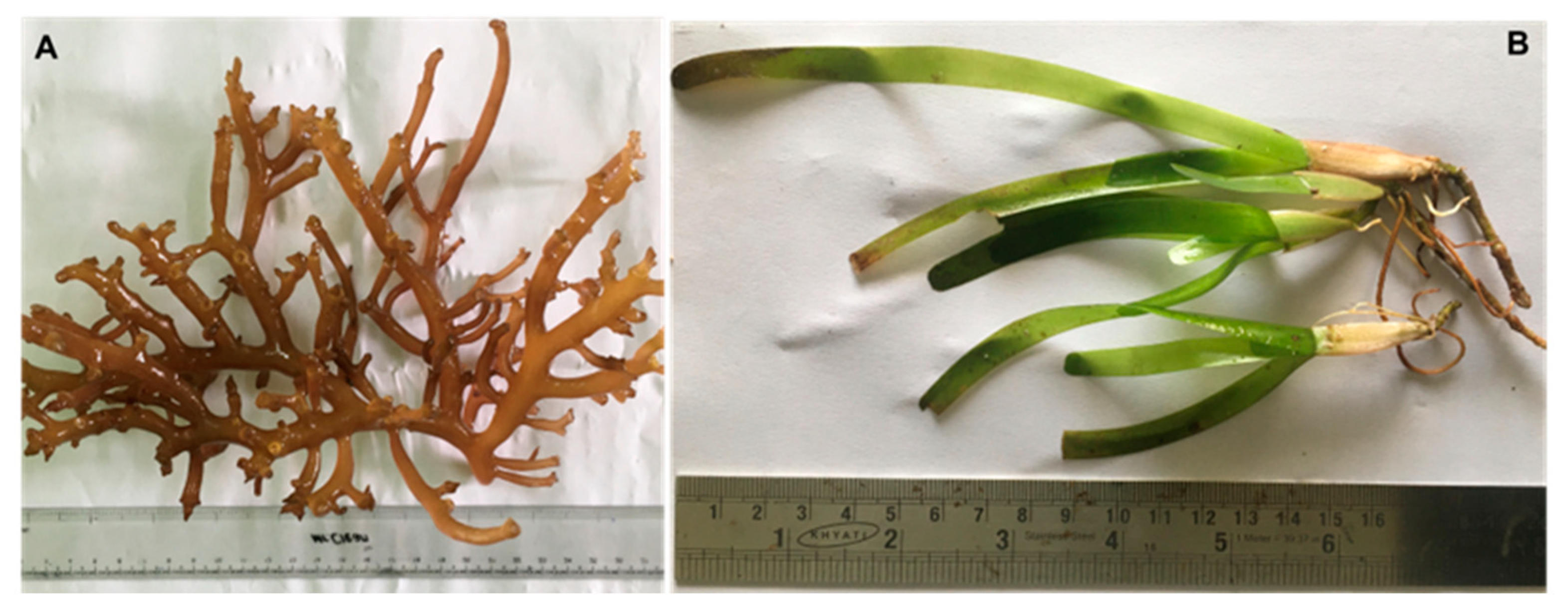
2.1. Collection, Identification and Processing

The red seaweed K. alvarezii and the seagrass C. serrulata were obtained from Thondi coastal waters (Latitude: 9°44″ N and Longitude: 79°00″ E) in Palk Bay, India. Freshly collected seaweed (K. alvarezii) and seagrass (C. serrulata) (Figure 1) were cleaned thoroughly in seawater and transported to the laboratory in a portable cooler that was protected from sunlight within 1 h of collection. The epiphytes, necrotic parts, muds, dust and other debris were removed via thorough washing with fresh water and double-distilled water. Next, they were shade-dried in an oven (Memmert, Roentgen, Germany) at 25 ± 2 °C for one week, ground into fine powder and stored at room temperature in an airtight container (Tarsons, Chennai, India) until analysis. The collected seaweed and seagrass were identified according to those standards established in the standard manual of Rao [22].

2.2. Preparation of Extracts

The seaweed and seagrass extracts were made by adding 5 g of dried seaweed or seagrass powder into 50 mL of three different solvents—chloroform, ethanol and distilled water—in a conical flask and placing the solution in a dark bottle in light agitation (AGIMATIC-N, J.P. SELECTA S. A, Barcelona, Spain) for 7 days. After that, the extracts were filtered through Whatman No. 1 filter papers and sterile cotton wools, and the supernatants were stored at 4 °C for future use [23,24].

2.3. Determination of Alkaloids

The alkaloid contents of K. alvarezii and C. serrulata were determined via the method proposed by Hikino et al. [25]. Next, 1 mL of test extract phosphate buffer (5 mL, pH 4.7) was added to 5 mL of bromocresol green solution, and the mixture was shaken vigorously added with 4 mL of chloroform. The extracts were collected in a 10-milliliter flask. The absorbance of the complex in chloroform was measured at 470 nm using a UV-Vis spectrophotometer (Shimadzu, Kyoto, Japan) against a prepared blank, as described above but without extract. Atropine (Sigma-Aldrich, St. Louis, MO, USA) was used as the standard, and the assay was compared to atropine equivalents.

2.4. Determination of Flavonoids

Total flavonoid content was determined via the aluminum chloride method [26] and using catechin (Sigma-Aldrich) as standard. Next, 1 mL of test sample and 4 mL of water were added to a volumetric flask (10-milliliter volume). After 5 min, 0.3 mL of 5% sodium nitrite and 0.3 mL of 10% aluminum chloride (Sisco Research Laboratories, Mumbai, India) were added. After 6 min of incubation at room temperature, 2 mL of 1-molarity sodium hydroxide (Sisco Research Laboratories) was added to the mixture. Afterwards, the final volume was brought to 10 mL via addition of distilled water. The absorbance of the reaction mixture was spectrophotometrically measured at 510 nm against a blank using a UV-Vis spectrophotometer (Shimadzu). The results obtained were expressed as catechin equivalents (mg catechin/g dried extract).

2.5. Determination of Tannins

The total tannin content extracts were determined according to the Julkunen–Titto [27] method. Firstly, 50-microliter extracts were mixed with 1.5 mL of 40% vanillin (Sisco Research Laboratories) (prepared with methanol), and 750 µL of HCl was then added. The solution was shaken vigorously and left at room temperature for 20 min in darkness. The absorbance of the mixtures was measured at 500 nm using a spectrophotometer (Shimadzu). A calibration curve was constructed using catechin (Sigma-Aldrich) in the range of 20–200 mg/L.

2.6. Determination of Phenolic Compounds

The total phenolic content in solvent extracts was determined using Folin–Ciocalteu’s reagent, as proposed by Sangeeta and Vrunda [28]. During the procedure, different concentrations of the extracts were mixed with 0.4 mL of Folin–Ciocalteu’s reagent (Sigma-Aldrich) (diluted 1:10 v/v). After 5 min, 4 mL of a sodium carbonate solution was added. The final volume of the tubes was brought to 10 mL by adding distilled water and left for 90 min at room temperature. The absorbance of the samples was measured against a blank sample at 750 nm using a spectrophotometer (Shimadzu). A calibration curve was constructed using 1,2-dihydroxybenzen (catechol) (Sigma-Aldrich) solutions as standards, and the total phenolic content of the extract was expressed in terms of the mg of catechol per g of dry weight.

2.7. Determination of Cardiac Glycosides

The cardiac glycoside content was determined using Buljet’s reagent based on the method reported by El-Olemy et al. [29]. Firstly, 1 g of the fine powder of K. alvarezii and C. serrulata was soaked in 10 mL of 70% MeOH for 2 h and filtered. The extract obtained was then purified using lead acetate and Na2HPO4 solution before the addition of freshly prepared Buljet’s reagent (containing 95 mL of aqueous Picric acid and 5 mL of 10% aqueous NaOH) (Sigma-Aldrich). The difference between the intensity of colors of the experiment and blank samples gives an absorbance of 217 nm using a spectrophotometer (Shimadzu), which was used to calculate the concentration of glycosides.

2.8. Determination of Steroids

The steroid content was determined by Ejikeme et al. [26]. Firstly, 1 mL of test extract of the steroid solution was transferred into 10-milliliter volumetric flasks. Sulfuric acid (Sisco Research Laboratories) (4 N, 2 mL) and iron (III) chloride (Sisco Research Laboratories) (0.5% w/v, 2 mL) were added, followed by potassium hexacyanoferrate (III) solution) (Sisco Research Laboratories) (0.5% w/v, 0.5 mL). The mixture was heated via a Memmert WTB water bath (Memmert, Schutzart, Germany) that was maintained at 70 ± 20 °C for 30 min, along with shaking, and afterwards diluted to the mark with distilled water. The absorbance was measured using a 780 nm spectrophotometer (Shimadzu) against the reagent blank.

2.9. Determination of Carbohydrates

Carbohydrate content was estimated based on the phenol-sulfuric acid method [30]. In brief, 200 mg of a powdered sample, which was weighed using an analytical balance (OHAUS GA200, Nänikon Switzerland), was hydrolyzed by adding 5 mL of 2.5 N HCl. The sample was kept in boiling water, and after 3 h, the solution was neutralized with solid Na2CO3 until effervescence ceased. The solution was made of up to 50 mL and centrifuged at 8000 rpm for 10 min in a centrifuge (Remi Lab World, Mumbai, India). Afterwards, the supernatant was aliquoted and brought up to 1 mL using deionized water, to which component 1 mL of phenol and 5 mL of 96% sulfuric acid (Sisco Research Laboratories) were previously added. After mixing the solution, it was kept in a water bath at 25 ± 1°C for 20 min. The absorbance was measured at 490 nm using a UV-Vis spectrophotometer (Shimadzu) against the reagent blank.

2.10. Ash Content

The ash content was determined using the method of Yemm and Willis [30]. Firstly, 2 g of each sample was taken and weighed accurately using a Cobos CB balance (Barcelona, Spain) in a clean silica dish. The dish was first heated over a low burner flame. Next the dish was transferred to a SNOL muffle furnace (Utena, Lithuania) maintained at 500–550 °C for 3–5 h. The ash residue obtained was then cooled in a desiccator and weighed on a balance. The percentage of total ash content was calculated via the following formula:
Total Ash Percent of plant sample (%) = [Weight of dry ash residue (g) ÷ Weight of plant sample (g)] × 100

2.11. Hydrogen Peroxide Radical Scavenging Activity

The antioxidant activity of seaweed and seagrass extracts was evaluated based on the hydrogen peroxide radical scavenging activity, as described by Ebrahimzadeh et al. [31]. The seaweed and seagrass extracts (100 µg/mL) were reacted with 0.6 mL of 40 mM H2O2 solution prepared in phosphate buffer (pH 7.4) (Sisco Research Laboratories). After incubation at 37 °C for 10 min, absorbance was measured at 230 nm using a UV-Vis spectrophotometer (Shimadzu). Phosphate buffer was used as the corresponding blank solution. A similar procedure was repeated using distilled water instead of the extract, which served as a control. Ascorbic acid (Sigma-Aldrich) (20–100 µg/mL) was used as a standard.

2.12. In Vitro Antibacterial Activity of Seaweed and Seagrass against Human Pathogenic Bacteria

The antibacterial activity of seaweed and seagrass extracts was evaluated via the well diffusion method using a Muller–Hinton agar (Hi-Media, Mumbai, India). Approximately 100 µL of 105 CFU/mL of diluted inoculum of bacterial culture was applied to the surface of Muller–Hinton agar plates. The Muller–Hinton agar well was made using a well borer under aseptic conditions and filled with K. alvarezii and C. serrulata extracts, and methanol served as a positive control. The plates were incubated at 37 °C for bacterial growth, and the antibacterial activity of the seaweed and seagrass samples was evaluated by measuring the zone of inhibition (mm) in relation to the tested pathogenic bacteria. All experiments were performed in triplicate, and the data are expressed as the mean values of the experiments.

2.13. Characterization of the Active Compound by Gas Chromatography-Mass Spectrometry (GC-MS)

The crude extracts of K. alvarezii and C. serrulata were loaded into a silica gel (Hi-Media) packed column (20 cm length and 2 cm diameter) and eluted using n-hexane: ethyl acetate (50:50 v/v) (Sigma-Aldrich). The fractions were analyzed via a gas chromatograph GC-2010 interfaced with a quadrupole mass spectrometer QP-2010 (Shimadzu, Japan) analyzer, which used an Rtx-PCB capillary column (60 m × 0.25 mm i.d., 0.25 mm film thickness, Resteck, Bellefonte, PA, USA). Helium with a purity of 99.99% was used as the carrier gas at a flow rate of 1 mL/min. Next, 1 mL of extract was injected in spilt mode using an autosampler (Shimadzu). The injector port, interface and ion source temperature were set at 250, 270 and 230 °C, respectively. The mass spectrometer was operated in electron ionization (EI) mode at 70 eV and at an emission current of 60 mA. Full scan data were obtained in a mass range of 50–500 m/z. Interpretation of mass spectrum data was performed using the National Institute Standard and Technology (NIST) database.

2.14. Statistical Analysis

All determinations were given in terms of the mean ± standard deviation (SD). The results obtained were compared via one-way analysis of variance (ANOVA). The significance of the difference between means was determined via Duncan’s multiple range test (p < 0.05) using SPPS version 14 (Chicago, IL, USA).

3. Results and Discussion

3.1. Phytochemical Analysis

Phytochemical analysis of K. alvarezii and C. serrulata revealed the presence of alkaloids (only in the case of K. alverazii), flavonoids, tannins, phenolic compounds, glycosides, steroids, carbohydrates and ashes. Among the six phytochemicals present in K. alvarezii, higher contents were found for phenolic compounds (3.39 ± 0.41 mg/g) and tannins (2.94 ± 0.41 mg/g). Both phenolic compounds and tannins have important roles as bioactive compounds. Phenols have important antioxidant, antimicrobial, anti-inflammatory and anticancer activities [32,33], whereas tannins are reported to have antiviral, antibacterial and antioxidant activities [34,35]. Among the five phytochemicals present in C. serrulata, the highest contents were found for glycosides (2.47 ± 0.41 mg/g) and flavonoids (2.11 ± 1.40 mg/g) (Table 1). With respect to glycosides, it was reported that they have antioxidants and anti-inflammatory activities, which find application in the prevention and managements of several human diseases [34]. Flavonoids also make up an important phytochemical group due to their antimicrobial, antiviral, antioxidant and spasmolytic activities [35]. These constituents significantly contribute to the biological activity of seaweeds and seagrass [36]. Similar observations were also made by other works [37,38], which found tannins, flavonoids, phenolic compounds, carotenoids and polysaccharides in both seaweed and seagrasses.
In the present study, K. alvarezii showed a higher tannin content (2.94 ± 0.41 mg catechin equivalent (CAE)/g) than C. serrulata (1.94± 0.85 mg CAE/g). Similarly, Deyad and Ward [35] reported similar tannin content in the brown seaweed Dictyota dichotoma (2.12 ± 0.45 mg CAE/g), whereas Domettila et al. [32] reported a higher presence of tannins in the red seaweed Sargassum wightii (27.54 ± 0.54 mg CAE/g). In previous studies [9], the presence of tannins in C. serrulata (264.71 mg/mL tannic acid equivalence) was reported. Similarly, another work reported the presence of tannins in the seagrass Syringodium isoetifolium (80.65 ± 5.64 mg CAE/g [39]. Tannins are polyphenols, which have a large influence on the nutritive value of humans and animals due to their antimicrobial, anti-inflammatory, and astringent activities [9].
Flavonoid content was similar in both K. alvarezii and C. serrulata, although in global terms, it was found in lower amounts than in previous works. Vaghela et al. [40] found a much higher flavonoid content (15.26 ± 0.95 mg CAE 100 g−1). Similarly, Smadi et al. [41] reported the flavonoid content of C. nodosa to be 3.98 ± 0.03 mg CAE/g, which is comparatively higher than the results of the present study.
K. alvarezii had an alkaloid content of 1.91 ± 0.58 mg CAE/g. Similarly, Domettila et al. [32] showed an alkaloid content of 1.32 ± 0.02 mg CAE/g in Ulva reticulata. Previously, Alghazeer et al. [42] reported the alkaloid content in the brown algae species Cystoseira compressa and Sargassum hornschuchii to be 4125.00 ± 180.28 mg/g DW and 3708.33 ± 152.75 mg/g DW, respectively. Alkaloids have been proven to have antiplasmodic, antimicrobial, and cytotoxic properties [42].
The phenolic compound content in seaweeds is, in part, responsible for their scavenging activity, which protects them against lipid oxidation [43]. In this work, K. alvarezii showed a higher phenolic content (3.39 ± 0.45 mg gallic acid equivalents (GAE)/g) than C. serrulata (1.01 ± 0.39 mg GAE/g). Previously, other authors reported a significantly higher content of phenolic compounds in both K. alvarezii (3.14 ± 0.14 mg GAE/g) [44] and Kappaphycus striatum (7.24 ± 0.21 mg GAE/g) [45]. Regarding C. serrulata, the results obtained in the current work were also significantly lower than those reported by Libin et al. [17] for C. serrulata (2.98 ± 0.12 mg GAE/g) and Cynodocea rotundata (2.04 ± 0.1) [46]. The phenolic contents of seaweed and seagrass depend on the solvent used to analyze the extraction process, environment, habitat and biomass.
The presence of steroids in seaweed K. alvarezii (2.51 ± 0.15 mg/g) was higher than that recorded in seagrass C. serrulata (1.60 ± 0.24 mg/g). Previously, another study showed that the presence of steroids in seaweed C. elongata was 2.27 ± 0.26 mg/g [47]. Kumar et al. [48] also reported the presence of steroids in Champai parvula (24.30 ± 0.11 mg/g). Previously, the presence of steroids in Himanthalia elongata (2.64 ± 2.21 mg/g) was reported [49]. In previous studies, Kannan et al. [50] reported the presence of steroids in C. rotundata (2.37 ± 1.27 mg/g). Similarly, Tango et al. [51] also reported the presence of steroids in the seagrass Haludole pinifolia (5.62 ± 0.76 mg/g). Steroids isolated from seaweed and seagrass have medicinal values, such as antihelmintic, antioxidant, antimicrobial and antiviral activities [52].
K. alvarezii showed a glycoside content of 1.88 ± 0.11 mg/g, while in C. serrulate, glycoside content was reported to be 2.47 ± 0.28 mg/g. Previously, Kumar et al. [48] reported the presence of glycosides in seaweed Cymodocea parvula (35.33 ± 0.14 mg/g). Similarly, Prabakaran et al. [52] reported the presence of glycosides in Chorella vulgaris (5.75 ± 0.23 mg/g). Deyad and Ward [35] also reported the presence of glycosides in the seaweed D. dichotoma (2.14 ± 0.15 mg/g). A previous work performed by Regalado et al. [53] reported the presence of glycosides in Thalassia testudinum (4.61 ± 1.60 mg/g). Glycosides are well known for being able to lower blood pressure in humans [48].
With respect to carbohydrates and ash content, the carbohydrate content of K. alvarezii was 2.57 ± 1.89 mg/g DW, while that of C. serrulata was 1.44 ± 1.75 mg/g DW. The wide variation in the carbohydrate content observed between seaweed and seagrass might be due to the influence of different factors, such as salinity, temperature and sunlight intensity. Regarding ash, K. alvarezii had a higher ash content (8.5 ± 0.95 g/100 g) than C. serrulata (6.9 ± 0.49 g/100 g). High ash content showed the presence of appreciable amounts of diverse minerals found in both seaweed and seagrass.

3.2. Antioxidant Activity

Antioxidant effectiveness is measured by monitoring the inhibition of oxidation of a suitable substrate [15]. In biological systems, antioxidant effectiveness is classified into two groups: evaluation of lipid peroxidation and measurement of free radical scavenging ability [31]. The in vitro antioxidant activiies of K. alvarezii and C. serrulata extracts were evaluated based on hydrogen peroxide radical scavenging activity, and K. alvarezii had higher scavenging activity (27.9 ± 0.1%) than C. serrulata (22.1 ± 0.1%). Regarding K. alvarezii, the results obtained were higher than those previously reported by other authors, such as Farah et al. [37] or Chew et al. [54], who reported lower (18.34 ± 0.57% and 11.8 ± 5.7%, respectively) 2,2-Diphenyl-1-picrylhydrazyl (DPPH) scavenging activity. Regarding C. serrulata, the DPPH scavenging activity results obtained were lower than those obtained by Kannan et al. [50] (61.85 ± 0.95%) regarding free radical scavenging activity from the same seagrass species, though higher than those results reported by Rengasamy et al., [33] (6.65 ± 0.12%) for other Cymodocea species, such as C. rotundata.

3.3. Antimicrobial Activity

The antibacterial activity of both K. alvarezii and C. serrulata were investigated using chloroform extracts based on those reported by Pusparaj et al. [14], who reported that the best inhibitory effects of K. alvarezii were reported using chloroform extracts. The antibacterial activities of both K. alvarezii and C. serrulata depend on the presence of bioactive compounds, phenolic content and free radical scavenging activity [55]. In all cases, inhibitory activities against the five pathogenic bacteria investigated were detected (Table 2). The higher inhibitory activity was observed in K. alvarezii (26 ± 0.03 mm) against Bacillus subtilis, as well as in the case of C. serrulate, which exhibited maximum inhibitory activity (26 ± 0.08 mm) against Vibrio parahaemolyticus. The chloroform extract of K. alvarezii showed maximum activity of 26 ± 0.03 mm against B. subtilis at 100 µg/mL, and C. serrulata showed maximum activity of 26 ± 0.08 mm against V. parahaemolyticus at 100 µg/mL and minimum activity of 22 ± 0.01 mm and 20 ± 0.04 mm against Vibrio alginolyticus at 100 µg/mL in both K. alvarezii and C. serrulata, respectively (Table 2).
Jaswir et al. [55] reported maximum inhibitory activity (12 ± 1.02 mm) against B. subtilis using the methanolic extract of K. alvarezii. Similarly, Pusparaj et al. [14] reported the antibacterial activity of K. alvarezii against six human pathogens: Staphylococcus aureus, B. subtilis, Lactobacillus acidophilus, Pseudomonas aeruginosa, Escherichia coli and Proteus mirabillis. He also reported that the best activity was recorded in chloroform extracts. Kumar et al. [56] studied the antibacterial activity of C. serrulata against four fish-borne pathogens, namely Bacillus cereus, B. subtilis, E. coli and Micrococcus luteus, and reported that C. serrulata was effective against several Bacillus species.

3.4. Presence of Bioactive Compounds

The GC-MS running time for the n-hexane:ethyl acetate (50:50 v/v) extracts of K. alvarezii and C. serrulata was 30 min. The target mass ions (m/z) and retention times (min) of all identified compounds in K. alvarezii and C. serrulata are shown in Table 3 and Table 4. The results show that K. alvarezii extracts contained 94 different bioactive compounds, including phenol, decane, dodecane, hexadecane, vanillin, heptadecane, diphenylamine, benzophenone, octadecanoic acid, dotriacontane and benzene (Table 3). On the other hand, C. serrulata was found to contain 104 different bioactive compounds, including tetradecane, dodecanal, diphenylamine, heptadecane, phytol, butanoic acid, 2-hydroxy-, ethyl ester, dodecane and benzene (Table 4). These compounds were responsible for the antioxidant and antibacterial activities of both K. alverazii and C. serrulata.
Datchanamurthy et al. [57] reported that red algae (Acoathophora deilei) contain major common components, such as hexadecanoic acid methyl ester, dibutyl phthalate, 2-ethyl butyric acid, octadecyl ester, 9-octadecanoic acid, methyl ester and 1,2-benzendicarboxylic acid. Similarly, Anitha et al. [36] also studied the presence of phenols, hexadecanoic acid, n-hexadecanoic acid, tridecanoic acid, n-nonadecanoic acid and benzene reported to be present in red algae (Gracilaria cervicornis). Pushpabharathi et al. [9] reported that nine bioactive components were present in seagrass (C. serrulata): hexahydofarnesyl acetone, hexadecanoic acid, methyl ester, n-hexadecanoic acid, tetradecanoic acid, pentadecanoic acid, cholestesta 4,6 dien 3-ol and stigmasterol.

4. Conclusions

The red seaweed K. alvarezii and seagrass C. serrulata examined in the present study were found to possess rich sources of phytochemicals. The antioxidant properties of both seaweed and seagrass reveal that they have appreciable levels of protection against free radicals.
GC-MS analysis revealed the presence of large active metabolites (94 in the case of K. alvarezii, and 104 in the case of C. serrulate), such as phenol, decane, dodecane, hexadecane, vanillin, heptadecane, diphenylamine, benzophenone, octadecanoic acid, dotriacontane and benzene, in both red seaweed and seagrass. In view of the results obtained, both K. alvarezii and C. serrulata could be employed as potential marine-sourced drugs and may be used in the pharmaceutical and food processing industries as sources of ingredients with appreciable medicinal value. Since both red seaweed and seagrass were found to be good sources of essential phytochemicals, their commercial value can be enhanced by marketing them to consumers as value-added products.

Author Contributions

Conceptualization, S.P.; formal analysis, D.D. and A.A.; investigation, D.D. and S.P.; data curation, S.P.; literature data collection, A.L.-S. and S.P.; writing—original draft, A.A. and D.D.; writing—review and editing, A.d.C.M. and A.L.-S. supervision, S.P. and J.M.M.L. All authors have read and agreed to the published version of the manuscript.

Funding

The authors thank the European Regional Development Fund (FEDER), grant ED431C 2018/05, for covering the cost of publication.

Institutional Review Board Statement

Not applicable.

Informed Consent Statement

Not applicable.

Data Availability Statement

The data used to support the findings of this study can be made available by the corresponding author upon request.

Acknowledgments

The authors (D.D and S. P) acknowledge RUSA–2.0 [F.24–51/2014–U, Policy (TNMulti–Gen), Department of Education, Government of India] for providing infrastructure facilities. A.L.S. wishes to acknowledge her predoctoral fellowship from Campus Terra (Lugo).

Conflicts of Interest

The authors declare no conflict of interest.

References

  1. Ebrahimi, B.; Baroutian, S.; Li, J.; Zhang, B.; Ying, T.; Lu, J. Combination of marine bioactive compounds and extracts for the prevention and treatment of chronic diseases. Front. Nutr. 2023, 9, 1047026. [Google Scholar] [CrossRef]
  2. Cheung, R.C.F.; Ng, T.B.; Wong, J.H.; Chen, Y.; Chan, W.Y. Marine natural products with anti-inflammatory activity. Appl. Microbiol. Biotecnol. 2016, 100, 1645–1666. [Google Scholar] [CrossRef]
  3. Miranda, J.M.; Trigo, M.; Barros-Velazquez, J.; Aubourg, S.P. Effect of an icing medium containing the alga Fucus spiralis on the microbiological activity and lipid oxidation in chilled megrim (Lepidorhombus whiffiagonis). Food Cont. 2016, 59, 290–297. [Google Scholar] [CrossRef] [Green Version]
  4. Lopez-Santamarina, A.; Miranda, J.M.; Mondragón, A.C.; Lamas, A.; Cardelle-Cobas, A.; Franco, C.M.; Cepeda, A. Potential use of marine seaweeds as prebiotics: A review. Molecules 2020, 25, 1004. [Google Scholar] [CrossRef] [Green Version]
  5. Kim, D.H.; Mahomoodally, M.F.; Sadeer, N.B.; Seok, P.G.; Zengin, G.; Palaniveloo, K.; Khalil, A.A.; Rauf, A.; Rengasamy, K.R.R. Nutritional and bioactive potential of seagrasses: A review. S. Afr. J. Bot. 2021, 137, 216–227. [Google Scholar] [CrossRef]
  6. Anjum, A.; Aruna, G.; Noorjahan, C.M. Phytochemical analysis and antibacterial activity of selected seaweeds from coast of Mandapam, Tamilnadu. Indian J. Appl. Microbiol. 2014, 17, 50–58. [Google Scholar]
  7. Ragunath, C.; Santhosh Kumar, Y.A.; Kanivalan, I.; Radhakrishnan, S. Phytochemical screening and GC-MS analysis of bioactive constituents in the methanolic extract of Caulerpa racemosa (Forssk.) J. Agardh and Padina boergesenii Allender & Kraft. Curr. Appl. Sci. Technol. 2020, 20, 380–393. [Google Scholar]
  8. Balachandran, P.; Anson, S.M.; Ajay, K.T.V.; Parthasarathy, V. Preliminary phytochemical analysis of the ethanolic extract of brown Seaweed Sargassum wightii. Int. J. Res. Pharm. Sci. 2016, 7, 154–156. [Google Scholar]
  9. Pushpabharathi, N.; Jayalakshmi, M.; Amudha, P.; Vanitha, V. Identification of bioactive compounds in Cymodocea serrulata seagrass by Gas Chromatography–Mass Spectroscopy. Asian J. Pharm. Clin. Res. 2018, 11, 317–320. [Google Scholar]
  10. Govindasamy, C.; Arulpriya, M.; Anantharaj, K.; Ruban, P.; Srinivasan, R. Seasonal variations in seagrass biomass and productivity in Palk Bay, Bay of Bengal, India. Int. J. Biodivers. Conserv. 2013, 5, 408–417. [Google Scholar]
  11. Zulkifli, L.; Muksin, Y.D.; Hartanto, P.; Desimarlina, Y.; Idrus, A.A.; Syukur, A. Phytochemical profiles and ethnomedicine preliminary studies on seagrass species in the Southern Coast of Lombok Island Indonesia. Environ. Earth Sci. 2021, 913, 012102. [Google Scholar] [CrossRef]
  12. Manivannan, K.; Kathiga Devi, G.; Ananthararaman, P.; Balasubramanian, T. Antimicrobial potential of selected brown seaweeds from vedalia coastal waters, Gulf of Mannar. Asian Pac. J. Trop. Biomed. 2011, 1, 114–120. [Google Scholar] [CrossRef] [Green Version]
  13. Chin, Y.X.; Mi, Y.; Cao, W.X.; Lim, P.E.; Xue, C.H.; Tang, Q.J. A pilot study on anti-obesity mechanisms of Kappaphycus alvarezii: The role of native κ-carrageenan and the leftover sans-carrageenan fraction. Nutrients 2019, 11, 1133. [Google Scholar] [CrossRef] [Green Version]
  14. Pushparaj, A. Antibacterial activity of Kappaphycus alvarezii and Ulva lactuca extracts against human pathogenic bacteria. Int. J. Curr. Microbiol. Appl. Sci. 2014, 3, 432–436. [Google Scholar]
  15. Bharathi, N.P.; Jayalakshmi, M.; Amudha, P.; Vanitha, V. Phytochemical screening and in vitro antioxidant activity of the seagrass Cymodocea serrulata. Indian J. Mar. Sci. 2019, 48, 1216–1221. [Google Scholar]
  16. Libin, B.; Sankar, T.V.; Chandramohana, K.N. Changes in phenolic compounds in seagrasses against changes in the ecosystem. J. Pharmacogn. Phytochem. 2017, 6, 742–747. [Google Scholar]
  17. Adharini, R.; Setyawan, A.R.; Suadi, S.; Jayanti, A.D. Comparison of nutritional composition in red and green strains of Kappaphycus alvarezii cultivated in Gorontalo Province, Indonesia. J. Appl. Phycol. 2020, 31, 725–730. [Google Scholar] [CrossRef]
  18. Prasad, M.P.; Shekhar, S.; Babhulkar, A.P. Antibacterial activity of seaweed (Kappaphycus) extracts against infectious pathogens. Afr. J. Biotechnol. 2013, 12, 2968–2971. [Google Scholar]
  19. Rupert, R.; Rodrigues, K.F.; Thien, V.Y.; Lym Yong, W.T. Carrageenan from Kappaphycus alvarezii (Rodophyta, Soliariaceae): Metabolism, structure, production, and application. Front. Plant. Sci. 2022, 13, 859635. [Google Scholar] [CrossRef]
  20. Dilipan, E.; Arulbalachandran, D. Genetic diversity of seagrass Cymodocea species as an ecological indicator on the Palk Bay Coast, India. Ecol. Genet Genom. 2022, 23, 100119. [Google Scholar] [CrossRef]
  21. Sanchez-Arcos, C.; Paris, D.; Mazzella, V.; Mutalipassi, M.; Costantini, M.; Buia, M.C.; von Elert, E.; Cutignano, A.; Zupo, V. Responses of the macroalga Ulva prolifera Müller to ocean acidifications revealed by complementary NMR- and MS-based omics approaches. Mar. Drugs 2023, 20, 743. [Google Scholar] [CrossRef]
  22. Rao, M. Key for identification of economical important seaweeds. Cent. Mar. Fish. Res. Inst. Bull. 1987, 41, 116. [Google Scholar]
  23. Harborne, J.B. Phytochemical Methods—A Guide to Modern Techniques of Plant Analysis; Chapman and Hall: London, UK, 1998; Volume 5, pp. 21–27. [Google Scholar]
  24. Arulkumar, A.; Thomas, R.; Paramasivam, S. Phytochemical composition, in vitro antioxidant, antibacterial potential, and GC-MS analysis of red seaweeds (Gracilaria corticata and Gracilaria edulis) from Palk Bay, India. Biocatal. Agric. Biotechnol. 2018, 15, 63–71. [Google Scholar] [CrossRef]
  25. Hikino, H.; Kiso, Y.; Wagner, H.; Fiebig, M. Antihepatotoxic actions of flavonolignans from Silybum marianum. Planta Med. 1984, 50, 248–250. [Google Scholar] [CrossRef]
  26. Ejikeme, C.M.; Ezeonu, C.S.; Eboatu, A.N. Determination of physical and phytochemical constituents of some tropical timbers indigenous to Niger Delta area of Nigeria. Eur. Sci. J. 2014, 10, 247–270. [Google Scholar]
  27. Julkunen-Titto, R. Phenolic constituents in the leaves of northern willows. Methods for the analysis of certain phenolics. J. Agric. Food Chem. 1985, 33, 213–217. [Google Scholar] [CrossRef]
  28. Sangeeta, S.; Vrunda, V. Quantitative and qualitative analysis of phenolic and flavonoid content in Moringa oleifera. Pharmacogn. Res. 2016, 8, 16–21. [Google Scholar]
  29. El-Olemy, M.M.; Al-Muhtadi, F.J.; Afifi, A.F.A. Experimental Phytochemistry: A Laboratory Manual; King Saud University Press: Riyadh, Saudi Arabia, 1994; pp. 21–27. [Google Scholar]
  30. Yemm, E.W.; Willis, J. The estimation of carbohydrates in plant extracts by anthrone. Biochem. J. 1954, 57, 508–514. [Google Scholar] [CrossRef] [Green Version]
  31. Ebrahimzadeh, M.A.; Nabavi, S.M.; Nabavi, S.F.; Bahramian, F.; Bekhradnia, A.R. Antioxidant and free radical scavenging activity of H. officinalis L. var. angustifolius, V. odorata, B. hyrcana and C. speciosum. Pak. J. Pharm. Sci. 2010, 23, 29–34. [Google Scholar]
  32. Domettila, C.; Joselin, J.; Jeeva, S. Phytochemical analysis on some south Indian seaweeds. J. Chem. Pharm. Res. 2013, 5, 275–278. [Google Scholar]
  33. Rengasamy, R.R.K.; Arumugam, R.; Perumal, A. Seagrasess as potential source of medicinal food ingredients: Nutritional analysis and multivariate approach. Biomed. Prev. Nutr. 2013, 3, 375–380. [Google Scholar] [CrossRef]
  34. Kytidou, K.; Artola, M.; Overkleeft, H.S.; Aerts, J.M.F.G. Plant glycosides and glycosidades: A treasure-trove for therapeutics. Front. Plant. Sci. 2020, 11, 357. [Google Scholar] [CrossRef] [Green Version]
  35. Deyad, M.; Ward, F. Qualitative and quantitative analysis of phytochemical studies on brown seaweed, Dictyota dichotoma. Int. J. Eng. Develop. Res. 2016, 4, 674–678. [Google Scholar]
  36. Anitha, K.G.; Arputha, G.; Muthubala, G.; Susithra, R.; Mullaivendhan, M.; Anandham, R. GC-MS Analysis of bioactive compounds of seaweed extracts collected from seashore of Manalmelkudi (Pudukkottai dist., Tamilnadu), responsible for antifungal activity. Int. J. Curr. Microbiol. App. Sci. 2019, 8, 2319–7706. [Google Scholar] [CrossRef]
  37. Sheela, D.; Uthayakumari, F. GC-MS analysis of bioactive constituents from coastal sand dune taxon—Sesuvium portulacastrum (L.). Biosci. Dis. 2013, 4, 47–53. [Google Scholar]
  38. Mahabaleshwara, K.; Chandrasekhar, N.; Govindappa, M. Phytochemical investigations of methanol leaf extracts of Randia spinosa using column chromatography, HPTLC and GC-MS. Nat. Prod.Chem. Res. 2016, 4, 2. [Google Scholar]
  39. Kalaivani, P.; Kavitha, D.; Amudha, P. In vitro antioxidant activity and phytochemical composition of Syringodium isoetifolium. Res. J. Pharm. Technol. 2021, 14, 6201–6206. [Google Scholar]
  40. Vaghela, P.; Das, A.K.; Trivedi, K.; Vijay, K.G.; Shinde, P.; Ghosh, A. Characterization and metabolomics profiling of Kappaphycus seaweed extract. Algal Res. 2022, 66, 1022774. [Google Scholar] [CrossRef]
  41. Smadi, A.; Civatta, M.L.; Bitam, F.; Carbone, M.; Villani, G.; Gavagnin, M. Flavonoids and phenolic compounds from the Cymodocae nodosa. Plant Med. 2018, 84, 704–709. [Google Scholar]
  42. Alghaweer, R.; Azwai, S.; Garbaj, A.M.; Amr, A.; Elghmasi, S.; Sidati, M.; Yudiati, E.; Kubbat, M.G.; Eskandrani, A.A.; Shamlan, G.; et al. Alkaloids rich extracts from brown algae against multidrug-resistant bacteria by distinctive mode of action. Arab. J. Sci. Eng. 2021, 47, 179–188. [Google Scholar] [CrossRef]
  43. Chan, P.T.; Matanjun, P.; Yasir, S.M.; Tan, T.S. Antioxidant activities and polyphenolics of various solvent extracts of red seaweed, Gracilaria changii. J. Appl. Phycol. 2015, 27, 2377–2386. [Google Scholar] [CrossRef]
  44. Farah, D.; Abdullah, A.; Shahrul, H.; Chan, K. Antioxidant activity of red algae Kappaphycus alvarezii and Kappaphycus striatum. Int. Food Res. J. 2015, 22, 1977–1984. [Google Scholar]
  45. Araújo, P.G.; Nardelli, A.E.; Fujii, M.T.; Chow, F. Antioxidant properties of different strains of Kappaphycus alvarezii (Rhodophyta) farmed on the Brazilian coast. Phycologia 2020, 53, 272–279. [Google Scholar] [CrossRef]
  46. Dumay, O.; Costa, J.; Desiobert, J.M.; Pergent, G. Variations in the concentrations of phenolic compounds in the seagrass Cymodocae rotundata under conditions of competition. Phytochemistry 2004, 65, 3211–3220. [Google Scholar]
  47. Devi, K.N. Antibacterial activity of seagrass species of Cymodocea serrulata against chosen bacterial fish pathogens. Ann. Biol. Res. 2011, 2, 88–93. [Google Scholar]
  48. Kumar, V.; Murugesan, S.; Bhuvaneswari, S. Phytochemical analysis of red alga Champia parvula (C. Agardh) collected from Mandapam coast of Tamil Nadu, India. Int. J. Adv. Pharm. 2015, 4, 15–20. [Google Scholar]
  49. Sanchez, M.; Lopez, D.L.; Seiro, J.P.L. An HPLC method for the quantification of sterols in edible seaweeds. Biomed. Chromatogram. 2004, 18, 183–190. [Google Scholar] [CrossRef]
  50. Kannan, R.R.; Arumugam, R.; Lyapparaj, P.; Thangaradjou, T.; Anantharaman, P. In vitro antibacterial, cytotoxicity and haemolytic activities and phytochemical analysis of seagrasses from the Gulf of Mannar, South India. Food Chem. 2013, 136, 1484–1489. [Google Scholar] [CrossRef]
  51. Tango, E.; Canencia, O.; Del Rosario, R.M. Phytochemical screening and proximate composition of the seagrass Halodule pinifolia of the coastal waters of Carmen, Agusan Del Norte, Philippines. Int. J. Modern Pharm. Res. 2021, 5, 75–80. [Google Scholar]
  52. Prabakaran, G.; Moovendhan, M.; Arumugam, A.; Matharasi, A.; Dineshkumar, R.; Sampathkumar, P. Quantitative analysis of phytochemical profile in marine microalgae Chlorella vulgaris. Int. J. Pharm. Biol. Sci. 2018, 8, 562–565. [Google Scholar]
  53. Regalado, E.L.; Menendez, R.; Valdés, O.; Morales, R.A.; Laguna, A.; Thomas, O.P.; Hernandez, Y.; Nogueiras, C.; Kijjoa, A. Phytochemical analysis and antioxidant capacity of BM-21, a bioactive extract rich in polyphenolic metabolites from the Sea Grass Thalassia testudinum. Nat. Prod. Commun. 2011, 7, 47–50. [Google Scholar] [CrossRef]
  54. Chew, Y.L.; Lim, Y.Y.; Omar, M.; Khoo, K.S. Antioxidant activity of three edible seaweeds from two areas in South East Asia. LWT-Food Sci. Tecnol. 2008, 41, 1067–1072. [Google Scholar] [CrossRef]
  55. Jaswir, I.; Tawakalit Tope, A.H.; Raus, R.A.; Monsur, H.A.; Ramli, N. Study on antibacterial potentials of some Malaysian brown seaweeds. Food Hydrocoll. 2014, 42, 275–279. [Google Scholar] [CrossRef]
  56. Kumar, C.S.; Sarada, D.V.L.; Gideon, T.P.; Rengasamy, R. Antibacterial activity of three South Indian seagrasses, Cymodocea serrulata, Halophila ovalis and Zostera capensis. World J. Microbiol. Biotechnol. 2008, 24, 1989–1992. [Google Scholar] [CrossRef]
  57. Datchanamurthy, B.; Narayanamurthy, U.; Anandh, S.J.V. Preliminary phytochemical and GC-MS analysis of marine seaweed Acathophora deilei (Red alga). Biomed. Pharmacol. J. 2022, 15, 1695–1707. [Google Scholar]
Figure 1. Images of Kappaphycus alvarezii (A) and Cymodocea serrulata (B), respectively.
Foods 12 02811 g001
Table 1. Steroids, tannins, flavonoid, glycosides, alkaloids and phenolic compounds of Kappaphycus alvarezii and Cymodocea serrulata.
ParametersK. alveraziiC. serrulata
Alkaloids (ATE/g dry wt)1.91 ± 0.58 *-
Flavonoids (CAE/g dry wt)1.63 ± 0.732.11 ± 1.40
Tannins (CAE/g dry wt)2.94 ± 0.41 *1.94 ± 0.85
Phenolic compounds (GAE/g dry wt)3.39 ± 0.45 *1.01 ± 0.39
Glycosides (mg/g dry wt)1.88 ± 0.112.47 ± 0.28 *
Steroids (mg/g dry wt)2.51 ± 0.15 *1.60 ± 0.24
Carbohydrates (% DW)2.57 ± 1.891.44 ± 1.75
Ash (% DW)8.5 ± 0.956.9 ± 0.49
Antioxidant activity27.9 ± 0.122.1 ± 0.1
Values are means of three analyses of the extracts ± standard deviation (n = 3); CAE: catechin equivalent; GAE: gallic acid equivalents; ATE: atropine equivalent. * Average values are significantly higher than counterparts (p < 0.05).
Table 2. Antibacterial activity of K. alvarezii and C. serrulata in chloroform extract against human pathogenic bacteria.
Human Pathogenic BacteriaConcentration (µg/mL)Seaweed ExtractSeagrass Extract
Zone of Inhibition (mm)
Bacillus subtilis10026 ± 0.0325 ± 0.16
Klebsiella pneumoniae10023 ± 0.0122 ± 0.20
Vibrio alginolyticus10022 ± 0.0120 ± 0.04
Vibrio parahaemolyticus10024 ± 0.0226 ± 0.08
Vibrio harveyi10024 ± 0.1022 ± 0.01
Date were expressed as the mean ± SD values of triplicates (n = 3).
Table 3. List of compounds identified by analyzing the purified extracts of K. alvarezii using GC-MS analysis.
No.NameRetention Time (min)Base m/z
1Phenol4.11994.05
2Cyclopropyl methyl carbinol4.17058.05
3Decane4.34757.05
4Butanoic acid, 2-hydroxy-, ethyl ester4.42959.05
52-Methylpentyl formate4.45756.05
6Benzene, 1,4-dichloro-4.530145.95
7Cyclopentane, 1,2-dimethyl-, cis-4.56870.10
8Dodecane, 2,6,11-trimethyl-5.15857.05
9Undecane, 5-methyl-5.23757.10
10Ethane, hexachloro-5.395116.90
11Dodecane, 2,6,10-trimethyl-5.80357.05
123-Ethyl-3-methylheptane5.89057.05
13Naphthalene6.994128.10
14Dodecane7.20157.05
15Benzaldehyde, 2,5-dimethyl-7.450133.10
16Octadecanoic acid, phenyl ester7.51794.05
17Benzene, 1,3-bis(1,1-dimethylethyl)-7.986175.15
18Undecane, 2,4-dimethyl-8.09357.10
19Dodecane, 4,6-dimethyl-8.31757.05
20Hexadecane8.43757.10
21Formamide, N-phenyl-8.884121.05
22Dodecane, 2,6,10-trimethyl-8.94257.10
23Chloroxylenol9.762121.10
24Benzene, 1-cyclobuten-1-yl-9.844129.10
25Hexadecane9.90757.05
26Vanillin9.950151.05
27Heptadecane10.18657.05
28Dodecane, 2,6,10-trimethyl-10.69057.10
291-Dodecanol10.86455.05
30Nonadecane11.00585.10
31Decane, 1-bromo-2-methyl-11.04457.05
32Heneicosane11.11371.10
33Nonadecane11.15957.05
342,4-Di-tert-butylphenol11.351191.15
35Hexadecane11.65557.05
36Hexadecane12.35057.05
37Diphenylamine12.664169.15
38Benzophenone12.754105.05
393-Hydroxydiphenylamine13.216185.10
40Hexadecane, 2,6,10,14-tetramethyl-13.32857.10
41Heptadecane13.47657.05
42Dodecane, 2,6,10-trimethyl-13.54071.10
43Heneicosane13.59071.10
44Heneicosane13.68157.10
45Decane, 1-iodo-13.75171.10
46Heptadecane, 8-methyl-13.94071.10
47Heneicosane14.06171.10
48Tetradecanoic acid14.14857.05
49Formamide, N,N-diphenyl-14.487168.10
50Heneicosane14.54957.05
51p-(Benzylideneamino)phenol14.651196.10
52Isopropyl myristate14.83660.00
53Carbamic chloride, diphenyl-14.990196.10
542-Pentadecanone, 6,10,14-trimethyl-15.03657.05
55Phenoxazine15.211183.10
561,2-Benzenedicarboxylic acid, bis(2-methylpropyl) ester15.305149.05
57Heneicosane15.44557.05
58Hexadecane15.56957.05
59Tetrapentacontane15.73571.10
607,9-Di-tert-butyl-1-oxaspiro(4,5)deca-6,9-diene-2,8-dione15.83757.05
613-Hydroxydiphenylamine15.925185.10
62Benzoic acid, 2-benzoyl-, methyl ester16.008163.05
63n-Hexadecanoic acid16.22773.05
647,9-Di-tert-butyl-1-oxaspiro(4,5)deca-6,9-diene-2,8-dione16.41457.05
65Heneicosane16.54757.05
66Cyclic octaatomic sulfur16.95363.95
67Palmitic acid, TMS derivative17.018117.05
68Dotriacontane17.12557.05
69Tetrapentacontane17.47257.05
70Pentatriacontane17.56085.10
71Octadecane, 3-ethyl-5-(2-ethylbutyl)-17.63571.10
72Dotriacontane17.72357.05
73Dotriacontane17.82071.10
74Octadecanoic acid18.06373.05
75Tetrapentacontane18.12071.10
76Tetrapentacontane18.19371.10
77Heneicosane18.37457.05
78Tetracosane19.23157.10
79Tetrapentacontane19.60571.10
801-Heptadecanamine19.98585.10
81Heneicosane20.05657.05
82Benzenemethanamine, N-hydroxy-N-(phenylmethyl)-20.51991.05
839-Octadecenenitrile, (Z)-20.83355.05
84Bis(2-ethylhexyl) phthalate21.292149.05
8513-Docosenamide, (Z)-21.46159.05
869-Octadecenamide, (Z)-21.52059.05
87Dotriacontane21.67057.05
88Tetracontane22.65657.05
8913-Docosenamide, (Z)-23.72059.05
90Squalene24.33869.05
91Tetrapentacontane25.42957.05
9213-Docosenamide, (Z)-26.98359.00
93Dotriacontane27.13657.05
94Cholesterol28.552386.35
Table 4. List of compounds identified by analyzing the purified extracts of C. serrulata using GC -MS analysis.
No.NameR. Time (min)Base m/z
11-Trifluoroacetoxy-2-methylpentane3.11571.05
2Propanoic acid, 2-hydroxy-2-methyl-3.92959.05
3Cyclopropyl methyl carbinol4.16858.05
4Carbamic acid, 2-(dimethylamino)ethyl ester4.35458.05
5Butanoic acid, 2-hydroxy-, ethyl ester4.42759.05
62-Methylpentyl formate4.45571.05
71-Heptanol4.56570.10
8Octane, 3,3-dimethyl-4.65671.10
93-Heptanol, 4-methyl-4.78059.05
10Propane, 1,3-dichloro-4.81776.00
11Dodecane, 2,6,10-trimethyl-5.15657.05
12Dodecane, 4,6-dimethyl-5.23657.10
13Ethane, hexachloro-5.392116.90
14Dodecane, 2,6,10-trimethyl-5.80057.05
15Naphthalene6.993128.10
16Tetradecane7.26557.05
17(Z),(Z)-2,4-Hexadiene7.33277.00
18Decane, 2-methyl-7.39957.05
19Benzaldehyde, 2,4-dimethyl-7.455133.10
20Undecane, 4,8-dimethyl-7.51071.10
21Tridecane7.81757.05
22Tridecane7.89357.05
23Benzene, 1,3-bis(1,1-dimethylethyl)-7.983175.15
24Nonadecane8.09257.05
25Dodecane, 4,6-dimethyl-8.31671.10
26Nonadecane8.43157.05
27Dodecane, 4,6-dimethyl-8.50771.10
28Dodecane, 2,6,10-trimethyl-8.61571.10
29Dodecane, 4,6-dimethyl-8.94071.10
30Naphthalene, decahydro-1,4a-dimethyl-7-(1-methylethyl)-, [1S-(1.alpha.,4a.alpha.,7.alpha.,8a9.20057.05
31Benzene, 1-cyclobuten-1-yl-9.841129.10
32Hexadecane9.90357.05
33Dodecanal10.03957.05
34Heptadecane10.18457.05
35Cyclotetrasiloxane, octamethyl-10.471281.05
36Hexadecane10.57057.05
37Dodecane, 2,6,10-trimethyl-10.68771.10
38Heptane, 2,4-dimethyl-10.72085.10
39Hexadecane, 1-bromo-10.85257.05
40Undecane, 2,4-dimethyl-11.00585.10
41Octane, 2-methyl-11.03971.10
42Hexadecane11.11071.10
43Tetradecane11.15757.05
44Octadecane, 1-iodo-11.22057.05
452,4-Di-tert-butylphenol11.349191.15
46Hexadecane11.65357.10
47Dodecanoic acid11.91873.05
48Octadecane12.00857.10
49Hexadecane12.34657.10
501,4-Methanoazulen-9-ol, decahydro-1,5,5,8a-tetramethyl-, [1R-(1.alpha.,3a.beta.,4.alpha.,8a.12.42585.10
51Heptadecane12.51557.05
52Pentadecane, 4-methyl-12.59071.10
53Diphenylamine12.653169.10
54Heptadecane12.74957.05
55Hexadecane, 2,6,10,14-tetramethyl-13.05757.05
56Heneicosane13.19257.05
57Heneicosane13.24957.05
58Hexadecane13.33057.05
59Dodecane, 1-iodo-13.38557.05
60Heneicosane13.48357.05
61Hexadecane13.54071.10
62Heneicosane13.58871.10
63Hexadecane13.94557.05
64Heneicosane14.05871.10
65Pentacosane14.15657.05
663,5-di-tert-Butyl-4-hydroxybenzaldehyde14.250219.15
67Octadecane, 1-iodo-14.32057.05
68Heneicosane14.54657.05
69Octacosane14.64557.10
706-Octen-1-ol, 3,7-dimethyl-, acetate14.96668.05
71Heneicosane15.04257.05
721-Tetradecanamine15.15159.05
73Phytol15.21357.05
741,2-Benzenedicarboxylic acid, bis(2-methylpropyl) ester15.302149.05
75Phytol15.39757.05
76Hexadecane15.44557.05
77Hexadecane15.48957.05
78Tetracosane15.65057.10
79Dotriacontane15.69571.10
80Tetracosane15.738267.05
817,9-Di-tert-butyl-1-oxaspiro(4,5)deca-6,9-diene-2,8-dione15.83557.05
82Benzoic acid, 2-benzoyl-, methyl ester16.006163.05
83n-Hexadecanoic acid16.20273.05
84Dibutyl phthalate16.441149.05
85Heneicosane16.54457.05
86Palmitic acid, TMS derivative17.010117.10
87Heneicosane17.11557.10
88Dotriacontane17.24657.05
89Heneicosane17.47657.05
90Tetrapentacontane17.56457.05
91Phytol17.63171.10
92Tetracosane17.72557.05
93Tetrapentacontane17.81771.10
94Octadecanoic acid18.05657.05
95Tetrapentacontane18.11671.10
96Tetrapentacontane18.19071.10
97Docosane18.37157.05
984-Morpholinepropanamine18.495100.05
993-Isopropyl-2,5-piperazine-dione18.583114.10
100Heptadecane, 2-methyl-18.94557.05
101Tetrapentacontane18.99757.10
102Heneicosane19.22957.05
103Tetracosane19.45057.05
104Tetrapentacontane19.52971.10
Disclaimer/Publisher’s Note: The statements, opinions and data contained in all publications are solely those of the individual author(s) and contributor(s) and not of MDPI and/or the editor(s). MDPI and/or the editor(s) disclaim responsibility for any injury to people or property resulting from any ideas, methods, instructions or products referred to in the content.

