
Phytochemicals and Biological Activities of Barleria (Acanthaceae)


1
School of Life Sciences, Westville Campus, University of KwaZulu-Natal, Private Bag X54001, Durban 4000, South Africa
2
Plant Production Department, College of Food and Agriculture Sciences, King Saud University, P.O. Box 2460, Riyadh 11451, Saudi Arabia
3
Department of Horticulture, Faculty of Agriculture, Kafrelsheikh University, Kafr El-Sheikh 33516, Egypt
4
Department of Agronomy, Faculty of Agriculture, Suez Canal University, Ismailia 41522, Egypt
*
Author to whom correspondence should be addressed.
Plants 2022, 11(1), 82; https://doi.org/10.3390/plants11010082
Submission received: 27 October 2021
/
Revised: 16 December 2021
/
Accepted: 22 December 2021
/
Published: 28 December 2021
(This article belongs to the Special Issue Updates on African Traditional Medicinal Plants Research)
Abstract
:Plant species belonging to the family Acanthaceae are globally known to possess various medicinal properties and have cultural and economic importance in both traditional medicine and horticulture. They are important to both animals and humans and are used as food or for ornamental purposes worldwide. Barleria is the third largest genus in the family Acanthaceae. A few of the highly important and reported species of Barleria include B. prionitis, B. cristata, B. grandiflora, and B. lupulina. The flowers, leaves, stems, roots, and seed extracts of plants belonging to this genus are rich in bioactive compounds and have exhibited significant medicinal potential for the treatment of various ailments and infections. Evidence derived from several studies has demonstrated the antioxidant, antibacterial, antifungal, anti-inflammatory, anticancer, antidiabetic, antiulcer, hepatoprotective, analgesic, antiamoebic, antihelminthic, antiarthritic, antihypertensive, antiviral properties and toxicity of extracts, in addition inhibition of acetylcholinesterase activity and biosynthesis of nanoparticles, of the plant and seed extracts of species belonging to Barleria. Studies have reported that bioactive compounds such as flavonoids, quinones, iridoids, phenylethanoid glycosides, the immunostimulant protein “Sankaranin”, and antibiotics isolated from Barleria species are resposnsible for the above biological activities. Traditionally, the genus Barleria has significant medicinal potential; however, there is a scarcity of information on various species that are yet to be evaluated. This review provides a comprehensive report on existing literature, concerning the phytochemistry and biological activities of the genus Barleria.
1. Introduction
Traditional medicine is an ancient practice which is nearly as old as the existence of mankind. This declaration is backed by evidence obtained from studies of the older civilizations of human settlements where paleontologists discovered bunches of medicinal herbs among the fossilized remains of Neanderthal ancestors [1]. Previously, man depended solely on higher plants for medicine, and this dependence is still apparent in the present era [2,3,4,5]. Traditional preparations of plants continue to offer mankind novel remedies. Plants are rich in a diversity of secondary metabolites such as alkaloids, flavonoids, terpenoids and tannins which have been found to have antimicrobial properties [6,7,8,9]. Plant extracts have treated various infectious diseases throughout the history of mankind [10,11,12] by means of herbal preparations. These include concoctions, decoctions, infusions and teas [13]. Ancient texts of the Vedas and the Bible have described some of these traditional practices, using traditional herbs [14,15]. A great deal of conventional medicine have also originated from plant extracts, with some of the effective drugs being plant-based, such as aspirin from the bark of the willow tree [16].
Medicines manufactured by pharmaceutical companies are largely synthetic [17]. The fear for adverse side-effects and toxicity, has brought about further scientific investigations on the potential usage of medicinal plants [18]. The increasing use of medicinal plants in various cultures has prompted scientific studies into natural products. These studies [14,15] are aimed at evaluating whether various cultures traditional practices in using natural products are supported with evidence on their pharmacological effects or if their use is simply based on folklore [19]. Due to the growing interest in the use of traditional medicine, it is essential to meet some of the concerning challenges such as: the overall lack of research, evidence of safety, efficiency and high quality of natural products, lack of patenting rights of traditional medicines and, the need to maximize and integrate natural products as possible sources of remedies in primary health care [20,21]. Various techniques have been used in extracting compounds from medicinal plants for the production of drugs. These include the isolation of compounds from plants and other natural sources, molecular modelling, synthetic and combinatorial chemistry [22]. The significance of plants as one of the natural sources of medicines can never be over-emphasized, as approximately 25% of prescribed drugs worldwide originate from plants [23]. Fifty five categorized human diseases such as cancer, parasitic and microbial infections were researched by Newman et al. [24]. It was found that 87% of medications used for treatment were derived from natural products extracted from plants. Fabricant and Farnsworth [25] showed that 122 bioactive compounds from approximately 94 plant species were consumed as clinical drugs. Knowledge of the use of plants in traditional medicine is beneficial to healers and the pharmaceutical industry. Also, the validation of the ethnomedicinal traditional practiceusing new scientific approaches can benefit a large number of individuals [26]. There is a growing need in linking the phytochemical compounds of a medicinal plant with its pharmacological activity [27]. Many plant species of Acanthaceae possess great therapeutic potential, whilst some are unexplored to-date [28]. Plant species of this family play an important role to both man and animals as they are used for food, medicine or as ornamentals [29,30,31] and contain many essential secondary metabolites, some include, alkaloids, terpenoids, tannins, quinones and flavonoids [28]. Several plant species are being utilized for their ethnomedicinal properties based on their phytocompounds they acquire, with Barleria (Acanthaceae) being one of such genera. The genus Barleria belongs to the Acanthaceae family [32] (Figure 1). The maximum representation of Barleria is in Africa where the diversity exists in two centers, one in tropical east Africa (approximately 80 species) and the other in southern Africa (approximately 70 species) [33]. The name Barleria was provided by a French botanist and Dominican monk, Jacques Barrelier (1606–1673), who dedicated his spare time to botany [34]. Barleria is the third largest genus in the family Acanthaceae after Justicia and Ruellia [35,36,37], and it is the most species-rich genus in Barlerieae [38]. This genus includes approximately 300 species of shrubs and herbs that are distributed in the subtropical and tropical regions of the world [39,40,41,42,43]. The members of this genus have originated from the Far East of Japan, through southern Asia, Arabia, India, Kenya, Tanzania, Angola, Democratic Republic of the Congo, Namibia, Botswana, Mozambique, southern Africa, and Madagascar to as far West of Central America and Mexico [33,42]. Barleria is predominantly an “Old World genus” (a term used in the West to refer to Africa, Asia, and Europe), with its maximum species diversity being present in east tropical Africa followed by South Africa [33]. The distribution of Barleria throughout Africa is illustrated in Figure 2. In southern Africa, there are 70 species of Barleria of which approximately 65% are endemic to the region [43,44].
Barleria can be easily distinguished from other genera within Acanthaceae based on the following three features: (i) a four-partite calyx consisting of two outer large segments and two smaller inner segments, (ii) globular, honeycombed pollen, and (iii) the prevalence of double cystoliths located in the epidermal cells [33,40,45]. The fruits of Barleria are hygrochastic [46], implying that the opening of the fruit is initiated by moisture or water [47,48]. In Barleria, the cystoliths are always double and lie in two adjacent cells. These structures are scattered over the leaf lamina and often lie parallel on the midrib [49,50,51]. Several species of Barleria are known for their medicinal or ornamental values [37,52,53]. There have been studies reviewing the traditional, phytochemical and pharmacological properties of specific species of Barleria (e.g., B. lupulina and B. prionitis) [54,55,56], however none have comprehensively reported on species within Barleria using exisiting literature in a clear and concise manner. Therefore, this review is intended to elaborate on only the important and extensively studied species belonging to the genus Barleria, with an emphasis on their biological activities that have been published.
2. Phytochemical Evaluation of Barleria
2.1. Phytochemicals Isolated from Barleria
Plants possess the ability to synthesize various secondary metabolites, among which at least 57182 have already been isolated [57]. It is important to determine the relationship between the phytochemical compounds of a medicinal plant and its pharmacological activity. A few of the highly important species of Barleria include B. prionitis, B. cristata, B. grandiflora, and B. lupulina [42,52]. Several authors have reported that species belonging to this genus exert biological effects, including antibacterial, antifungal, anti-inflammatory, anticancer, antidiabetic, antiulcer, hepatoprotective, analgesic, antiamoebic, antihelminthic, antiarthritic, antihypertensive, and antiviral activities and inhibition of acetylcholinesterase activity [58,59,60,61,62,63,64,65]. Studies have reported that bioactive compounds such as flavonoids, quinones, iridoids, phenylethanoid glycosides, immunostimulant protein “sankaranin,” and antibiotics that are isolated from Barleria species are responsible for the abovementioned biological activities [66,67,68,69,70]. Jäger et al. [71] suggested that when bioactive compounds are detected in a plant species, it is possible that numerous species of the same genus contain active compounds of a similar nature. It has been reported that Barleria consists of various secondary metabolites that have been primarily isolated from the flowers, leaves, stems, roots, and seeds of the plant (Table 1). The important phytochemical compounds isolated from Barleria are iridoids, phenolic acids, phenylethanoid glycosides lignans, flavonoids, and phytosterols (Table 1).
2.1.1. Iridoids
Chemical compounds such as iridoids are monoterpenes that are biosynthesized from isoprene and are also identified to be precursors in the biosynthesis of alkaloids [96,97,98,99]. Like glycosides, iridoids are generally found in various medicinal plants and are most often bound to glucose [96,97]. Iridoids that are isolated and purified exhibit a broad spectrum of bioactivities, including antihepatotoxic, choleretic, hypoglycemic, cardiovascular, anti-inflammatory, antimutagenic, antitumor, antiviral, and analgesic activities [96,97]. Some important iridoid medicinal compounds found in Barleria include barlerin, shanzhiside methyl ester, ipolamiide, acetylbarlerin, phlorigidoside, lupulinoside, 7-methoxydiderroside, isoverbascoside, decaffeoylverbascoside, and 10-O-trans-coumaroyl-eranthemoside (Table 1).
2.1.2. Phenolic Compounds (Acids/Glycosides/Lignans/Neolignans)
Phenolic acids are natural compounds that are prevalent throughout the plant kingdom. They are involved in a variety of biological activities such as antimicrobial, anti-inflammatory, antioxidant, antidiabetic, hepatoprotective, and anticancer properties [98,100,101,102,103]. Phenolic acids can be categorized into hyrdoxybenzoic acids that contain seven carbon atoms and cinnamic acids that contain nine carbon atoms (C6-C3). Phenolic compounds derived from plants are different in their molecular structure and are typically characterized by their hydroxylated aromatic rings [104]. In several plants, phenolic compounds are polymerized into large molecules such as lignins and proanthocyanidins (condensed tannins). The antioxidant capacity of phenolic compounds has attracted the attention of researchers, as these compounds can reduce the risk of developing several diseases and protect the human body from free superoxide radicals [105]. The important phenolic acids found in Barleria include p-hydroxycinnamic acid, p-coumaric acid, α-tocopherol, melilotic acid, syringic acid, vanillic acid, and p-hydroxybenzoic acid (Table 1). The aromatic compound 4-hydroxy-trans-cinnamate derivative found in Barleria was isolated from B. cristata [70]. The phenolic glycosides found in this genus are barlerisides A and B (Table 1).
Lignans and neolignans are a group of large, naturally occurring phenols that are derived from the shikimic acid biosynthetic pathway and have a wide distribution within the plant kingdom [106]. Both class of compounds exhibit dimeric structures that are formed by a β-linkage between the two phenyl propane units and an altered degree of oxidation in the side chain [106]. One of the major ecological functions of lignans is protecting the plants that synthesize them against herbivores and microorganisms [107].
2.1.3. Flavonoids
Flavonoids are present in the leaves, flowers, and pollen of several plants and comprise a group of polyphenolic compounds [108]. Flavones, flavanones, flavonols, isoflavones, and anthocyanins are the major classes of flavonoids that have been reported to possess a broad spectrum of biological and therapeutic activities [109]. Studies have also reported that flavonoids or flavonoid-rich extracts exhibit antioxidant, anti-inflammatory, and antimicrobial activities [110,111,112,113,114]. Flavonoids play a vital role in inhibiting the activity of important enzymes in mitochondrial respiration and in protection against heart diseases [115]. This compound has the potential to prevent early stages of cancer due to its ability to scavenge free radicals [116]. A total of 14 flavonoids (Table 1) have been isolated from various plant parts of Barleria, including 6-O-α-l-rhamnopyranoside-3,7,3′-O-trimethylated-8-hydroxyquercetin, 6-O-α-l-rhamnopyranoside quercetagetin, 3-O-methylquercetin, gossypetin 8-methyl ether, quercetagetin, tamarixetin, gossypetin, quercetin, luteolin, 7-O-methylluteolin, apigenin 7-O-β-d-glucoside, 6-hydroxyflavone, apigenin 7-O-α-l-rhamnosyl-(1→6)-O-β-d-glucoside, scutellarin, and luteolin-7-O-β-d-glucoside.
2.1.4. Terpenoids
Terpenoids are the most frequent and structurally diverse organic compounds that are derived from five-carbon isoprene units [117]. Terpenoids are classified based on the number of isoprene units, such as hemiterpenoids (C5), monoterpenoids (C10), sesquiterpenoids (C15), diterpenoids (C20), sesterterpenoids (C25), and triterpenoids (C30) [117,118]. A large number of terpenoids are of plant origin and have several biological roles in higher plants [119,120]. In addition, four terpenoid compounds, oleanolic acid, balarenone, pipataline, and lupeol, were isolated from the aerial parts and the entire plant of Barleria (Table 1). The terpenoids isolated from plant extracts are known to possess antiviral, antifungal, antibacterial, anti-inflammatory, antihyperglycemic, anticancer, and insecticidal properties [121].
2.1.5. Phytosterols (Terpernoids)
Phytosterols are an important family of lipids that are typically found in plants and fungi and are essential to humans because of their nutritional and medicinal values. Phytosterols also function as precursors in the production of essential bioactive compounds such as steroidal glycoalkaloids, steroidal saponins, brassinosteroids, and phytoecdysteroids [122]. They are grouped into 24-ethylsterols and 24-methylsterols [123]. Some examples of 4-desmethylsterols that are abundantly found in most of the plants are campesterol, sitosterol, and stigmasterol [124]. Only two isolated phytosterols have been reported in B. prionitis, viz., 13,14-seco-stigmasta-5,14-diene-3-β–ol and β-sitosterol (Table 1).
2.1.6. Phenylethanoid Glycosides
Phenylethanoid glycosides are a group of aqueous-soluble compounds, with majority of them have been isolated from medicinal plants [125,126]. The general structure of phenylethanoid glycosides has one glucopyranoside unit linked to the phenethyl alcohol. This compound has chemotaxonomic relevance being considered with one additional chemotaxonomic marker in several families of Asterids, in particular when in co-occurrence with iridoids [127]. Phenylethanoid glycosides have been described to contain novel structures with diverse bioactivities [128,129]. Five phenylethanoid glycosides, viz., acteoside (synonym verbascoside), desrhamnosyl acteoside, poliumoside, forsythoside and barlerinoside, have been isolated from several species of Barleria. However, verbascoside (synonym acetoside), was isolated from B. acanthoides, B. prionitis, and B. strigosa (Table 1).
3. Biological Activities of Extracts, Fractions, and Isolated Compounds from Barleria
3.1. Antioxidant Properties
Antioxidants are defined as substances that inhibit or delay oxidative damage to a specific molecule [130]. Oxidative stress is a key contributor to various chronic diseases [131]. It implies a disruption in the imbalance between reactive oxygen species (ROS), free radicals (FR), and the endogenous antioxidant defense mechanisms [132]. When antioxidant molecules encounter single FR, they neutralize them by donating one of their own electrons, which in turn ends the carbon-stealing reaction [133,134]. The antioxidant defense mechanisms in plants are enzymatic and nonenzymatic. The enzymatic defense mechanism includes catalase (CAT), peroxidase (POX), and superoxide dismutase. These antioxidants effectively mitigate cell damage against ROS. The nonenzymatic antioxidant mechanism consists of carotenoids, vitamin C, vitamin E, and flavonoids [135,136,137]. There is substantial evidence indicating that FR cause oxidative damage to biomolecules (nucleic acids, lipids, and proteins), which eventually results in aging, atherosclerosis, diabetes mellitus, cancer, acquired immunodeficiency syndrome (AIDS), inflammation, and various degenerative diseases in humans [138]. Plants are a source of natural antioxidants, including phenols, flavonoids, ascorbic acid, and carotenoids. Ascorbic acid and β-carotene are one of the widely used antioxidants [139].
The reported antioxidant properties of various extracts and isolated compounds of Barleria are summarized in Table 2. Various methods have been used to evaluate the antioxidant activities of aqueous, acetone, ethanol, ethyl acetate, hydroalcoholic and methanolextracts and those of the isolated compounds barlerisides A and B, shanzhiside methyl ester, 6-O-trans-p-coumaroyl-8-O-acetylshanzhiside, methyl ester, barlerin, acetylbarlerin, 7-methoxydiderroside, and lupulinoside. Antioxidant activity was observed and reported in all plant extracts by several researchers using various assays. The most frequently investigated species within the genus is B. prionitis. Amoo et al. [98] examined the methanolic extracts of the different parts of B. prionitis using the 1,1-diphenyl-2-picrylhydrazyl (DPPH) scavenging assay and reported that the extracts exhibited free radical scavenging activity, with the EC50 values varying from 6.65 to 12 µg/mL. In addition, they evaluated the ferric reducing antioxidant power and the β-carotene bleaching rate of the extracts and found that the extracts reduced the ferric ion complex to the ferrous form and decreased the carotenoid bleaching rate. The findings of that study suggest the occurrence of antioxidant compounds in the methanol extracts, which are capable of donating electrons and hydrogen atoms in their reactions [98]. Moreover, Jaiswal et al. [140] evaluated the β-carotene bleaching potential, and the hydroxyl radical scavenging activity of the ethanolic extracts of B. prionitis. They found the highest β-carotene bleaching rate of 79.20% ± 1.26% compared to those of flower (62.16% ± 2.56%) and stem (48.31% ± 1.960%) extracts. The leaf extract exhibited good free radical scavenging activities compared to the other plant extracts, with the IC50 values being 336.15 ± 7.21 μg/mL for DPPH and 568.65 ± 6.11 μg/mL for the hydroxyl radical. Quercetin was used as the standard for DPPH and hydroxyl radicial with IC50 values of 0.021 ± 0.004 ug/mL and 0.072 ± 0.007 ug/mL, respectively. Various species within the genus Barleria exhibit excellent antioxidant properties. Therefore, the antioxidants found in Barleria plant extracts exhibiting free radical scavenging activities may play an important role as therapeutic agents in numerous diseases that are related to oxidative stress [141].
3.2. Antibacterial Activity
Infectious diseases are a serious concern in Africa [164]. One of the primary causes of ill health and death are bacterial infections [165,166,167]. The extensive use of antibiotics to treat bacterial infections has encouraged researchers to screen medicinal plants for antibacterial activity [168]. Plant species belonging to the genus Barleria (Acanthaceae) are known to exhibit exceptional antibacterial properties. Several studies have demonstrated the antibacterial activity of extracts and isolated compounds of Barleria (Table 3). The antibacterial activity of the various plant extracts has been evaluated against the Gram-positive bacteria Bacillus cereus, Bacillus pumilus, Bacillus sp., Bacillus subtilis, Enterococcus faecalis, Lactobacillus acidophilus, Lactobacillus rhamnosus, Lactobacillus sporogenes, Micrococcus luteus, Staphylococcus aureus, Staphylococcus epidermidis, Streptococcus mutans, and Streptococcus pyogenes and the Gram-negative bacteria Comamonas acidovorans, Citrobacter sp., Enterobacter aerogenes, Escherichia coli, Klebsiella pneumoniae, Pseudomonas aeruginosa, Pseudomonas fluorescens, Proteus mirabilis, Providencia sp., Pseudomonas sp, Proteus vulgaris, Salmonella paratyphi, Salmonella typhi, Shigella dysenteriae, Serratia marcescens, Vibrio cholera, and Xanthomonas oryzae. The most commonly investigated species for the antibacterial activity within the genus is B. prionitis. Amoo et al. [66] examined the minimum inhibitory concentration (MIC) of the petroleum ether, dichloromethane, and ethanol extracts of B. prionitis. Neomycin was used as a positive control against each bacterium.
These authors and found that these extracts exhibited a broad spectrum of antibacterial activity. The MIC values ranged from 781 to 3125 μg/mLfor B. subtilis, S. aureus, E. coli, and K. pneumoniae. These MIC values were compared to neomycin which ranged from 1000 to 1563 μg/mL. MIC values were (>100 μg/mL) from the tested extracts and were considered moderately effective when compared to neomycin, while others displayed low antibacterial activity. Findings from this study demonstrated the potential of B. prionitis as an antibacterial agent, while further studies are necessary. Furthermore, Aneja et al. [169] evaluated the antibacterial activity of the acetone, ethanol, methanol, and water extracts of B. prionitis bark. Ciprofloxacin served as the positive control. Their study results suggested that the methanolic bark extract (100 μg/mL) was the most effective against all four oral bacteria with varying inhibition zones (S. mutans (15.65 ± 0.57 mm), S. aureus (16.32 ± 0.57 mm), Pseudomonas sp. (19.32 ± 0.57 mm), and Bacillus sp. (28.65 ± 0.57 mm). Zones of inhibition values of the tested extract were compared to ciprofloxacin (100 μg/mL) which ranged from 27.32 ± 0.57 mm to 29.65 ± 0.57 mm. Therefore, the methanolic bark extract displayed promising antibacterial activity when compared to ciprofloxacin. Statistical analyses on the antibacterial activity of crude extracts are lacking in both studies, this should be further explored.
3.3. Antifungal Activity
Opportunistic fungal infections can become fatal to individuals with immunocompromised conditions [187], in particular those with cancer [188] and HIV/AIDS [189]. Management of these infections has become complex due to the limited number of cost effective antifungal agents, toxicity of the accessible agents, relapse of infections, and resistance to these commonly used agents [190,191]. Consequently, it has become critical to explore naturally occurring antifungal agents. Barleria, being one of such genera, has exhibited excellent antifungal properties. Numerous studies have validated the antifungal activity of the extracts and fractions of Barleria (Table 4). Aneja et al. [169] evaluated the antifungal activity of acetone, ethanol, and methanolic extracts of B. prionitis and found that the extracts significantly reduced the growth of fungi, with the maximum zone of inhibition being observed for Candida albicans (100 μg/mL) strain 1 (13.65 ± 0.57, 12.94 ± 1, and 15.31 ± 0.57 mm), C. albicans (100 μg/mL) strain 2 (16 ± 0, 11.31 ± 0.57, and 16.96 ± 1 mm), and Saccharomyces cerevisiae (100 μg/mL) (11.64 ± 0.57, 11.31 ± 0.57, and 13.95 ± 1 mm). Amphotericin-B (100 μg/mL) served as the positive control, with inhibition zones ranging from 11.94 ± 1 mm to 13 ± 0. Results of the tested extracts were similar to/ or higher then amphotericin-B, thus displaying siginificant antifungal activity.
Furthermore, Amoo et al. [98] demonstrated the fungicidal activity of extracts derived from different parts of B. prionitis against C. albicans. Amphotericin B was used as a positive control in this study. They reported a minimum fungicidal concentration range of 4700–6300 μg/mL for the extracts of stems and roots. Minimum fungicidal concentration (MFC) for amphotericin B was 0.193 µg/mL. Therefore, the tested extracts displayed low antifungal activity (<100 µg/mL) when compared to the positive control. Statistical analyses on the antifungal activity of crude extracts are lacking in both studies, this should be further explored.
3.4. Anti-Inflammatory Activity
Several deteriorating diseases such as shoulder tendonitis, gouty arthritis, rheumatoid arthritis, polymyalgia rheumatica, asthma, cancer, heart disease, and inflammatory bowel disease are related to inflammatory processes [93,198,199]. Scientific researchers and pharmaceutical companies have been showing a growing interest in identifying novel anti-inflammatory compounds in medicinal plants. This can potentially lead to the production of novel drugs in treating pain-related ailments with no side effects [200]. Several studies have validated the anti-inflammatory activity of the extracts and fractions of Barleria (Table 5). Amoo et al. [66] evaluated the anti-inflammatory activity of petroleum ether, dichloromethane, and ethanolic extracts using cyclooxygenase (COX)-1 and COX-2 assays. The positive control used was indomethacin, with a concentration of 5µM for COX-1 and 200µM for COX-2. They reported that petroleum ether extracts (leaf (72.5% ± 1.26%) and root (77.2% ± 1.41%) and dichloromethane extracts (leaf (79.7% ± 1.55%)) of B. prionitis exhibited promising activity (>70%) in COX-1 assay. Indomethacin inhibited prostaglandin synthesis in COX-1 assay with a value of 63.4 ± 1.98%. Moreover, in COX-2 assays, the root, petroleum ether (78.5% ± 1.90%), and dichloromethane extracts (70.4% ± 1.80%) of B. prionitis demonstrated the best activity (>70%). The nonpolar extracts (petroleum ether and dichloromethane) exhibited greater activity than ethanolic extract. Additionally, for COX-2 assay, indomethacin inhibited prostaglandin synthesis (73.6 ± 1.47%). Statistical analysis showed extracts had significantly different activity (p < 0.05). Overall, extracts, to some degree, presented good anti-inflammatory activity. Cos et al. [192] reported that compounds that are strong inhibitors of enzymes fail in vitro to settle against the entire organism, as their passage toward the cell membrane is restricted. In addition, Zschocke and Van Staden [201] explained that the activity exhibited by nonpolar extracts is of significant interest because the lipophilic compounds extracted from these solvents exhibit better resorption through the cell membrane. Overall, their study results demonstrated that the anti-inflammatory activity of these extracts is related to their inhibition of cyclooxygenase enzymes, following the inhibition of prostaglandin synthesis. Singh et al. [202] examined the anti-inflammatory activity of methanol–aqeuous fractions (TAF) of B. prionitis on different acute and chronic animal test models. They observed that the iridoid-enriched fraction demonstrated activity against carrageenan-, histamine-, and dextran-induced inflammation models. Ibuprofen served as a standard for authenticity of the experiment. Marked inhibitory effect was exhibited by TAF in a dose-dependent manner on carrageenan-induced edema (normal rats), with the ED50 values being 89.70 and 143.51 mg/kg (11.93–44.56%) in adrenalectomized rats. Ibuprofen exhibited inhibition with a value of 54.03 ± 2.51%. The oral administration of TAF inhibited histamine- and dextran-induced edema, with the ED50 values being 333.52 mg/kg (12.16–36.14%) and 467.19 mg/kg (12.35–34.05%), respectively. Additionally, the standard drug displayed inhibition with a value of 41.23 ± 2.55%. Therefore, the tested extract displayed promising anti-inflammatory activity when compared to the standard drug.
3.5. Anticancer Activity
Worldwide, cancer has been considered as the most critical disease in humans due to its high morbidity and mortality rates [210]. Radiotherapy, surgery, and chemotherapyare the primary therapies used to treat cancer. Although these therapies have saved the lives of several patients with cancer, the severe side effects and the high relapse rates have rendered them only moderately effective to control and in certain cases cure cancers. Therefore, there is an urgent need to develop more diverse and effective therapies from several sources [210]. Compared with synthetic chemotherapeutic drugs, natural chemicals derived from plants are relatively less toxic and possess high target specificity. Therefore, the potential usage of medicinal plants as anticancer drugs is important. In this regard, Barleria has demonstrated significant potential for anticancer activity, with several studies reporting the potent activity of extracts and isolated compounds against tumor cell lines (Table 6). In addition to B. prionitis, B. cristata and B. grandiflora have been frequently reported to exhibit potent anticancer activities (Table 6). El-Halawany et al. [211] examined the anticancer effects of phenolic compounds (verbascoside, isoverbascoside, dimethoxyverbascoside, p-hydroxybenzoic acid, and apigenin-7-O-glucoside) isolated from B. cristata. They found that preliminary treatment of Hepa-1c1c7 cells with 3.125 μM of the tested isolated compounds inhibited the cytotoxic effect caused by menadione. Sulforaphane (5 μM) served as the positive vehicle control. Among the tested compounds, the best results were observed for isoverbascoside, which potently induced the activity of the enzyme in a dose-dependent manner. Isoverbascoside exhibited the strongest effect in protecting Hepa-1c1c7 cells against the toxicity of menadione (quinone substrate for NQO1), causing an 8.8-fold induction of NQO1 activity at 25 μM (compared with vehicle control activity level). The anticancer activity of the various phenolic compounds and controls used in this study is lacking statistical analyses, this may be a limitation which should be further explored. In addition, Manglani et al. [212] evaluated the anticancer activity of the leaf extracts of B. grandiflora on various normal and cancerous cell lines such as human lung cancer cells (A-549), Dalton’s lymphoma ascites (DLA tumor cells), and African green monkey kidney (Vero) normal cells. Standard drug 5-Flurouracil (20 mg/kg) was the positive control in this study. They found that alcoholic leaf extracts exhibited cytotoxic effects against A-549 (IC50 values (alcoholic extract 143.4 μg/mL, aqueous extract 210.8 μg/mL) and DLA (IC50 values (alcoholic extract 137.2 μg/mL, aqueous extract 217.8 μg/mL). The anti tumor activity of the alcoholic extract against DLA tumor bearing mice was assessed in vivo. The tumor volume, and viable cell count were significantly (p < 0.01) increased and non viable cell count had significantly (p < 0.01) declined in DLA control animals, when compared with normal control animals. The administration of the alcoholic extract in vivo, at 200 and 400 mg/kg significantly (p < 0.01) decreased the tumor volume and viable cell count. Overall, the alcoholic extract was potent to the Vero cell line, witha IC50 value of148.7 μg/mL, while the aqueous was less potent, with a IC50 value of 52.6 μg/mL. Their study showed that the alcoholic extracts were less toxic to human cells and exhibited significant in vitro and in vivo antitumor activity against DLA cells.
3.6. Antidiabetic Activity
Based on folkloric claims, people with diabetes have been treated orally with various medicinal plants or their extracts since ancient times [216]. Hypoglycemic synthetic agents can produce severe side effects, including liver and kidney function disturbances and hematological coma [217]. Therefore, the search for more safe and effective antidiabetic agents in plants has continued to be a critical area for research. Barleria species have also demonstrated antidiabetic activities as shown in Table 7. According to Singh et al. [218], oral administration of ethanolic seed extract (200 mg/kg) from B. cristata for 7 days decreased blood glucose levels in a model of alloxan-induced diabetes in rats. The control group received normal saline only. Statistical analysis is lacking in this study. Therefore, further studies should evaluate the active compounds responsible for the anti-diabetic displayed in B. cristata. Furthermore, Vasanth et al. [147] investigated the ethanol and petroleum ether leaf extracts of B. cristata for their antidiabetic activity and found that both extracts exhibited dose-dependent increases in the inhibitory activities of α-glucosidase (inhibition: ethanol extract 47% and petroleum ether extract 44%) and α-amylase (inhibition: ethanol extract 67% and petroleum ether extract 61%) at a concentration of 100 μL. Butylated hydroxytoluene served as the positive control. Overall, the best results were obtained with ethanol extracts that demonstrated the maximum in vitro antidiabetic activity compared with petroleum ether extract. It is recommended that further investigations should evaluate other compounds as positive controls (i.e., acarbose). Reema and Pradeep [219] reported about the antidiabetic properties of B. prionitis. The control group received distilled water. They observed a reduction in glycosylated hemoglobin (p < 0.01) and blood glucose (p < 0.01) levels in alloxan-induced diabetic rats treated with the ethanolic leaf extract. A further observation in their study was an increase in liver glycogen and serum insulin levels but a decrease in body weight. In experimental animals, the root ethanolic extract (200 mg/dL) exhibited a moderate but nonsignificant antidiabetic activity. Further studies should elucidate its mechanism in detail. The above-described results thus confirm the antidiabetic potential of the various species of Barleria, however further studies should be conducted to for further validation.
3.7. Antiulcer Activity
Gastric hyperacidity is a common problem that affects millions of individuals worldwide due to an imbalance between protective and aggressive factors [226]. Peptic ulcers are generally treated using proton pump inhibitors, H2 receptor antagonists, and antimuscarinics. However, the majority of these agents produce adverse effects such as impotence, arrhythmia, gynecomastia, hypersensitivity, and hematopoietic disorders [227]. Therefore, it is crucial to explore plants containing natural antiulcer and antioxidant compounds that can be used as safer treatment alternatives with less side effects. Several studies have demonstrated the antiulcer activity of extracts of Barleria (Table 8). Kumar and Singh [52] investigated the antiulcer activity of the methanolic leaf extracts of B. prionitis. They reported a statistically significant reduction (p = 0.05) of ulcer index in the treated animals in comparison with control groups in both models. Ranitidine (50 mg/kg) served as the positive control in this study exhibited significant protection, p ˂ 0.01. Substantial changes were observed only in the total acidity at a dose of 500 mg/kg, and changes were significant in the levels of aspartate aminotransferase (AST) and alanine aminotransferase (ALT) at both doses in the ethanol-induced gastric ulcer model. Further studies are required to isolate compounds from these extracts and elucidate their mechanism of action. Jaiswal et al. [228] examined the gastroprotective effect of iridoid fractions obtained from the leaves of B. prionitis against various gastric ulcer models in rats. They observed that the fractions exhibited a dose-dependent ulcer-protective effect in ulcer models induced by pylorus ligation (PL) (18.67–66.26% protection), aspirin (24.65–63.25% protection), cold-restraint stress (CRS) (20.77–59.42% protection), and ethanol (16.93–77.04% protection). Ranitidine and sucralfate were used as the positive control.The iridoid fractions derived from B. prionitis demonstrated antiulcerogenic properties (200 mg·kg−1) by decreasing the acid-pepsin secretions in rat models of gastric ulcer [228]. The fractions reduced the ulcer index by significantly decreasing the lipid peroxidation product (p < 0.01–0.001) in comparison to the control, and superoxide dismutase activity (p < 0.01–0.001) and increasing the catalase activity in the CRS-induced model.
3.8. Hepatoprotective Activity
Liver diseases (acute and chronic) are a global concern [231], and their treatment is difficult to achieve because none of the available drugs have been effective in stimulating liver function or aiding the liver to regenerate hepatic cells [232,233]. In addition, hepatotoxic chemicals cause damage to liver cells by accelerating lipid peroxidation and other oxidative injuries [234,235,236,237,238]. Hence, due to increasing incidences of chemically induced hepatotoxicity, there is a demand for safe protective agents [233]. Conseqeuntly, it is essential to explore alternative drugs from plant sources that are safe and efficient in treating liver diseases. Therefore, several medicinal plants, especially within the genus Barleria, have been screened for hepatoprotective activity by various researchers (Table 9). For instance, Balaji et al. [239] investigated the hepatoprotective activity of the ethanolic leaf extracts of B. cristata against CCl4 (0.7 mL/kg, i.p)-induced hepatic damage in Wistar albino rats (at dose levels of 100–200 mg/kg). Silymarin was administered as the positive control in this study. The ethanolic extract significantly (p < 0.001) decreased the serum levels of specific liver enzymes such as alanine aminotransferase, aspartate aminotransferase, and alkaline phosphatase and total protein, total bilirubin, triglyceride, and cholesterol levels. They used a known hepatoprotective drug, silymarin (25 mg/kg), for comparison that displayed significant activity (p < 0.001). The ethanolic extract did not cause any mortality in the Wistar rats (up to a dose level of 200 mg/kg). Overall, their study results indicated that the ethanolic extract exhibited hepatoprotective properties, which may be due to the presence of flavonoids and alkaloids [239].
Singh et al. [240] also evaluated the iridoid-enriched fractions obtained from the ethanol–aqueous leaf and stem extracts of B. prionitis for hepatoprotective activity in Charles Foster rats and Swiss albino mice. These fractions exhibited hepatoprotective activity in several chronic and acute animal test models. Silymarin; Liv-52 and stimuliv were used as positive controls to compare the results. After a single dose of drug administration, the oral LD50 value was found to be >3000 mg/kg, with no signs of deformities or mortality (for a duration of 15 days). However, the intraperitoneal LD50 was found to be 2530 ± 87 mg/kg in mice. Although the maximum tolerance dose is high, the safety evaluation of animal models displayed no signs of abnormalities or any mortality. Statistical analysis showed a significant difference between control and the drug treated groups. The extracts provided significant hepatoprotection against hepatotoxicity induced by galactosamine, carbon tetrachloride, and paracetamol. Overall, their study reported significant and concentration-dependent hepatoprotective activity of the iridoid-enriched fractions, as most of the altered hepatic parameters in experimental rodents (liver damage) were reversed. Further studies should be conducted to assess the extracts safety. Hence, extracts derived from Barleria have hepatoprotective properties that can serve as an effective treatment for acute hepatic diseases.
3.9. Analgesic Activity
Unbearable and long-term pain is one of the primary causes for poor quality of life, and therefore, several researchers are investigating the mechanisms and causes of pain and are exploring novel drugs in plants to reduce pain with less to no side effects. Although analgesic drugs are available and efficient in reducing pain, their repetitive application can cause several side effects such as tolerance and physical dependence [244,245]. Regarding the genus Barleria, the literature reports only one study conducted by Begum et al. [246], who investigated the effects of ethanol, chloroform, and petroleum ether extracts of the aerial parts of B. prionitis in Swiss albino mice at doses of 200 and 400 mg/kg. Overall, their study indicated that all the three extracts demonstrated significant analgesic effects in mice, with inhibition proportions of 30.36%, 59.40%, and 33.70% when tested at 400 mg/kg.
3.10. Antiamoebic Activity
A common intestinal infection occurring in humans in developing countries is amoebiasis, which is caused by the protozoan parasite Entamoeba histolytica. Trophozoites of E. histolytica invade the intestinal mucosa, resulting in dysentery, and thereafter sporadically migrate to the liver, triggering abscesses [247,248]. Although the drugs used in the treatment of amoebiasis are effective, they induce various side effects such as nausea, stomatitis, gastrointestinal discomfort, and vomiting [249,250,251,252,253]. Therefore, it is essential to identify new compounds in plants possessing antiamoebic activity that are safe for human usage. Till date, only one study has been conducted on the antiamoebic activity of a species of Barleria. Sawangiaroen et al. [254] evaluated the antiamoebic activities of the chloroform leaf and stem extracts of B. lupulina at a concentration of 1000 μg/mL against the E. histolytica strain (HM1:IMSS). Metronidazole served as the positive control. They observed that the chloroform extract derived from B. lupulina stem exhibited the best antiamoebic activity (IC50 78.5 μg/mL) against E. histolytica then when compared to the leaf extract. The stem extract was classified as active, with an IC50 value of < 100 μg/mL [254]. The IC50 of a standard drug, metronidazole, was 1.1 μg/mL.
3.11. Antihelminthic Activity
Helminths are parasitic worms that are infectious to humans in developing countries [255]. These worms reside in the gastrointestinal tract and can burrow into the liver and other organs. Infected individuals excrete helminth eggs in their fecal matter, causing the contamination of soil in areas with poor sanitation [256]. The drugs used to treat these infections have common side effects such as vomiting, nausea, abdominal pain, allergic reactions, expulsion of ascaris from mouth or nose, body ache, and fever [257]. Consequently, the search for plants exhibiting antihelminthic activity with no side effects is critical. There is a scarcity of research on the genus Barleria exploring the antihelmintic activity of its plant extracts; Table 10 displays the few studies investigating this activity reported in the literature. Chander et al. [258] examined the antihelmintic activity of B. buxifolia water and ethanolic leaf extracts against Pheretima posthuman worms. They found that the ethanolic extract at 100 mg/mL produced a significant effect (p < 0.001) compared with the water extract. The water extract caused a dose-dependent paralysis that varied from loss of motility, loss of response to stimuli, and ultimately progressed to death. In the P. posthuma worms, the ethanolic extract took 37.75 ± 2.06 min for paralysis and 89.00 ± 1.82 min for death, whereas the duration for the water extract was 64.00 ± 2.16 min for paralysis and 150.50 ± 2.64 min for death. Chavan et al. [259] also evaluated the antihelmintic activity (paralysis and time of death) of whole water and ethanolic extracts of B. prionitis against P. posthuma. They reported that both water and ethanolic extracts significantly demonstrated paralysis (p < 0.01) in worms at lower doses (50, 75, and 100 mg/mL) and resulted in death at a high concentration of 100 mg/mL compared with albenadazole (standard).
3.12. Antiarthritic Activity
Rheumatoid arthritis is an autoimmune disease characterized by synovial membrane inflammation, pain, peripheral joint inflammation, destruction of articular tissue, and joint movement restriction [262,263,264]. This disease can affect an individual’s ability to perform daily tasks and causes premature death [265]. Irrespective of the progress made in the management of this disease, the treatments fail to generate long-term benefits, thus resulting in adverse effects such as renal morbidity, gastrointestinal ulcers, hematological toxicity, and cardiovascular complications [266,267]. This necessitates identifying alternative methods that cause less to no adverse effects. Therefore, it is essential to explore drugs from plant sources that exhibit antiarthritic activity. Table 11 summarizes the reported antiarthritic properties of various extracts and fractions of Barleria. A study conducted by Choudhary et al. [268] investigated the antiarthritic potential of ethyl acetate fractions from the leaves of B. prionitis against Freund’s complete adjuvant-induced chronic arthritis and formaldehyde-induced acute nonimmunological arthritis in rats. They reported significant inhibition of edema in Sprague Dawley rats in acute and chronic models. Diclofenac sodium served as the positive control in this study. The fraction used at a dose of 250 mg/kg exhibited potent and significant (p ≤ 0.05–0.01) inhibition of paw edema. Ethyl acetate fraction was found to decrease the histopathological changes induced by Freund’s complete adjuvant. Further studies are required to carry out the isolation of active constituents of the fraction responsible for the above activity. Overall, their study results disclosed the potential use of B. prionitis fraction in protecting the synovial membrane through hematinic parameters, thus demonstrating promising antiarthritic activity.
3.13. Antihypertensive Activity
Hypertension, also defined as high blood pressure, is an ailment in which blood vessels persistently increase the blood pressure of an individual [272]. This ailment contributes to the burden of premature mortality, heart diseases, disability, stroke, and kidney failure. Although several conventional antihypertensive drugs are used for hypertension treatment, they have adverse side effects such as extreme tiredness, dizziness, cramps, dehydration, and abnormal heart rate [273]. Therefore, researchers are focusing on herbal drugs as a source of treatment. Moreover, it is important to examine plants and their derivatives for antihypertensive activity. In this context, the methanolic leaf extracts of B. prionitis at doses of 200 and 400 mg/kg were found to exhibit antihypertensive effects and displayed 103 ± 2.54, 100.5 ± 2.74, and 105.5 ± 2.35 mm Hg of diastolic blood pressure and 136.5 ± 2.51, 146 ± 2.21, and 143 ± 3.11 mm Hg of systolic blood pressure after a 6-week treatment period [274].
3.14. Antiviral Activity
Viral infections are the primary causes of diseases because of their complexity and diversity. This makes it difficult to counteract their diffusion and effects, which often result in pandemic events [275]. Moreover, the increased frequency of global travel, urbanization, and migration have rendered virus outbreaks a challenging issue for public health, specifically when antiviral therapies and vaccines are not available [276]. In addition, the unsuccessful rate of numerous conventional drugs against viral infections and the onset of viral resistances have resulted in a growing interest in plants for promising antiviral agents [277]. Yoosook et al. [58] analyzed the leaf extracts of B. lupulina for intracellular activities against HSV-2 and five clinical HSV-2 isolates. Acyclovir was used as a positive control for anti-HSV in this study. Their study results demonstrated that the extracts exhibited activity against all the five clinical HSV-2 isolates. Further studies should be conducted using different assays on clinical isolates and not only standard strains of the virus. Chen et al. [87] also reported about the isolation of iridoid glycosides (6-O-trans-p-coumaroyl-8-O-acetylshanzhiside methyl ester and its cis isomer) from the methanolic extracts of B. prionitis, and these extracts were found to exhibit potent in vitro activity against the respiratory syncytial virus (EC50 2.46 μg/mL, IC50 42.2 μg/mL) [54].
3.15. Inhibition of Acetylcholinesterase Activity
Acetylcholine is a neurotransmitter at all parasympathetic, preganglionic autonomic, and sympathetic postganglionic nerve endings, as well as at the neuromuscular junction and at some central nervous system synapses. Acetylcholinesterase (AchE) inhibitors comprise several compounds of diverse structures and have the ability to inhibit the acetylcholine neurotransmitter [278,279]. AchE inhibitors are the most common drugs used in the treatment of diseases such as Parkinson’s, Alzheimer’s, senile dementia, and ataxia [280]. However, drugs such as rivastigmine, galantamine, and donepezil have limitations for medical use due to their adverse side effects [281]. Therefore, it is necessary to explore the plant kingdom for drugs that may inhibit acetylcholinesterase. The various extracts and isolated compounds of Barleria with reported acetylcholinesterase inhibitory activity are summarized in Table 12.
Amoo et al. [98] evaluated the acetylcholinesterase inhibitory activity of the methanolic extract of B. prionitis and found that it exhibited a dose-dependent inhibition action. The positive control used in this study was galanthamine. The AChE inhibition activities by galanthamine at 0.5, 1.0 and 2 μM were 49.24, 59.81 and 77.03%, respectively. At a higher concentration of extract (625 μg/mL), the leaf and stem of B. prionitis demonstrated greater inhibitory activity than its root extract. Kosmulalage et al. [86] also reported about the isolation of various compounds from the ethanolic extract of B. prionitis and their potential in inhibiting acetylcholinesterase. Balarenone, along with lupeol, pipataline, and 13,14-seco-stigmasta-5,14-diene-3-α-ol, isolated from ethanolic extract demonstrated moderate inhibitory activity against AChE [86]. Three distinct derivatives of pipataline, viz., 8-amino-7-hydroxypipataline, 7,8-epoxypipataline, and 7,8-dibromopipataline, were further synthesized to evaluate their inhibitory potential against acetylcholinesterase. Among the tested compounds, the best results were observed with 8-amino-7-hydroxypipataline, which exhibited significant acetylcholinesterase inhibitory activity with an IC50 value of 36.8 μM. Therefore, plant species within the genus Barleria demonstrate significant potential in inhibiting acetylcholinesterase activity.
3.16. Toxicology/Safety of Extracts of Barleria
Narmadha and Devaki [282] evaluated the acute toxicity and effective dose determination of the ethanolic leaf extract of B. cristata L. in wistar albino rats. Based on their body weight (250, 500, 1000 and 2,000 mg/kg), the ethanolic leaf extract were administered orally as a single dose to rats. Results showed that the administration of the ethanolic leaf extract at all doses (up to 2000 mg kg) did not produce any sign of acute toxicity or instant death in rats while tested during the period of observation. Singh et al. [240] evaluated the induced hepatotoxicity of the ethanol-water extract of the leaves and stems of B. prionitis in various experimental models, CCl4, D-GalN and paracetamol. In the safety evaluation study the oral LD50 was found to be >3000 mg/kg, with no signs of mortality after a single dose of drug administration. Kumari et al. [42] determined the toxicity of the methanol leaf and stem extracts by selecting different concentration of doses administered to albino rat (% mortality by using standard test). No mortality of albino rats (200, 400 and 600 mg/kg body weight) was recorded in both treatments of extracts. There is a scarcity of information on the toxicology and safety of extracts of Barleria, thus further studies are required.
4. Synthesis of Silver Nanoparticles from Plant Extracts of Species within Barleria
Nanotechnology is an emerging field that focuses on the synthesis and application of small materials known as nanoparticles (<100 nm) [283,284,285,286,287]. The physical properties of nanoparticles such as their size, shape, morphology, and their large surface-area-to-volume ratio have optimized their activity in various fields such as chemistry, medicine, and agriculture [288,289,290]. Significant development has been made in the study of metal-derived nanomaterials for their therapeutic and biomedical applications [291]. The development of multiple drug-resistant microorganisms poses a worldwide threat to public health [292]. Inappropriate use of antibiotics allow microorganisms to develop mutations, thereby making them resistant to conventional biocides [285,292,293]. Treatment of diseases caused by drug-resistant pathogens can lead to increased rates of morbidity and mortality [294,295,296]. Therefore, there is a need for extensive research in nanotechnology for identifying an effective treatment against drug-resistant bacteria [297].
Synthesis of nanoparticles from plants has received considerable attention due to their efficient use as reducing and capping agents of metals and their broad range of pharmacological applications [298]. Plants are widely available and less toxic, making this technique environmentally friendly and cost effective [299,300]. Medicinal plants are an abundant source of biologically active compounds. It is assumed that the bioreduction of nanoparticles using plant extracts is merely due to the presence of phytochemicals such as flavones, organic acids, polyphenols, and quinones [301,302]. The most frequently used metal nanoparticle for synthesizing plant constituents is silver [303]. Silver nanoparticles (AgNPs) are extremely toxic to multidrug-resistant bacteria [304].
Table 13 summarizes the biological activities of synthesized AgNPs from various extracts of Barleria. Govindarajan and Benelli [305] examined the toxicity of AgNPs synthesized from B. cristata leaf extracts against the larvae of Aedes albopictus (LC50 value 12.46 μg/mL), Culex tritaeniorhynchus (LC50 value 13.49 μg/mL), and Anopheles subpictus (LC50 value 15.01 μg/mL) (vectors of mosquitoes). The synthesized AgNPs demonstrated acute toxicity at low dosages against the various larvae of mosquitoes [305]. Overall, their study results emphasized that AgNPs synthesized from B. cristata are promising and ecofriendly agents that can be used against the vectors of mosquito. In addition, Gomathi et al. [306] reported that AgNPs synthesized from the leaf extracts of B. cristata exhibited potent antimicrobial activity. The nanoparticles demonstrated extremely promising antibacterial activity against E. coli and S. aureus that were inhibited considerably [306]. These studies have shown that the phytochemical compounds present in leaf extracts could serve as reducing and capping agents of silver nitrate (AgNO3), a frequently used precursor in AgNP synthesis. Medicinal plants are considered as a promising biological route for the synthesis of biocompatible metal nanoparticles. There is a scarcity of scientific information on the synthesis of AgNPs from plants extracts of species within Barleria. Therefore, it is necessary to screen more plant extracts for the biosynthesis of AgNPs as these particles have promising use in the nanotechnology industry and can be used as an affordable, environmentally friendly alternative to conventional medicine.
5. Advantages and Challenges
To our knowledge, this review represents the first detailed report summarizing the phytochemical analysis of species belonging to the genus Barleria and correlating the pharmacological effects with its most important compounds. Information from this review combines reported literature on various species within Barleria, thus providing baseline information on the potential usage of the extracts. This review may offer as a model for studies trying to scientifically explain medicinal plants effects. Traditionally, the genus Barleria has significant medicinal potential; however, there is a scarcity of information on the clinical and food applications on species within the genus and a lack of scientific information on the biological activities of several species. The safety efficacy of several plants are not documented and need to be further validated. These are all aspects which deserve further validation.
6. Conclusions
This review describes a comprehensive account of the phytochemical constituents and biological activities of plants belonging to the genus Barleria. Several bioactive compounds isolated from Barleria species, such as iridoids, phenolics, flavonoids, terpenoids, phytosterols and phenylethanoid glycosides possess various biological properties of medicinal importance. Moreover, both extracts and bioactive compounds from Barleria have demonstrated several biological activities, including antioxidant, antibacterial, antifungal, anti-inflammatory, anticancer, antidiabetic, antiulcer, hepatoprotective, analgesic, antiamoebic, antihelmintic, antiarthritic, antihypertensive, antiviral, and acetylcholinesterase activity inhibition properties and the ability to synthesize silver nanoparticles. Further investigations are recommended to explore more about the species within Barleria to identify new therapeutic compounds or drug leads, as most of them have not yet been subjected to chemical and biological assessment. Therefore, further research on the bioactive compounds and pharmacological activities of plants within this genus will provide a basic understanding of the importance of these species as medicinal plants and a potential source of novel and useful drugs.
Author Contributions
Conceptualization, methodology, S.G. and Y.N.; investigation and data curation, S.G., Y.N. and Y.H.D.; writing—original draft preparation, S.G. and Y.N.; writing—review and editing, Y.H.D. and S.E.-H.; supervision, Y.N. and Y.H.D. All authors have read and agreed to the published version of the manuscript.
Funding
Authors extend their appreciation to the Deanship of Scientific Research at King Saud University for funding this work through research group NO (RGP-1438-012).
Institutional Review Board Statement
Not applicable.
Informed Consent Statement
Not applicable.
Data Availability Statement
Not applicable.
Acknowledgments
Authors extend their appreciation to the Deanship of Scientific Research at King Saud University for funding this work through research group NO (RGP-1438-012). Authors acknowledge the National Research Foundation, South Africa.
Conflicts of Interest
The authors declare no conflict of interest.
References
- Mayeng, I. Relationship between the sources of traditional and western medicine. In Indigenous Knowledge and Its Uses in Southern Africa; Normann, H., Synman, I., Cohen, M., Eds.; The Human Sciences Research Council Publishers: Pretoria, South Africa, 1996; pp. 45–50. [Google Scholar]
- Balandrin, M.; Kinghorn, A.; Farnsworth, N. Plant-derived natural products in drug discovery and development: An overview. ACS Symp. Ser. 1993. [Google Scholar] [CrossRef] [Green Version]
- Jackson, M. A Global History of Medicine; Oxford University Press: Oxford, UK, 2018. [Google Scholar]
- Walsh, J.J. Medieval Medicine, 1st ed.; BoD-Books on Demand: Norderstedt, Germany, 2018. [Google Scholar]
- Kerdel-Vegas, F. Medical Paradoxes: Contradictions in Modern Medicine; Troubador Publishing Ltd.: Kibworth, UK, 2019. [Google Scholar]
- Cowan, M.M. Plant products as antimicrobial agents. Clin. Microbiol. Rev. 1999, 12, 564–582. [Google Scholar] [CrossRef] [Green Version]
- Lewis, K.; Ausubel, F.M. Prospects for plant derived antibacterials. Nat. Biotechnol. 2006, 24, 1504–1507. [Google Scholar] [CrossRef] [PubMed]
- Rai, M.; Agarkar, G.; Rathod, D. Multiple applications of endophytic Colletotrichum species occurring in medicinal plants. In Novel Plant Bioresources: Applications in Food, Medicine and Cosmetics; Gurib-Fakin, A., Ed.; Wiley: Chichester, UK, 2014; pp. 227–236. [Google Scholar] [CrossRef]
- Umashankar, D.D. Plant secondary metabolites as potential usage in regenerative medicine. J. Phytopharmacol. 2020, 9, 270–273. [Google Scholar] [CrossRef]
- Rabe, T.; Van Staden, J. Antibacterial activity of South African plants used for medicinal purposes. J. Ethnopharmacol. 1997, 56, 81–87. [Google Scholar] [CrossRef]
- Buwa, L.V.; Van Staden, J. Antibacterial and antifungal activity of traditional medicinal plants used against venereal diseases in South Africa. J. Ethnopharmacol. 2006, 103, 139–142. [Google Scholar] [CrossRef]
- Singh, A.; Mishra, A.; Chaudhary, R.; Kumar, V. Role of herbal plants in prevention and treatment of parasitic diseases. J. Sci. Res. 2020, 64, 50–58. [Google Scholar] [CrossRef]
- Van Wyk, B.E.; Wink, M. Medicinal Plants Of the World; Briza Publications: Pretoria, South Africa, 2004; p. 480. [Google Scholar]
- Hoareau, L.; Edgar, D.J. Medicinal plants: Are-emerging health aid. Plant Biotechnol. 1999, 2, 57–70. [Google Scholar] [CrossRef]
- Shaila, M.; Begum, N. Ancient farming methods of seed storage and pest management practices in India—A Review. Plant Arch. 2021, 21, 499–509. [Google Scholar]
- Vickers, A.; Zollman, C.; Lee, R. Herbal medicine. West. J. Med. 2001, 175, 125–128. [Google Scholar] [CrossRef]
- Vlieghe, P.; Lisowski, V.; Martinez, J.; Khrestchatisky, M. Synthetic therapeutic peptides: Science and market. Drug Discov. 2010, 15, 40–56. [Google Scholar] [CrossRef] [PubMed]
- Wood, M. The Book of Herbal Wisdom: Using Plants as Medicines; North Atlantic Books: Berkeley, CA, USA, 2017. [Google Scholar]
- Sparg, S.G.; Van Staden, J.; Jäger, A.K. Pharmacological and phytochemical screening of two Hyacinthaceae species: Scilla natalensis and Ledebouria ovatifolia. J. Ethnopharmacol. 2002, 80, 95–101. [Google Scholar] [CrossRef]
- Gamaniel, K.S.; Jsselmuiden, C.I. Ethical challenges posed by herbal traditional medicines research. In Proceedings of the 8th Global Forum for Health Research, Mexico City, Mexico, 16–20 November 2004. [Google Scholar]
- Muhammad, B.Y.; Awaisu, A. The need for enhancement of research, development, and commercialization of natural medicinal products in Nigeria: Lessons from the Malaysian experience. Afr. J. Tradit. Complement. Altern. Med. 2008, 5, 120–130. [Google Scholar]
- Balunas, M.J.; Kinghorn, A.D. Drug discovery from medicinal plants. Life Sci. 2005, 78, 431–441. [Google Scholar] [CrossRef]
- Rates, S.M.K. Plants as source of drugs. Toxicon 2001, 39, 603–613. [Google Scholar] [CrossRef]
- Newman, D.J.; Cragg, G.M.; Snader, K.M. Natural products as sources of new drugs over the period 1981-2002. J. Nat. Prod. 2003, 66, 1022–1037. [Google Scholar] [CrossRef] [PubMed]
- Fabricant, D.S.; Farnsworth, N.R. The value of plants used in traditional medicine for drug discovery. Environ. Health Perspect. 2001, 109, 69–75. [Google Scholar] [CrossRef]
- Cragg, C.M.; Newman, D.J.; Snader, M. Natural products in drug discovery and development. J. Nat. Prod. 1997, 60, 52–60. [Google Scholar] [CrossRef]
- Vidhya, R.; Udayakumar, R. Gas chromatography-Mass spectrometry (GC-MS) analysis of ethanolic extracts of Aerva lanata (L.). Int. J. Biochem. Res. 2015, 7, 192–203. [Google Scholar] [CrossRef]
- Khan, I.; Jan, S.A.; Shinwari, Z.K.; Ali, M.; Khan, Y.; Kumar, T. Ethnobotany and medicinal uses of folklore medicinal plants belonging to family Acanthaceae: An updated review. J. Biol. Med. 2017, 1, 34–38. [Google Scholar]
- Fongod, A.G.N.; Modjenpa, N.B.; Veranso, M.C. Ethnobotany of Acanthaceae in the Mount Cameroon region. J. Med. Plant Res. 2013, 7, 2859–2866. [Google Scholar] [CrossRef]
- Koekemoer, M.; Steyn, H.M.; Bester, S.P. Guide to Plant Families of Southern Africa, Strelitzia 31; South African National Biodiversity Institute: Pretoria, South Africa, 2014. [Google Scholar]
- Kar, A.; Pandit, S.; Mukherjee, K.; Bahadur, S.; Mukherjee, P.K. Safety assessment of selected medicinal food plants used in Ayurveda through CYP450 enzyme inhibition study. J. Sci. Food Agric. 2017, 97, 333–340. [Google Scholar] [CrossRef]
- Makholela, T.; Van der Bank, H.; Balkwill, K. A preliminary study of allozyme variation in three rare and restricted endemic Barleria greenii (Acanthaceae) populations. Biochem. Syst. Ecol. 2003, 31, 141–154. [Google Scholar] [CrossRef]
- Balkwill, M.J.; Balkwill, K. A preliminary analysis of distribution patterns in a large, pantropical genus, Barleria L. (Acanthaceae). J. Biogeogr. 1998, 25, 95–110. [Google Scholar] [CrossRef]
- Pooley, E. A Field Guide to Wild Flowers KwaZulu-Natal and the Eastern Region, 1st ed.; Natal Flora Publication Trust: Durban, South Africa, 2005. [Google Scholar]
- Grant, W.F. A cytogenetic study in the Acanthaceae. Brittonia 1955, 8, 121–149. [Google Scholar] [CrossRef]
- Balkwill, M.J.; Balkwill, K. Delimitation and infra-generic classification of Barleria (Acanthaceae). Kew Bull. 1997, 52, 535–573. [Google Scholar] [CrossRef]
- Kumar, H.; Agrawal, R.; Kumar, V. Barleria cristata: Perspective towards phytopharmacological aspects. J. Pharm. Pharmacol. 2018, 70, 475–487. [Google Scholar] [CrossRef] [PubMed] [Green Version]
- Darbyshire, I.; Tripp, E.A.; Chase, F.M. A taxonomic revision of Acanthaceae tribe Barlerieae in Angola and Namibia. Part 1. Kew Bull. 2019, 74, 1–85. [Google Scholar] [CrossRef] [Green Version]
- Mabberley, D.J. Mabberley’s Plant-Book: A Portable Dictionary of Plants, their Classification and Uses, 3rd ed.; Cambridge University Press: Cambridge, UK, 2008. [Google Scholar]
- Darbyshire, I. Barleria . In Flora of Tropical East Africa. Acanthaceae (Part 2); Beentje, H.J., Ed.; Royal Botanic Gardens: Kew, UK, 2010; pp. 325–442. [Google Scholar]
- Darbyshire, I.; Vollesen, K.; Ensermu, K. Acanthaceae, part 2. In Flora Zambesiaca; Timberlake, J.R., Martins, E.S., Eds.; Royal Botanic Gardens: Richmond, UK, 2015; p. 304. [Google Scholar]
- Kumari, R.; Kumar, S.; Kumar, A.; Goel, K.K.; Dubey, R.C. Antibacterial, antioxidant and immuno-modulatory properties in extracts of Barleria lupulina Lindl. BMC Complement. Altern. Med. 2017, 17, 1–11. [Google Scholar] [CrossRef] [PubMed] [Green Version]
- Al-Hakimi, A.S.; Faridah, Q.Z.; Abdulwahab, A.S.; Latiff, A. Pollen and seed morphology of Barleria L.(Barlerieae: Ruellioideae: Acanthaceae) of Yemen. S. Afr. J. Bot. 2018, 116, 185–191. [Google Scholar] [CrossRef]
- Singh, Y.; Baijnath, H.; Condy, G. Barleria elegans. In Flowering Plants of Africa; Grobler, A., Condy, G., Eds.; South African National Biodiversity Institute: Pretoria, South Africa, 2015; pp. 136–142. [Google Scholar]
- Champluvier, D. New and overlooked Acanthaceae taxa from D.R. Congo, Rwanda and Burundi: (1) the genus Barleria. Plant Ecol. Evol. 2011, 144, 82–95. [Google Scholar] [CrossRef]
- Hughes, M.; Moller, M.; Edwards, T.J.; Bellstedt, D.U.; De Villiers, M. The impact of pollination syndrome and habitat on gene flow: A comparative study of two Streptocarpus (Gesneriaceae) species. Am. J. Bot. 2007, 94, 1688–1695. [Google Scholar] [CrossRef]
- Bremekamp, C.E.B. On the opening mechanism of the Acanthaceous fruit. S. Afr. J. Sci. 1926, 23, 488–491. [Google Scholar]
- Martínez-Berdeja, A.; Ezcurra, E.; Torres, M. Morphological variability in propagules of a desert annual as a function of rainfall patterns at different temporal and spatial scales. Funct. Ecol. 2015, 29, 1260–1267. [Google Scholar] [CrossRef]
- Obermeijer, A.A. A revision of the South African species of Barleria. Ann. Transvaal Mus. 1933, 15, 123–180. [Google Scholar]
- Bhogaonkar, P.Y.; Lande, S.K. Anatomical Characterization of Barleria prionitis Linn.: A well-known medicinal herb. Biol. Forum Int. J. 2012, 4, 1–5. [Google Scholar]
- Tripp, E.A.; Fekadu, M. Comparative leaf and stem anatomy in selected species of Ruellieae (Acanthaceae) representative of all major lineages. Kew Bull. 2014, 69, 1–8. [Google Scholar] [CrossRef]
- Kumar, V.; Singh, S. Gastroprotective activity of methanol leaves extract of Barleria prionitis Linn. on ethanol and indomethacin induced ulcer in rats. Br. J. Pharm. Res. 2013, 3, 817–829. [Google Scholar] [CrossRef]
- Tamboli, F.A.; More, H.N. Evaluation of antiulcer and antioxidant activity of Barleria gibsoni Dalz. leaves. Pharmacogn. Res. 2016, 8, 226–230. [Google Scholar] [CrossRef] [Green Version]
- Banerjee, S.; Banerjee, S.; Jha, G.K.; Bose, S. Barleria prionitis L.: An illustrative traditional, phytochemical and pharmacological: A review. J. Nat. Prod. 2021, 11, 258–274. [Google Scholar] [CrossRef]
- Banerjee, S.; Banerjee, S.; Jha, G.K.; Bose, S. Conspectus of phytoconstituents and pharmacological activities of Barleria lupulina Lindl.: A Review. Curr. Tradit. Med. 2021, 7, 325–334. [Google Scholar] [CrossRef]
- Sudheer, W.N.; Praveen, N. Phytochemical, pharmacological and tissue culture studies of some important species of the genus Barleria L. (Acanthaceae)—A review. Plant Sci. Today 2021, 8, 491–500. [Google Scholar] [CrossRef]
- Jain, C.; Khatana, S.; Vijayvergia, R. Bioactivity of secondary metabolites of various plants: A review. Int. J. Pharm. Sci. Res. 2019, 10, 494–498. [Google Scholar] [CrossRef]
- Yosook, C.; Panpisutchai, Y.; Chaichana, S.; Santisuk, T.; Reutrakul, V. Evaluation of anti-HSV-2 activities of Barleria lupulina and Clinacanthus nutans. J. Ethnopharmacol. 1999, 67, 179–187. [Google Scholar] [CrossRef]
- Wang, B.U.; Wu, M.; Perchellet, E.M.; Mcilvain, C.J.; Sperfslage, B.J.; Huang, X.; Tamura, M.; Stephany, H.A.; Hua, D.H.; Perchellet, J.P. Asynthetic triptycene bisquinone which blocks nucleoside transport and induces DNA fragmentation, retains its cytotoxic efficacy in daunorubicin-resistant HL-60 cell lines. Int. J. Oncol. 2001, 19, 1169–1178. [Google Scholar] [CrossRef] [PubMed]
- Jassim, S.A.A.; Naji, A.M. Novel antiviral agents: A medicinal plant perspective. J. Appl. Microbiol. 2003, 95, 412–427. [Google Scholar] [CrossRef] [PubMed] [Green Version]
- Suba, V.; Murugesan, T.; Arunachalam, G.; Mandal, S.C.; Saha, B.P. Anti-diabetic potential of Barleria lupulina extract in rats. Phytomedicine 2004, 11, 202–205. [Google Scholar] [CrossRef] [PubMed]
- Suba, V.; Murugesan, T.; Pal, M.; Mandal, S.C.; Saha, B.P. Antiulcer activity of methanol fraction of Barleria lupulina Lindl. in animal models. Phytother. Res. 2004, 18, 925–929. [Google Scholar] [CrossRef] [PubMed]
- Suba, V.; Murugesan, T.; Kumaravelrajan, R.; Mandal, S.C.; Saha, B.P. Antiinflammatory, analgesic and antiperoxidative efficacy of Barleria lupulina Lindl. extract. Phytother. Res. 2005, 19, 695–699. [Google Scholar] [CrossRef]
- Chomnawang, M.T.; Surassmo, S.; Nukoolkarn, V.S.; Gritsanapan, W. Antimicrobial effects of Thai medicinal plants against acne-inducing bacteria. J. Ethnopharmacol. 2005, 101, 330–333. [Google Scholar] [CrossRef]
- Shukla, S.; Gunjegaokar, S.M. Pharmacognostical and pharmacological profiling of Barleria prionitis Linn. J. Biol. Sci. Med. 2018, 4, 41–50. [Google Scholar]
- Amoo, S.O.; Finnie, J.F.; Van Staden, J. In vitro pharmacological evaluation of three Barleria species. J. Ethnopharmacol. 2009, 121, 274–277. [Google Scholar] [CrossRef] [PubMed]
- Ata, A.; Kalhari, K.S.; Samarasekara, R. Chemical constituents of Barleria prionitis and their enzyme inhibitory and free radical scavenging activities. Phytochem. Lett. 2009, 2, 37–40. [Google Scholar] [CrossRef]
- Jeyasankar, A.; Chinnamani, T. Effect of fractions of Barleria buxifolia and their biological activity against economically important lepidopteron pests. Int. J. Nat. Sci. 2017, 5, 43–49. [Google Scholar]
- Chetan, C.; Suraj, M.; Maheshwari, C.; Rahul, A.; Priyanka, P. Screening of antioxidant activity and phenolic content of whole plant of Barleria prionitis Linn. Int. J. Res. Ayurveda Pharm. 2011, 2, 1313–1319. [Google Scholar]
- Chowdhury, N.; Al-Hasan, A.; Tareq, F.S.; Ahsan, M.; Azam, A.Z. 4-Hydroxy-trans-cinnamate derivatives and triterpene from Barleria cristata. Dhaka Univ. J. Pharm. Sci. 2014, 12, 143–145. [Google Scholar] [CrossRef]
- Jäger, A.K.; Hutchings, A.; Van Staden, J. Screening of Zulu medicinal plants for prostaglandin-synthesis inhibitors. J. Ethnopharmacol. 1996, 52, 95–100. [Google Scholar] [CrossRef]
- Karim, A.; Noor, A.T.; Malik, A.; Qadir, M.I.; Choudhary, M.I. Barlerisides A and B, new potent superoxide scavenging phenolic glycosides from Barleria acanthoides. J. Enzym. Inhib. Med. Chem. 2009, 24, 1332–1335. [Google Scholar] [CrossRef] [PubMed]
- Karim, A.; Noor, A.T.; Malik, A. Structure of barlericin, the neolignan diglycoside from Barleria acanthoides. J. Asian Nat. Prod. Res. 2010, 12, 714–718. [Google Scholar] [CrossRef]
- Salib, J.Y.; Nabila, H.S.; Helana, N.M.; Emad, F.E. Antibacterial activity of Barleria cristata bark extracts. J. Appl. Sci. Res. 2013, 9, 2156–2159. [Google Scholar]
- Hemalatha, K.; Hareeka, N.; Sunitha, D. Chemical constituents isolated from leaves of Barleria cristata Linn. Int. J. Pharma Bio Sci. 2012, 3, 609–615. [Google Scholar]
- Ei-Mawla, A.; Ahmed, A.S.; Ibraheim, Z.Z.; Ernst, L. Phenylethanoid glycosides from Barleria cristata L. callus cultures. Bull. Pharm. Sci. Assiut Univ. 2005, 28, 199–204. [Google Scholar] [CrossRef]
- Gololo, S.S.; Bassey, K.; Olivier, M.T.; Agyei, N.M.; Shai, L.J.; Masoko, P.; Gamedze, M.; Mogale, M.A. Isolation of an Iridoid glycoside compound from the leaves of Barleria dinteri collected from Zebediela sub-region in Limpopo province, South Africa. J. Pharm. Sci. 2017, 9, 1368. [Google Scholar]
- Damtoft, S.; Jensen, S.R.; Nielsen, B.J. Structural revision of barlerin and acetyl barlerin. Tetrahedron Lett. 1982, 23, 4155–4156. [Google Scholar] [CrossRef]
- Byrne, L.T.; Sasse, J.M.; Skelton, B.W.; Suksamrarn, A.P.I.C.H.A.R.T.; White, A.H. The minor iridoid glucosides of Barleria lupulina: Isolation, crystal structure and plant growth-inhibiting properties of 6-O-acetylshanzhiside methyl ester. Aust. J. Chem. 1987, 40, 785–794. [Google Scholar] [CrossRef]
- Tuntiwachwuttikul, P.; Pancharoen, O.; Taylor, W.C. Iridoid glucosides of Barleria lupulina. Phytochemistry 1998, 49, 163–166. [Google Scholar] [CrossRef]
- Kanchanapoom, T.; Kasai, R.; Yamasaki, K. Iridoid glucosides from Barleria lupulina. Phytochemistry 2001, 58, 337–341. [Google Scholar] [CrossRef] [Green Version]
- Lans, C.; Harper, T.; Georges, K.; Bridgewater, E. Medicinal and ethnoveterinary remedies of hunters in Trinidad. BMC Complement. Altern. Med. 2001, 1, 1–17. [Google Scholar] [CrossRef] [Green Version]
- Suksamrarn, S.; Wongkrajang, K.; Kirtikara, K.; Suksamrarn, A. Iridoid glucosides from the flowers of Barleria lupulina. Planta Med. 2003, 69, 877–879. [Google Scholar] [CrossRef] [PubMed]
- Widyowati, R.; Tezuka, Y.; Miyahara, T.; Awale, S.; Kadota, S. Alkaline phosphatase (ALP) enhancing iridoid glucosides from the Indonesian medicinal plant Barleria lupulina. Nat. Prod. Commun. 2010, 5, 1934578X1000501101. [Google Scholar] [CrossRef] [Green Version]
- Yadav, S.A.; Ramalingam, S.; Jebamalairaj, A.; Subban, R.; Sundaram, K.M. Biochemical fingerprint and pharmacological applications of Barleria noctiflora Lf leaves. J. Complement. Integr. Med. 2016, 13, 365–376. [Google Scholar] [CrossRef]
- Kosmulalage, K.S.; Zahid, S.; Udenigwe, C.C.; Akhtar, S.; Ata, A.; Samarasekera, R. Glutathione S-transferase, acetylcholinesterase inhibitory and antibacterial activities of chemical constituents of Barleria prionitis. Z. Naturforsch. B. 2007, 62, 580–586. [Google Scholar] [CrossRef]
- Chen, J.L.; Blanc, P.; Stoddart, C.A.; Bogan, M.; Rozhon, E.J.; Parkinson, N.; Ye, Z.; Cooper, R.; Balick, M.; Nanakorn, W.; et al. New iridoids from the medicinal plant Barleria prionitis with potent activity against respiratory syncytial virus. J. Nat. Prod. 1998, 61, 1295–1297. [Google Scholar] [CrossRef] [PubMed]
- Singh, K.A.M.I.N.I.; Gupta, R.S. Antifertility activity of β-sitosterol isolated from Barleria prionitis (L.) roots in male albino rats. Int. J. Pharm. Pharm. Sci. 2016, 8, 88–96. [Google Scholar]
- Mabry, T.; Markham, K.R.; Thomas, M.B. The Systematic Identification of Flavonoids; Springer Science & Business Media: Heidelberg, Germany, 1970; p. 55. [Google Scholar] [CrossRef]
- Taneja, S.C.; Tiwari, H.P. Structure of two new iridoids from B. prionitis. Tetrahedron Lett. 1975, 24, 1995–1998. [Google Scholar] [CrossRef]
- Daniel, M. Medicinal Plants: Chemistry and Properties; Science Publishers: Hauppauge, NY, USA, 2006. [Google Scholar]
- Gupta, H.M.; Saxena, V.K. A new acylated luteolin-7-O-β-Dglucoside from the roots of Barleria prionitis (Linn.). Natl. Acad. Sci. Lett. 1984, 7, 187–189. [Google Scholar]
- Daniel, M.; Sabnis, S.D. Chemosystematics of some Indian members of the Acanthaceae. Proc. Plant Sci. 1987, 97, 315–323. [Google Scholar] [CrossRef]
- Kanchanapoom, T.; Noiarsa, P.; Ruchirawat, S.; Kasai, R.; Otsuka, H. Phenylethanoid and iridoid glycosides from the Thai medicinal plant, Barleria strigosa. Chem. Pharm. Bull. 2004, 52, 612–614. [Google Scholar] [CrossRef] [Green Version]
- Harraz, F.M.; El-Halawany, A.M.; El Gayed, S.H.; Abdel-Sattar, E. Iridoid glycosides from Barleria trispinosa. Nat. Prod. Res. 2009, 23, 903–908. [Google Scholar] [CrossRef]
- Didna, B.; Debnath, S.; Harigaya, Y. Naturally occurring iridoids. A review, Part 1. Chem. Pharm. Bull. 2007, 55, 159–222. [Google Scholar] [CrossRef] [Green Version]
- Tundis, R.; Loizzo, M.R.; Menichini, F.; Statti, G.A.; Menichini, F. Biological and pharmacological activities of iridoids: Recent developments. Mini Rev. Med. Chem. 2008, 8, 399–420. [Google Scholar] [CrossRef] [PubMed]
- Amoo, S.O.; Ndhlala, A.R.; Finnie, J.F.; Van Staden, J. Antifungal, acetylcholinesterase inhibition, antioxidant and phytochemical properties of three Barleria species. S. Afr. J. Bot. 2011, 77, 435–445. [Google Scholar] [CrossRef] [Green Version]
- Salim, V.; Yu, F.; Altarejos, J.; De Luca, V. Virus-induced gene silencing identifies Catharanthus roseus 7-deoxyloganic acid-7-hydroxylase, a step in iridoid and monoterpene indole alkaloid biosynthesis. Plant J. 2013, 76, 754–765. [Google Scholar] [CrossRef] [PubMed]
- Marcucci, M.C.; Ferreres, F.; Garcla-Viguera, C.; Bankova, V.S.; De Castro, S.L.; Dantas, A.P.; Valente, P.H.M.; Paulino, N. Phenolic compounds from Brazilian propolis with pharmacological activities. J. Ethnopharmacol. 2001, 74, 105–112. [Google Scholar] [CrossRef]
- Polya, G. Biochemical Targets of Plant Bioactive Compounds: A Pharmacological Reference Guide to Sites of Action and Biological Effects; CRC Press: Boca Raton, FL, USA, 2003. [Google Scholar] [CrossRef]
- Hosseinimehr, S.J.; Pourmorad, F.; Shahabimajd, N.; Shahrbrandy, K.; Hosseinzadeh, R. In vitro antioxidant activity of Polygonium hyranicum, Centaurea depressa, Sambusus ebulus, Mentha spicata and Phytolacca americana. Pak. J. Biol. Sci. 2007, 10, 637–640. [Google Scholar] [CrossRef] [Green Version]
- Saibabu, V.; Fatima, Z.; Khan, L.A.; Hameed, S. Therapeutic potential of dietary phenolic acids. Adv. Pharmacol. Sci. 2015, 2015, 1–10. [Google Scholar] [CrossRef] [PubMed] [Green Version]
- Mandal, S.M.; Chakraborty, D.; Dey, S. Phenolic acids act as signaling molecules in plant-microbe symbioses. Plant Signal. Behav. 2010, 5, 359–368. [Google Scholar] [CrossRef] [PubMed] [Green Version]
- Halliwell, B. Antioxidants in human health and disease. Annu. Rev. Nutr. 1996, 16, 33–50. [Google Scholar] [CrossRef]
- Bernini, R.; Gualandi, G.; Crestini, C.; Barontini, M.; Bel, M.C.; Ore, S.; Willför, P.; Eklund, P.; Saladino, R. A novel and efficient synthesis of highly oxidized lignans by a methyltrioxorhenium/hydrogen peroxide catalytic system. Studies on their apoptogenic and antioxidant activity. Bioorg. Med. Chem. 2009, 17, 5676–5682. [Google Scholar] [CrossRef] [PubMed]
- Teponno, R.B.; Kusari, S.; Spiteller, M. Recent advances in research on lignans and neolignans. Nat. Prod. Rep. 2016, 33, 1044–1092. [Google Scholar] [CrossRef] [Green Version]
- Larson, R.A. The antioxidants of higher plants. Phytochemistry 1988, 27, 969–978. [Google Scholar] [CrossRef]
- Jucá, M.M.; Filho, F.M.S.C.; De Almeida, J.C.; Mesquita, D.D.S.; Barrig’a, J.R.D.M.; Dias, K.C.F.; Barbosa, T.M.; Vasconcelos, L.C.; Leal, L.K.A.M.; Ribeiro, J.E.; et al. Flavonoids: Biological activities and therapeutic potential. Nat. Prod. Res. 2020, 34, 692–705. [Google Scholar] [CrossRef] [PubMed]
- Burda, S.; Oleszek, W. Antioxidant and antiradical activities of flavonoids. J. Agric. Food Chem. 2001, 49, 2774–2779. [Google Scholar] [CrossRef]
- Havsteen, B.H. The biochemistry and medical significance of the flavonoids. Pharmacol. Ther. 2002, 96, 67–202. [Google Scholar] [CrossRef]
- Tunalier, Z.; Kosar, M.; Küpeli, E.; Çalis, I.; Baser, K.H.C. Antioxidant, anti-inflammatory, anti-nociceptive activities and composition of Lythrum salicaria L. extracts. J. Ethnopharmacol. 2007, 110, 539–547. [Google Scholar] [CrossRef] [PubMed]
- Pattanayak, S.P.; Sunita, P. Wound healing, anti-microbial and antioxidant potential of Dendrophthoe falcata (L.f) Ettingsh. J. Ethnopharmacol. 2008, 120, 241–247. [Google Scholar] [CrossRef]
- Wu, P.; Ma, G.; Li, N.; Deng, Q.; Yin, Y.; Huang, R. Investigation of in vitro and in vivo antioxidant activities of flavonoids rich extract from the berries of Rhodomyrtus tomentosa (Ait.) Hassk. Food Chem. 2015, 173, 194–202. [Google Scholar] [CrossRef]
- Aust, O.; Sies, H.; Stahl, W.; Polidori, M.C. Analysis of lipophilic antioxidants in human serum and tissues: Tocopherols and carotenoids. J. Chromatogr. A 2001, 936, 83–93. [Google Scholar] [CrossRef]
- Cushnie, T.; Lamb, A.J. Antimicrobial activity of flavonoids. Int. J. Antimicrob. Agents. 2005, 26, 343–356. [Google Scholar] [CrossRef]
- Zwenger, S.; Basu, C. Plant terpenoids: Applications and future potentials. Biotechnol. Mol. Biol. 2008, 3, 001–007. [Google Scholar]
- Prakash, V. Terpenoids as cytotoxic compounds: A perspective. Pharmacogn. Rev. 2018, 12, 166–176. [Google Scholar] [CrossRef]
- Yu, F.; Utsumi, R. Diversity, regulation, and genetic manipulation of plant mono-and sesquiterpenoid biosynthesis. Cell. Mol. Life Sci. 2009, 66, 3043–3052. [Google Scholar] [CrossRef] [PubMed]
- Dudareva, N.; Klempien, A.; Muhlemann, J.K.; Kaplan, I. Biosynthesis, function and metabolic engineering of plant volatile organic compounds. New Phytol. 2013, 198, 16–32. [Google Scholar] [CrossRef] [PubMed]
- Thoppil, R.J.; Bishayee, A. Terpenoids as potential chemopreventive and therapeutic agents in liver cancer. World J. Hepatol. 2011, 3, 228–249. [Google Scholar] [CrossRef] [PubMed]
- Moreau, R.A.; Nyström, L.; Whitaker, B.D.; Winkler-Moser, J.K.; Baer, D.J.; Gebauer, S.K.; Hicks, K.B. Phytosterols and their derivatives: Structural diversity, distribution, metabolism, analysis, and health-promoting uses. Prog. Lipid Res. 2018, 70, 35–61. [Google Scholar] [CrossRef] [PubMed]
- Zhang, X.; Lin, K.; Li, Y. Highlights to phytosterols accumulation and equilibrium in plants: Biosynthetic pathway and feedback regulation. Plant Physiol. Biochem. 2020, 155, 637–649. [Google Scholar] [CrossRef] [PubMed]
- dos Santos, M.A.Z.; Roehrs, M.; de Pereira, C.M.P.; Freitag, R.A.; de Bairros, A.V. Analysis of phytosterols in plants and derived products by gas chromatography—A short critical review. Austin Chromatogr. 2014, 1, 01–04. [Google Scholar]
- Lee, S.R.; Clardy, J.; Senger, D.R.; Cao, S.; Kim, K.H. Iridoid and phenylethanoid glycosides from the aerial part of Barleria lupulina. Rev. Bras. De Farmacogn. 2016, 26, 281–284. [Google Scholar] [CrossRef]
- Sena Filho, J.G.; Nimmo, S.L.; Xavier, H.S.; Barbosa-Filho, J.M.; Cichewicz, R.H. Phenylethanoid and lignan glycosides from polar extracts of Lantana, a genus of verbenaceous plants widely used in traditional herbal therapies. J. Nat. Prod. 2009, 72, 1344–1347. [Google Scholar] [CrossRef]
- Jensen, S.R. Systematic implications of the distribution of iridoids and other chemical compounds in the Loganiaceae and other families of the Asteridae. Ann. Mo. Bot. Gard. 1992, 284–302. [Google Scholar] [CrossRef]
- Jimenez, C.; Riguera, R. Phenylethanoid glycosides in plants: Structure and biological activity. Nat. Prod. Rep. 1994, 11, 591–606. [Google Scholar] [CrossRef]
- Xue, Z.; Yang, B. Phenylethanoid glycosides: Research advances in their phytochemistry, pharmacological activity and pharmacokinetics. Molecules 2016, 21, 991. [Google Scholar] [CrossRef] [PubMed]
- Yildirim, A.; Oktay, M.; Bulaloulu, V. The antioxidant activity of the leaves of Cydonia vulgaris. Turk. J. Med. Sci. 2001, 31, 23–27. [Google Scholar]
- Ames, B.N.; Shigenaga, M.K.; Hagen, T.M. Oxidants, antioxidants, and the degenerative diseases of aging. Proc. Natl. Acad. Sci. USA 1993, 90, 7915–7922. [Google Scholar] [CrossRef] [Green Version]
- McCord, J.M. The evolution of free radicals and oxidative stress. Am. J. Med. 2000, 108, 652–659. [Google Scholar] [CrossRef]
- Sa’nchez-Moreno, C.; Larrauri, J.A.; Saura-Calixto, F. A procedure to measure the antiradical efficiency of polyphenols. J. Sci. Food Agric. 1998, 76, 270–276. [Google Scholar] [CrossRef]
- Hyldgaard, M.; Mygind, T.; Meyer, R.L. Essential oils in food preservation: Mode of action, synergies, and interactions with food matrix components. Front. Microbial. 2012, 3, 1–24. [Google Scholar] [CrossRef] [Green Version]
- Jacob, R.A. The integrated antioxidant system. Nutr. Res. 1995, 15, 755–766. [Google Scholar] [CrossRef]
- Willcox, J.K.; Ash, S.L.; Catignani, G.L. Antioxidants and prevention of chronic disease. Crit. Rev. Food Sci. Nutr. 2004, 44, 275–295. [Google Scholar] [CrossRef] [PubMed]
- Stepien, P.; Klobus, G. Antioxidant defense in the leaves of C3 and C4 plants under salinity stress. Physiol. Plant. 2005, 125, 31–40. [Google Scholar] [CrossRef]
- Choi, C.W.; Kim, S.C.; Hwang, S.S.; Choi, B.K.; Ahn, H.J.; Lee, M.Y.; Park, S.H.; Kim, S.K. Antioxidant activity and free radical scavenging capacity between Korean medicinal plants and flavonoids by assay-guided comparison. Plant Sci. 2002, 163, 1161–1168. [Google Scholar] [CrossRef]
- McCall, M.R.; Frei, B. Can antioxidant vitamins materially reduce oxidative damage in humans? Free Radic. Biol. Med. 1999, 26, 1034–1053. [Google Scholar] [CrossRef]
- Jaiswal, S.K.; Dubey, M.K.; Das, S.; Verma, A.R.; Rao, C.V. A comparative study on total phenolic content, reducing power and free radical scavenging activity of aerial parts of Barleria prionitis. Int. J. Phytomedicine 2010, 2. [Google Scholar] [CrossRef]
- Ramchoun, M.; Harnafi, H.; Alem, C.; Benlys, M.; Elrhaffari, L.; Amrani, S. Study on antioxidant and hypolipidemic effects of polyphenol rich extract from Thymus vulgaris and Lavendula multifida. Pharmacogn. Res. 2009, 1, 106–112. [Google Scholar]
- Amoo, S.O.; Van Staden, J. Pharmacological properties and in vitro shoot production of Barleria argillicola–A critically endangered South African species. S. Afr. J. Bot. 2013, 85, 87–93. [Google Scholar] [CrossRef] [Green Version]
- Sujatha, A.P.; Doss, A.; Muthukumarasamy, S.; Mohan, V.R. Study of antioxidant activity of Barleria courtrallica. Res. J. Life Sci. Bioinform. Pharm. Chem. Sci. 2018, 4, 513–521. [Google Scholar]
- Amutha, K.; Doss, D.V.A. Identification and antimicrobial activity of saponin fraction from the leaves of Barleria cristata L. Int. J. Pharm. Sci. Res. 2012, 3, 4040–4044. [Google Scholar]
- Narmadha, R.; Devaki, K. In vitro antioxidant activity and in vitro aglucosidase and a-amylase inhibitory activity of Barleria Cristata L. Res. J. Pharm. Biol. Chem. Sci. 2012, 3, 780–788. [Google Scholar]
- Pathy, M.; Sharma, T.; Bhatnagar, S. Barleria cristata: A comparative analysis of phytochemical, cytotoxic and antioxidant activities of leaf and bark extracts. Eur. J. Pharm. Med. Res. 2015, 5, 586–593. [Google Scholar]
- Vasanth, S.; Bupesh, G.; Vijayakumar, T.S.; Balachandar, V.; Gunasekaran, D.R. Evaluation of in vitro antidiabetic and antioxidant potential of Barleria cristata leaves extracts. Asian J. Pharm. Clin. Res. 2018, 11, 287–290. [Google Scholar] [CrossRef] [Green Version]
- Gololo, S.S.; Mogale, M.A.; Agyei, N.M.; Shai, L.J. Phytochemical, antioxidant and antibacterial screening of the leaves of Barleria dinteri (Oberm), Grewia flava (DC) and Jatropha lagarinthoides (Sond). J. Chem. Pharm. Res. 2016, 8, 56–60. [Google Scholar]
- Sawarkar, H.A.; Khadabadi, S.S.; Wandhare, M.D.; Farooqui, I.A.; Deokate, U.A. The antioxidant activity of the leaves of Barleria grandiflora dalz.(acanthaceae). Ethnobot. Leaflets. 2009, 13, 443–449. [Google Scholar]
- Sriram, S.; Sasikumar, C.G. Therapeutic Effect of Barleria montana Wight & Nees Leaf Extract Inflammation and Ulcer in Albino Wistar Rats. Ph.D. Thesis, Bharathidasan Universit, Tamil Nadu, India, 2017. [Google Scholar]
- Banu, S.; Arunachalam, G.; Jayaveera, K.N.; Ashoka Babu, V.L.; Premakumari, K.B. Estimation of total phenolic content and in vitro antioxidant activity of Barleria Montana. Der Pharm. Lett. 2011, 3, 178. [Google Scholar]
- Mathew, J.; Arora, K.M.; Mazumdar, A.; Kumar, G.; Karthik, L.; Rao, K.V.B. Evaluation of phytochemical composition and antioxidant activity of aqueous extract of Barleria mysorensis and Furcraea foetida leaves. Res. J. Pharm. Technol. 2012, 5, 1503–1508. [Google Scholar]
- Yadav, S.A.; Raj, A.J.; Sathishkumar, R. In vitro antioxidant activity of Barleria noctiflora L. f. Asian Pac. J. Trop. Biomed. 2012, 2, S716–S722. [Google Scholar] [CrossRef]
- Manjula, M.S.; Ganthi, A.S. In-vitro antioxidant and anti-inflammatory potential of ethanol extracts (root and aerial parts) of Barleria noctiflora. Ann. Plant Sci. 2018, 7, 1997–2001. [Google Scholar] [CrossRef] [Green Version]
- Arumugam, S.; Natesan, S.; Ganesan, S.; Kanagarajan, S. In vitro screening of various extract of Barleria noctiflora for their antioxidant and free radical scavenging activity. Int. J. Pharm. Phytopharmacol. Res. 2015, 5, 41–49. [Google Scholar]
- Kapoor, A.; Shukla, S.; Kaur, R.; Kumar, R.; Lehra, K.S.; Kapoor, S. Preliminary Phytochemical Screening and antioxidant activity of whole plant of Barleria prionitis linn. Int. J. Adv. Pharm. Biol. Chem. 2014, 3, 410–419. [Google Scholar]
- Ranade, R.; Jain, A.; Joshi, N. Estimation of phenolic compounds by RP-HPLC and antioxidant activity in leaf and stem extracts of Barleria prionitis L. Int. J. Pharm. Sci. Res. 2016, 7, 2445. [Google Scholar]
- Sharma, P.; Sharma, G.N.; Shrivastava, B.; Jadhav, H.R. Evaluation of antioxidant potential of Barleria prionitis leaf and stem. Am. J. Phytomed. Clin. Ther. 2014, 2, 177–186. [Google Scholar]
- Kumar, U.; Ahmed, F.; Khanojia, P.; Kukreja, K.; Kumari, S.; Bhat, R.A. Exploration of antioxidant and antibacterial activity of Barleria prionitis linn. Int. J. Curr. Microbiol. Appl. Sci. 2013, 2, 585–591. [Google Scholar]
- Sawarkar, H.A.; Kashyap, P.P.; Kaur, C.D. RBC Haemolysis prevention and antioxidant activity of Barleria prionitis. Chiang Mai J. Sci. 2018, 45, 888–896. [Google Scholar]
- Shukla, R. Study of phytochemical investigation and in vitro antioxidant potential of hydroalcoholic extract of Barleria prionitis. Int. J. Pharm. Sci. Drug Res. 2019, 7, 73–78. [Google Scholar]
- Deepak, M.; Sulaiman, C.; Balachandran, I.; Chandran, K.P.S. Identification of medicinally active flavonoids, phenolic compounds and terpenoids from traditional healing plant Barleria strigosa and its antioxidant activity. Asian J. Green Chem. 2021, 5, 12–22. [Google Scholar]
- Prapalert, W.; Santiarworn, D.; Liawruangrath, S.; Liawruangrath, B.; Pyne, S.G. Two phenylethanoid glycosides, Parvifloroside A and B, isolated from Barleria strigosa. Chiang Mai J. Sci. 2017, 44, 168–175. [Google Scholar]
- Van Vuuren, S.; Muhlarhi, T. Do South African medicinal plants used traditionally to treat infections respond differently to resistant microbial strains? S. Afr. J. Bot. 2017, 112, 186–192. [Google Scholar] [CrossRef]
- Elbashiti, T.A.; Elmanama, A.A.; Masad, A.A. The antibacterial and synergistic effects of some Palestinian plant extracts on Escherichia coli and Staphylococcus aureus. Funct. Plant Biol. 2011, 5, 57–62. [Google Scholar]
- Ncube, B.; Finnie, J.; Van Staden, J. In vitro antimicrobial synergism within plant extract combinations from three South African medicinal bulbs. J. Ethnopharmacol. 2012, 139, 81–89. [Google Scholar] [CrossRef]
- Islam, R.; Rahman, M.S.; Rahman, S.M. GC-MS analysis and antibacterial activity of Cuscuta reflexa against bacterial pathogens. Asian Pac. J. Trop. Dis. 2015, 5, 399–403. [Google Scholar] [CrossRef]
- Srivastava, P.; Upreti, D.K.; Dhole, T.N.; Srivastava, A.K.; Nayak, M.T. Antimicrobial property of extracts of Indian lichen against human pathogenic bacteria. Interdiscip. Perspect. Infect. Dis. 2013, 2013, 1–6. [Google Scholar] [CrossRef] [PubMed] [Green Version]
- Aneja, K.R.; Joshi, R.; Sharma, C. Potency of Barleria prionitis L. bark extracts against oral diseases causing strains of bacteria and fungi of clinical origin. N. Y. Acad. Sci. 2010, 3, 5–12. [Google Scholar]
- Bency, A.; Lohidas, J.; Murugan, M. Phytochemical studies and antibacterial activity of Barleria acuminata Nees (Acanthaceae). J. Pharmacogn. Phytochem. 2018, 7, 1909–1911. [Google Scholar]
- Sulthana, B.S.; Honey, E.; Anasuya, B.; Gangarayudu, H.; Reddy, M.J.; Girish, C. Investigation of anti-bacterial activity of different extracts of Barleria cristata leaves. Int. J. Health Sci. 2017, 7, 90–95. [Google Scholar]
- Myint, S.; Moe, Z.M.; Khaing, M.M. Morphological characters of Barleria cristata L. and Barleria prionitis L. and their antimicrobial activities. J. Myanmar. Acad. Arts. Sci. 2020, 18, 183–191. [Google Scholar]
- Sawarkar, H.A.; Kashyap, P.P.; Pandey, A.K.; Singh, M.K.; Kaur, C.D. Antimicrobial and cytotoxic activities of Barleria prionitis and Barleria grandiflora: A comparative study. Bangladesh J. Pharmacol. 2016, 11, 802–809. [Google Scholar] [CrossRef]
- Kumari, R.; Dubey, R.C. Phytochemical analysis and antibacterial and cytotoxic properties of Barleria lupulina Lindl. extracts. J. Plant Pathol. Microbiol. 2016, 7, 1–6. [Google Scholar] [CrossRef] [Green Version]
- Moin, S.; Babu, S.S.; Mahalakshmipriya, A. In vitro callus production and antibacterial activity of Barleria lupulina lindl. Asia Pac. J. Mol. Biol. Biotechnol. 2012, 20, 59–64. [Google Scholar]
- Sarmad, M.; Mahalakshmipriya, A.; Senthil, K. Chemical composition and in-vitro antimicrobial activity of Barleria lupulina essential oil. J. Herbs Spices Med. Plants. 2012, 18, 101–109. [Google Scholar] [CrossRef]
- Natarajan, D.; Gomathi, M.; Yuvarajan, R. Phytochemical and antibacterial evaluation of Barleria montana Nees. (MOUNTAIN Barleria). Asian J. Pharm. Clin. Res. 2012, 5, 44–46. [Google Scholar]
- Tulliballi, S.; Seru, G. Phytochemical investigation and evaluation of hepatoprotective and antimicrobial activities on the aerial parts of Barleria montana (Acanthaceae). Rasayan J. Chem. 2013, 6, 102–106. [Google Scholar] [CrossRef]
- Sridharan, S.; Chinnagounder, S. Evaluation of antimicrobial activity and GC-MS profiling of Barleria montana. J. Pharm. Res. 2012, 5, 2921–2925. [Google Scholar]
- Gangopadhyaym, A.; Malakar, J.; Ghosh, A.; Pramanik, G.; Karmakar, S. Comparative antibacterial study of Barleria prionitis Linn. leaf extracts. Int. J. Pharm. Biol. Arch. 2012, 3, 391–393. [Google Scholar]
- Diwan, P.D.; Gadhikar, Y.A. Assessment of phytochemical composition and antibacterial activity of different extracts of Barleria prionitis leaves against oral microflora to improve dental hygiene. Asian J. Pharm. Clin. Res. 2012, 5, 182–184. [Google Scholar]
- Amit, K.; Shiwani, S.; Rajesh, K.; Rajinder, K.; Singh, L.K.; Shilpa, K. Pharmacognostical, preliminary phytochemical screening and antimicrobial studies of leaves of Barleria prionitis Linn. Int. J. Pharmacogn. Phytochem. Res. 2014, 6, 369–378. [Google Scholar]
- Patel, B.K.; Chandel, B.S.; Chauhan, H.C.; Patel, K.B.; Parth, F.M.; Patel, M.V.; Patel, S.I.; Pandya, R.P.; Shah, J.D. Evaluation of antibacterial activities of Barleria Prionitis Linn. Afr. J. Microbiol. Res. 2015, 9, 1840–1848. [Google Scholar] [CrossRef] [Green Version]
- Kumari, P.; Yadav, P.; Arya, A.; Kumar, S. In vitro callus production and anti-bacterial activity of Barleria prionitis linn. against dental caries pathogens. Int. J. Bot. Res. 2013, 3, 1–6. [Google Scholar]
- Chavan, C.B.; Shinde, U.V.; Hogade, M.; Bhinge, S. Screening of in-vitro antibacterial assay of Barleria proinitis LINN. J. Herb. Med. Toxicol. 2010, 4, 197–200. [Google Scholar]
- Manapradit, N.; Poeaim, S.; Charoenying, P. Cytotoxicity and antimicrobial activities of leaf extracts from Barleria strigosa. Int. J. Agric. Technol. 2015, 11, 551–561. [Google Scholar]
- Jankowska, M.; Lemańska, M.; Trocha, H.; Gesing, M.; Smiatacz, T. Opportunistic infections in HIV-positive patients hospitalized in the Clinic of Infectious Diseases AMG. Przegl. Epidemiol. 2001, 55, 125–128. [Google Scholar]
- Awasthi, K.; Das, A.; Prasad, T. Detection of Multidrug-Resistant Fungal Infections in Cancer Patients. In Molecular Markers in Mycology; Singh, B., Gupta, V., Eds.; Springer: Cham, Switzerland, 2017; pp. 335–352. [Google Scholar] [CrossRef]
- Denning, D.W. Minimizing fungal disease deaths will allow the UNAIDS target of reducing annual AIDS deaths below 500 000 by 2020 to be realized. Philos. Trans. Biol. Sci. 2016, 371, 1–10. [Google Scholar] [CrossRef] [Green Version]
- Hamza, O.J.; van den Bout-van, C.J.; Matee, M.I.; Moshi, M.J.; Mikx, F.H.; Selemani, H.O.; Mbwambo, Z.H.; Van der Ven, A.J.; Verweij, P.E. Antifungal activity of some Tanzanian plants used traditionally for the treatment of fungal infections. J. Ethnopharmacol. 2006, 108, 124–132. [Google Scholar] [CrossRef]
- Doddanna, S.J.; Patel, S.; Sundarrao, M.A.; Veerabhadrappa, R.S. Antimicrobial activity of plant extracts on Candida albicans: An in vitro study. Indian J. Dent. Res. 2013, 24, 401–405. [Google Scholar] [CrossRef]
- Chellathai, D. Evaluation of antibacterial and antifungal activity of Barleria cristata—An in vitro study. World. J. Pharm. Res. 2015, 2, 1253–1258. [Google Scholar]
- Kumari, S.; Jain, P.; Sharma, B.; Kadyan, P.; Dabur, R. In vitro antifungal activity and probable fungicidal mechanism of aqueous extract of Barleria grandiflora. Appl. Biochem. Biotechnol. 2015, 175, 3571–3584. [Google Scholar] [CrossRef] [PubMed]
- Sawarkar, H.A.; Kashyap, P.P.; Kaur, C.D.; Pandey, A.K.; Biswas, D.K.; Singh, M.K.; Dhongade, H.K. Antimicrobial and TNF-α Inhibitory Activity of Barleria prionitis and Barleria grandiflora: A Comparative Study. Indian J. Pharm. Sci. 2016, 50, 409–417. [Google Scholar] [CrossRef] [Green Version]
- Panchal, P.; Singh, K. Antimicrobial activity of Barleria prionitis on pathogenic strains. Int. J. Curr. Pharm. Res. 2015, 7, 73–75. [Google Scholar]
- Ghule, B.V.; Yeole, P.G. In vitro and in vivo immunomodulatory activities of iridoids fraction from Barleria prionitis Linn. J. Ethnopharmacol. 2012, 141, 424–431. [Google Scholar] [CrossRef] [PubMed]
- Singh, A.; Pathak, V.M.; Navneet. Screening of antimicrobial potential of Barleria prionitis Linn aerial parts against common respiratory tract pathogens. Int. J. Curr. Microbiol. Appl. Sci. 2016, 5, 542–549. [Google Scholar] [CrossRef] [Green Version]
- Iwalewa, E.O.; McGaw, L.J.; Naidoo, V.; Eloff, J.N. Inflammation: The foundation of diseases and disorders. A review of phytomedicines of South African origin used to treat pain and inflammatory conditions. Afr. J. Biotechnol. 2007, 6, 2868–2885. [Google Scholar] [CrossRef] [Green Version]
- Fawole, O.A.; Amoo, S.O.; Ndhlala, A.R.; Light, M.E.; Finnie, J.F.; Van Staden, J. Anti-inflammatory, anticholinesterase, antioxidant and phytochemical properties of medicinal plants used for pain-related ailments in South Africa. J. Ethnopharmacol. 2010, 127, 235–241. [Google Scholar] [CrossRef]
- Cos, P.; Vlietinck, A.J.; Berghe, D.V.; Maes, L. Anti-infective potential of natural products: How to develop a stronger in vitro ‘proof-of-concept’. J. Ethnopharmacol. 2006, 106, 290–302. [Google Scholar] [CrossRef]
- Zschocke, S.; Van Staden, J. Cryptocarya species—substitute plants for Ocotea bullata? A pharmacological investigation in terms of cyclooxygenase-1 and -2 inhibition. J. Ethnopharmacol. 2000, 71, 473–478. [Google Scholar] [CrossRef]
- Singh, B.; Bani, S.; Gupta, D.K.; Chandan, B.K.; Kaul, A. Anti-inflammatory activity of ‘TAF’ an active fraction from the plant Barleria prionitis Linn. J. Ethnopharmacol. 2003, 85, 187–193. [Google Scholar] [CrossRef]
- Gambhire, M.N.; Wankhede, S.S.; Juvekar, A.R. Antiinflammatory activity of aqueous extract of Barleria cristata leaves. J. Young Pharmacist . 2009, 1, 220–224. [Google Scholar] [CrossRef] [Green Version]
- Gambhire, M.; Juvekar, M.; Juvekar, A.; Wankhede, S.; Sakat, S. Evaluation of anti-inflammatory and radical scavenging activity of an aqueous extract of Barleria cristata leaves. Planta Med. 2009, 75, PJ166. [Google Scholar] [CrossRef]
- Senger, D.R.; Hoang, M.V.; Kim, K.H.; Li, C.; Cao, S. Anti-inflammatory activity of Barleria lupulina: Identification of active compounds that activate the Nrf2 cell defense pathway, organize cortical actin, reduce stress fibers, and improve cell junctions in microvascular endothelial cells. J. Ethnopharmacol. 2016, 193, 397–407. [Google Scholar] [CrossRef] [PubMed] [Green Version]
- Wanikiat, P.; Panthong, A.; Sujayanon, P.; Yoosook, C.; Rossi, A.G.; Reutrakul, V. The anti-inflammatory effects and the inhibition of neutrophil responsiveness by Barleria lupulina and Clinacanthus nutans extracts. J. Ethnopharmacol. 2008, 116, 234–244. [Google Scholar] [CrossRef] [PubMed]
- Sridharan, S.; Venkatramani, M.; Janakiraman, K.; Pemiah, B. Barleria montana Wight and Nees- A promising natural anti-inflammatory agent against formalin induced inflammation. Int. J. Pharm. Pharm. Sci. 2015, 7, 80–84. [Google Scholar]
- Khadse, C.D.; Kakde, R.B. Anti-inflammatory activity of aqueous extract fractions of Barleria prionitis L. roots. Asian J. Plant Sci. Res. 2011, 1, 63–68. [Google Scholar]
- Ghule, B.V.; Kotagale, N.R.; Patil, K.S. Inhibition of the pro-inflammatory mediators in rat neutrophils by shanzhiside methyl ester and its acetyl derivative isolated from Barleria prionitis. J. Ethnopharmacol. 2020, 249, 112374. [Google Scholar] [CrossRef]
- Jemal, A.; Bray, F.; Center, M.M.; Ferlay, J.; Ward, E.; Forman, D. Global cancer statistics. CA Cancer J. Clin. 2011, 61, 69–90. [Google Scholar] [CrossRef] [PubMed] [Green Version]
- El-Halawany, A.M.; Abdallah, H.M.; Hamed, A.R.; Khalil, H.E.; Almohammadi, A.M. Phenolics from Barleria cristata var. Alba as carcinogenesis blockers against menadione cytotoxicity through induction and protection of quinone reductase. BMC Complement. Altern. Med. 2018, 18, 1–7. [Google Scholar] [CrossRef] [Green Version]
- Manglani, N.; Vaishnava, S.; Dhamodaran, P.; Sawarkar, H. In vitro and in vivo anticancer activity of leaf extract of Barleria grandiflora. Int. J. Pharm. Pharm. Res. 2014, 6, 70–72. [Google Scholar]
- Tamboli, F.A.; More, H.N. Inhibitory Effects of successive solvent extracts of Barleria gibsoni Dalz. on the proliferation of MDA MB 4355 (Human Breast Cancer) and Hep G2 (Liver Cancer Cell line). Asian J. Pharm. Res. 2015, 5, 183–185. [Google Scholar] [CrossRef]
- Kumari, R.; Kumar, S. Cytotoxicity and mitochondrial-mediated apoptosis induced by ethanolic leaf extract of Barleria lupulina Lindl. in human leukemia cells via reactive oxygen species generation. Preprints 2020, 2020050019. [Google Scholar] [CrossRef]
- Panchal, P.; Meena, S.; Singh, K.; Sharma, N. Anticancer and antimicrobial potential of Barleria prionitis leaves ethanol extract. Int. J. Pharm. Pharm. Sci. 2018, 10, 100. [Google Scholar] [CrossRef]
- Akhtar, F.M.; Ali, M.R. Study of the anti-diabetic effect of a compound medicinal plant prescription in normal and diabetic rabbit. J. Pak. Med. Assoc. 1980, 34, 239–244. [Google Scholar]
- Larner, J. Insulin and oral hypoglycemic drug, Glucogan. In The Pharmacological Basis of Therapeutics, 7th ed.; Gilman, A.G., Goodman, L.S., Rall, I.W., Murad, F., Eds.; Macmillan: New York, NY, USA, 1985; pp. 1490–1516. [Google Scholar]
- Singh, R.; Rajasree, P.H.; Sankar, C. Screening for anti-diabetic activity of the ethanolic extract of Barleria cristata seeds. Int. J. Pharm. Biol. 2012, 3, 2044–2047. [Google Scholar]
- Reema, D.; Pradeep, B. A study of the antidiabetic activity of Barleria prionitis Linn. Indian J. Pharmacol. 2010, 42, 70–73. [Google Scholar] [CrossRef] [Green Version]
- Ezzat, S.M.; Abdel-Sattar, E.; Harraz, F.M.; Ghareib, S.A. Antihyperglycemic and antihyperlipidemic effects of the methanol extracts of Cleome ramosissima Parl., Barleria bispinosa (Forssk.) Vahl. and Tribulus macropterus Boiss. Bull. Fac. Pharm. Cairo Univ. 2014, 52, 1–7. [Google Scholar] [CrossRef] [Green Version]
- Shyam, T.; Ganapaty, S. Evaluation of antidiabetic activity of methanolic extracts from the aerial parts of Barleria montana in streptozotocin induced diabetic rats. J. Pharmacogn. Phytochem. 2013, 2, 187–192. [Google Scholar]
- Arumugam, S.; Natesan, S.K. Hypoglycemic effects of Barleria noctiflora fractions on high fat fed with low dose Streptozotocin induced type-2 diabetes in rats. Int. J. Pharm. Pharm. Sci. 2016, 8, 193–200. [Google Scholar]
- Manjula, M.S.; Ganthi, A.S. In-vitro anti-diabetic activity of root and aerial parts of Barleria noctiflora Lf (Acanthaceae). Ann. Plant Sci. 2018, 7, 1073–1075. [Google Scholar]
- Arumugam, S.; Natesan, S.K.; Ganesan, S.; Kanagarajan, S. Wound healing activity of ethyl acetate fraction of Barleria noctiflora in experimentally induced diabetic rats. J. Pharm. Res. 2016, 15, 6–9. [Google Scholar] [CrossRef]
- Geetha, M.; Wahi, A.K. Antidiabetic activity of Barleria prionitis Linn. J. Nat. Remedies 2001, 1, 64–66. [Google Scholar]
- Alkofahi, A.; Atta, A.H. Pharmacological screening of the anti-ulcerogenic effects of some Jordanian medicinal plants in rats. J. Ethnopharmacol. 1999, 67, 341–345. [Google Scholar] [CrossRef]
- Al-Snafi, A.E. Arabian medicinal plants possessed gastroprotective effects-plant based review (part 1). IOSR J. Pharm. 2018, 8, 77–95. [Google Scholar]
- Peskar, B.M.; Maricic, N.E.N.A.D. Role of prostaglandins in gastroprotection. Dig. Dis. Sci. 1998, 43, 23S–29S. [Google Scholar] [PubMed]
- Khadeerunnisa, S.; Kumar, S.N.; Rajaram, C.; Manohar, R.; Reddy, K.R. Evaluation of antiulcer activity of methanolic extract of Barleria buxifolia in experimental rats. Res. J. Pharm. Technol. 2020, 13, 533–537. [Google Scholar] [CrossRef]
- Jaiswal, S.K.; Dubey, M.K.; Das, S.; Rao, C.V. Gastroprotective effect of the iridoid fraction from Barleria prionitis leaves on experimentally-induced gastric ulceration. Chin. J. Nat. Med. 2014, 12, 738–744. [Google Scholar] [CrossRef]
- Choudhary, M.; Kumar, V.; Singh, S. Gastro protective potential of chloroform leaves extract of Barleria prionitis Linn.: From traditional use to scientific approach. Adv. Chem. Biochem. Sci. 2014, 1, 1–11. [Google Scholar]
- Lee, C.H.; Park, S.W.; Kim, Y.S.; Kang, S.S.; Kim, J.A.; Lee, S.H.; Lee, S.M. Protective mechanism of glycyrrhizin on acute liver injury induced by carbon tetrachloride in mice. Biol. Pharm. Bull. 2007, 30, 1898–1904. [Google Scholar] [CrossRef] [Green Version]
- Chattopadhyay, R.R. Possible mechanism of hepatoprotective activity of Azadirachta indica leaf extract: Part II. J. Ethnopharmacol. 2003, 89, 217–219. [Google Scholar] [CrossRef]
- Jain, M.; Kapadia, R.; Jadeja, R.N.; Thounaojam, M.C.; Devkar, R.V.; Mishra, S.H. Protective role of standardized Feronia limonia stem bark methanolic extract against carbon tetrachloride induced hepatotoxicity. Ann. Hepatol. 2012, 11, 935–943. [Google Scholar] [CrossRef]
- Recknagel, R.O. A new direction in the study of carbon tetrachloride hepatotoxicity. Life Sci. 1983, 33, 401–408. [Google Scholar] [CrossRef]
- Wendel, A.; Feuerstein, S.; Konz, K.H. Acute paracetamol intoxication of starved mice leads to lipid peroxidation in vivo. Biochem. Pharmacol. 1979, 28, 2051–2055. [Google Scholar] [CrossRef]
- Dianzani, M.U.; Muzia, G.; Biocca, M.E.; Canuto, R.A. Lipid peroxidation in fatty liver induced by caffeine in rats. Int. J. Tissue React. 1991, 13, 79–85. [Google Scholar] [PubMed]
- Subramaniam, S.; Khan, H.B.H.; Elumalai, N.; Lakshmi, S.Y.S. Hepatoprotective effect of ethanolic extract of whole plant of Andrographis paniculata against CCl4—Induced hepatotoxicity in rats. Comp. Clin. Pathol. 2015, 24, 1245–1251. [Google Scholar] [CrossRef]
- Balaji, P.; Kishore, G.; Verma, Y. In-vivo hepatoprotective activity of Barleria cristata L. ethanolic leaf extracts against CCl4 induced hepatic injury in wistar rats. Pharm. Glob. 2013, 4, 1–6. [Google Scholar]
- Singh, B.; Chandan, B.K.; Prabhakar, A.; Taneja, S.C.; Singh, J.; Qazi, G.N. Chemistry and hepatoprotective activity of an active fraction from Barleria prionitis Linn. in experimental animals. Phytother. Res. 2005, 19, 391–404. [Google Scholar] [CrossRef] [PubMed]
- Tabassum, S.S.; Rajaram, C.; Kumar, S.N.; Manohar, R.; Reddy, K.R. Evaluation of hepatoprotective activity of the methanolic extract of Barleria Cuspidata against CCl4 induced liver damage in experimental rats. Res. J. Pharm. Technol. 2020, 13, 538542. [Google Scholar] [CrossRef]
- Lakshman Kumar, D.; Sravani, M.; Venkatesh, P.; Hepcy Kala Rani, D.; Purushothaman, M.; Sucharitha, P.A.M. Hepatoprotective activity of aqueous ethanolic extract of aerial parts of Barleria gibsoni on pct-induced hepatotoxicity in rats. World J. Pharm. Res. 2015, 4, 1973–1980. [Google Scholar]
- Banu, S.; Arunachalam, G.; Jayaveera, K.N.; Babu, V.A.; Kumar, V. Hepatoprotective activity of methanolic extract of Barleria montana leaves in ethanol treated rats. Asian Pac. J. Trop. Dis. 2012, 2, S748–S752. [Google Scholar] [CrossRef]
- Gilron, I.; Coderre, T.J. Emerging drugs in neuropathic pain. Expert Opin. Emerg. Drugs 2007, 1, 113–126. [Google Scholar] [CrossRef] [PubMed]
- Manchikanti, L.; Boswell, M.V.; Hirsch, J.A. Lessons learned in the abuse of pain-relief medication: A focus on healthcare costs. Expert review of neurotherapeutics. Expert Rev. Neurother. 2013, 13, 527–544. [Google Scholar] [CrossRef] [PubMed] [Green Version]
- Begum, A.; Venkatesh, S.; Bolleddu, R.; Alvala, R.; Jaya, D. Mechanistic evaluation of antinociceptive effects of bioactive guided fractions of Barleria prionitis. Int. J. Pharm. Sci. Nanotechnol. 2017, 10, 3715–3718. [Google Scholar] [CrossRef]
- Martinez-Palomo, A. The pathogenesis of amoebiasis. Parasitol. Today 1987, 3, 111–118. [Google Scholar] [CrossRef]
- Samie, A.; ElBakri, A.; AbuOdeh, R. Amoebiasis in the tropics: Epidemiology and Pathogenesis In Current Topics in Tropical Medicine; Rodríguez-Morales, A., Ed.; InTech: Rijeka, Croatia, 2012; pp. 201–226. [Google Scholar] [CrossRef] [Green Version]
- Kapoor, K.; Chandra, M.; Nag, D.; Paliwal, J.K.; Gupta, R.C.; Saxena, R.C. Evaluation of metronidazole toxicity: A prospective study. Int. J. Clin. Pharmacol. Res. 1999, 19, 83–88. [Google Scholar]
- Hanna, R.M.; Dahniya, M.H.; Badr, S.S.; El-Betagy, A. Percutaneous catheter drainage in drug-resistant amoebic liver abscess. Trop. Med. Int. Health 2000, 5, 578–581. [Google Scholar] [CrossRef]
- Upcroft, P.; Upcroft, J.A. Drug targets and mechanisms of resistance in the anaerobic protozoa. Clin. Microbiol. Rev. 2001, 14, 150–164. [Google Scholar] [CrossRef] [Green Version]
- Bansal, D.; Sehgal, R.; Chawla, Y.; Mahajan, R.C.; Malla, N. In vitro activity of antiamoebic drugs against clinical isolates of Entamoeba histolytica and Entamoeba dispar. Ann. Clin. Microbiol. Antimicrob. 2004, 3, 27. [Google Scholar] [CrossRef] [PubMed] [Green Version]
- Toumi, S.; Hammouda, M.; Essid, A.; Medimagh, L.; Slamia, L.B.; Laouani-Kechrid, C. Metronidazole-induced reversible cerebellar lesions and peripheral neuropathy. Med. Mal. Infect. 2009, 39, 906–908. [Google Scholar] [CrossRef]
- Sawangjaroen, N.; Phongpaichit, S.; Subhadhirasakul, S.; Visutthi, M.; Srisuwan, N.; Thammapalerd, N. The anti-amoebic activity of some medicinal plants used by AIDS patients in southern Thailand. Parasitol. Res. 2006, 98, 588–592. [Google Scholar] [CrossRef]
- Hotez, P.J.; Brindley, P.J.; Bethony, J.M.; King, C.H.; Pearce, E.J.; Jacobson, J. Helminth infections: The great neglected tropical diseases. J. Clin. Investig. 2008, 118, 1311–1321. [Google Scholar] [CrossRef] [Green Version]
- Idika, I.K.; Okonkwo, E.A.; Onah, D.N.; Ezeh, I.O.; Iheagwam, C.N.; Nwosu, C.O. Efficacy of levamisole and ivermectin in the control of bovine parasitic gastroenteritis in the sub-humid savanna zone of southeastern Nigeria. Parasitol. Res. 2012, 111, 1683–1687. [Google Scholar] [CrossRef] [PubMed]
- Tripathi, K.D. Essentials of Medical Pharmacology, 6th ed.; Jaypee Brothers Medical Publishers Ltd.: New Delhi, India, 2008. [Google Scholar]
- Chander, P.A.; Sri, H.Y.; Sravanthi, N.B.; Susmitha, U.V. In vitro anthelmintic activity of Barleria buxifolia on Indian adult earthworms and estimation of total flavonoid content. Asian Pac. J. Trop. Dis. 2014, 4, 233–235. [Google Scholar] [CrossRef]
- Chavan, C.B.; Hogade, M.G.; Bhinge, S.D.; Kumbhar, M.; Tamboli, A. In vitro anthelmintic activity of fruit extract of Barleria prionitis Linn. against Pheretima posthuma. Int. J. Pharm. Pharm. Sci. 2010, 2, 49–50. [Google Scholar]
- Tamboli, F.A.; More, H.N. Anthelmintic activity of leaves extract of Barleria gibsoni Dalz. against Pheretima posthuma. J. Pharmacogn. Phytochem. 2016, 5, 250. [Google Scholar]
- Kaur, R.; Kaur, G.; Goyal, S.; Kapoor, A.; Kaur, T. Preliminary phytochemical screening and in vitro anthelmintic activity of whole plant extracts of Barleria prionitis Linn. against earth worms: Pheretima posthuma. World J. Pharm. Pharm. Sci. 2015, 4, 1340–1347. [Google Scholar]
- Paval, J.; Kaitheri, S.K.; Potu, B.K.; Govindan, S.; Kumar, R.S.; Narayanan, S.N.; Moorkoth, S. Anti-arthritic potential of the plant Justicia gendarussa Burm F. Clinics 2009, 64, 357–362. [Google Scholar] [CrossRef] [Green Version]
- Banji, D.; Pinnapureddy, J.; Banji, O.J.F.; Kumar, A.R.; Reddy, K.N. Evaluation of the concomitant use of methotrexate and curcumin on Freund’s complete adjuvant-induced arthritis and haematological indices in rats. Indian J. Pharmacol. 2011, 43, 546–550. [Google Scholar] [CrossRef] [Green Version]
- Patil, M.V.K.; Kandhare, A.D.; Bhise, S.D. Anti-arthritic and anti-inflammatory activity of Xanthium srtumarium L. ethanolic extract in Freund's complete adjuvant induced arthritis. Biomed. Aging Pathol. 2012, 2, 6–15. [Google Scholar] [CrossRef]
- Murugananthan, G.; Kumar, S.G.; Sathya, C.P.; Mohan, S. Antiarthritic and anti-inflammatory constituents from medicinal plants. J. Appl. Pharm. Sci. 2013, 3, 161–164. [Google Scholar]
- Campbell, S.M. Rheumatoid arthritis: Current strategies. J. Hosp. Med. 1988, 34, 29–32. [Google Scholar]
- Nandi, P.; Kingsley, G.H.; Scott, D.L. Disease-modifying antirheumatic drugs other than methotrexate in rheumatoid arthritis and seronegative arthritis. Curr. Opin. Rheumatol. 2008, 20, 251–256. [Google Scholar] [CrossRef] [PubMed]
- Choudhary, M.; Kumar, V.; Gupta, P.K.; Singh, S. Anti-arthritic activity of Barleria prionitis Linn. leaves in acute and chronic models in Sprague Dawley rats. Bull. Fac. Pharm. Cairo Univ. 2014, 52, 199–209. [Google Scholar] [CrossRef] [Green Version]
- Mazumder, P.M.; Mondal, A.; Sasmal, D.; Arulmozhi, S.; Rathinavelusamy, P. Evaluation of antiarthritic and immunomodulatory activity of Barleria lupulina. Asian Pac. J. Trop. Biomed. 2012, 2, 1400–1406. [Google Scholar] [CrossRef]
- Basini, J.; Sathrapalli, S. November. In Vivo anti-arthritic activity of ethanolic extract of Barleria montana Nees leaves against chemical induced arthritis. In Conference on Drug Design and Discovery Technologies; Royal Society of Chemistry: London, UK, 2019; Volume 355, p. 143. [Google Scholar] [CrossRef]
- Sivakumar, G.; Sivakumar, G.A. Evaluation of Anti-arthritic activity of Methanolic extract of Barleria prionitis on CFA induced rats. Asian J. Pharm. Technol. 2019, 9, 159–164. [Google Scholar] [CrossRef]
- WHO. Global Atlas on Cardiovascular Disease Prevention and Control. World Health Organization in Collaboration with the World Heart Federation and the World Stroke Organization; WHO Press: Geneva, Switzerland, 2013. [Google Scholar]
- Singh, P.; Mishra, A.; Singh, P. Hypertension and herbal plant for its treatment: A review. Indian J. Res. Pharm. Biotechnol. 2015, 3, 2320–3471. [Google Scholar]
- Marya, B.H.; Bothara, S.B. Investigation of antihypertensive activity of leaves of Barleria prionitis in doca salt induced hypertensive rats. Int. J. Pharm. Sci. Rev. Res. 2013, 18, 17–19. [Google Scholar]
- Drexler, M. What You Need to Know About Infectious Disease; The National Academies Press: Washington, DC, USA, 2010. [Google Scholar]
- Neiderud, C.J. How urbanization affects the epidemiology of emerging infectious diseases. Infect. Ecol. Epidemiol. 2015, 5, 27060. [Google Scholar] [CrossRef] [PubMed]
- Irwin, K.K.; Renzette, N.; Kowalik, T.F.; Jensen, J.D. Antiviral drug resistance as an adaptive process. Virus Evol. 2016, 2, 1–10. [Google Scholar] [CrossRef] [Green Version]
- Rosini, M.; Andrisano, V.; Bartolini, M.; Bolognesi, M.L.; Hrelia, P.; Minarini, A.; Tarozzi, A.; Melchiorre, C. Rational approach to discover multipotent anti-Alzheimer drugs. J. Med. Chem. 2005, 48, 360–363. [Google Scholar] [CrossRef]
- Khan, A.S. Flowering Plants: Structure and Industrial Products; John Wiley & Sons: Hoboken, NJ, USA, 2017. [Google Scholar] [CrossRef]
- Ahmad, W.; Ahmad, B.; Ahmad, M.; Iqbal, Z.; Nisar, M.; Ahmad, M. In vitro inhibition of acetylcholinesterase, butyrylcholinesterase and lipoxygenase by crude extract of Myricaria elegans Proc. R. Soc. Biol. Sci. 2003, 11, 1046–1049. [Google Scholar] [CrossRef] [Green Version]
- Ferreira, A.; Proença, C.; Serralheiro, M.L.M.; Araújo, M.E.M. The in vitro screening for acetylcholinesterase inhibition and antioxidant activity of medicinal plants from Portugal. J. Ethnopharmacol. 2006, 108, 31–37. [Google Scholar] [CrossRef] [PubMed]
- Narmadha, R.; Devaki, K. Toxicological evaluation and oral glucose tolerance test of ethanolic leaf extract of Barleria cristata L. in wistar albino rats. Int. J. Basic Clin. Pharmacol. 2013, 2, 742–746. [Google Scholar] [CrossRef]
- Albrecht, M.A.; Evans, C.W.; Raston, C.L. Green chemistry and the health implications of nanoparticles. Green Chem. 2006, 8, 417–432. [Google Scholar] [CrossRef]
- Mittal, J.; Batra, A.; Singh, A.; Sharma, M.M. Phytofabrication of nanoparticles through plants as nanofactories. Adv. Nat. Sci. Nanosci. Nanotechnol. 2014, 5, 1–10. [Google Scholar] [CrossRef]
- Sigamoney, M.; Shaik, S.; Govender, P.; Krishna, S.B.N. African leafy vegetables as bio-factories for silver nanoparticles: A case study on Amaranthus dubius C Mart. Ex Thell. S. Afr. J. Bot. 2016, 103, 230–240. [Google Scholar] [CrossRef]
- Devi, S.R.; Selvan, S.A.C. Greener synthesis and characterization of silver nanoparticles using Murraya koenigii leaf extract and its antibacterial activity. Int. J. Pharma Bio Sci. 2017, 8, 292–298. [Google Scholar] [CrossRef]
- Khatoon, N.; Mazumder, J.A.; Sardar, M. Biotechnological applications of green synthesized silver nanoparticles. J Nanosci. Curr. Res. 2017, 2, 1–8. [Google Scholar] [CrossRef]
- Iravani, S. Green synthesis of metal nanoparticles using plants. Green Chem. 2011, 13, 2638–2650. [Google Scholar] [CrossRef]
- Safavi, K. Evaluation of using nanomaterial in tissue culture media and biological activity. In Proceedings of the 2nd International Conference on Ecological, Environmental and Biological, Sciences, Bali, Indonesia, 13–14 October 2012. [Google Scholar]
- Vanaja, M.; Annadurai, G. Coleus aromaticus leaf extract mediated synthesis of silver nanoparticles and its bactericidal activity. Appl. Nanosci. 2012, 9, 1–7. [Google Scholar] [CrossRef] [Green Version]
- Liao, H.; Nehl, C.L.; Hafner, J.H. Biomedical applications of plasmon resonant metal nanoparticles. Future Med. 2006, 1, 201–208. [Google Scholar] [CrossRef] [PubMed] [Green Version]
- Rai, M.; Yadav, A.; Gade, A. Silver nanoparticles as a new generation of antimicrobials. Biotechnol. Adv. 2009, 27, 76–83. [Google Scholar] [CrossRef]
- Kim, J.S.; Kuk, E.; Yu, K.N.; Kim, J.H.; Park, S.J.; Lee, H.J.; Cho, M.H. Antimicrobial effects of silver nanoparticles. Nanomedicine 2007, 3, 95–101. [Google Scholar] [CrossRef]
- Huh, A.J.; Kwon, Y.J. Nanoantibiotics: A new paradigm for treating infectious diseases using nanomaterials in the antibiotic resistant era. J. Control. Release 2011, 156, 128–145. [Google Scholar] [CrossRef]
- Sousa, C.; Botelho, C.; Oliveira, R. Nanotechnology applied to medical biofilms control. In Science against Microbial Pathogens: Communicating Current Research and Technological Advances; Formatex Research Center: Badajoz, Spain, 2011. [Google Scholar]
- Bhatt, P.; Tandel, K.; Sheter, V.; Rathi, K.R. Burden of extensively drug-resistant and pandrug-resistant gram-negative bacteria at a tertiary-care centre. New Microbes New Infect. 2015, 8, 166–170. [Google Scholar] [CrossRef] [Green Version]
- Morones, J.R.; Elechiguerra, J.L.; Camacho, A.C.; Holt, K.; Kouri, J.B.; Ramirez, J.T.; Yacaman, M.J. The bactericidal effect of silver nanoparticles. Nanotechnology 2005, 16, 2346–2353. [Google Scholar] [CrossRef] [Green Version]
- Savithramma, N.; Rao, M.L.; Rukmini, K.; Devi, P.S. Antimicrobial activity of silver nanoparticles synthesized by using medicinal plants. Int. J. ChemTech Res. 2011, 3, 1394–1402. [Google Scholar]
- Lee, H.J.; Lee, G.; Jang, N.R.; Yun, J.H.; Song, J.Y.; Kim, B.S. Biological synthesis of copper nanoparticles using plant extract. Nanotechnology 2011, 1, 371–374. [Google Scholar]
- Khalil, A.T.; Ovais, M.; Ullah, I.; Ali, M.; Shinwari, Z.K.; Hassan, D.; Maaza, M. Sageretia thea (Osbeck.) modulated biosynthesis of NiO nanoparticles and their in vitro pharmacognostic, antioxidant and cytotoxic potential. Artif. Cells Nanomed. Biotechnol. 2018, 46, 838–852. [Google Scholar] [CrossRef] [Green Version]
- Chinnasamy, C.; Tamilselvan, P.; Karthik, V.; Karthik, B. Optimization and characterization studies on green synthesis of silver nanoparticles using response surface methodology. Adv. Nat. Appl. Sci. 2017, 11, 214–221. [Google Scholar]
- Maddila, S.; Hemalatha, K.P.J. Phytochemical screening and in vitro antimicrobial properties of crude leaf extracts of Wrightia tinctoria R.Br. Int. J. Curr. Microbiol. Appl. Sci. 2017, 6, 707–720. [Google Scholar] [CrossRef] [Green Version]
- Salunke, G.R.; Ghosh, S.; Kumar, R.S.; Khade, S.; Vashisth, P.; Kale, T.; Chopade, S.; Pruthi, V.; Kundu, G.; Bellare, J.R.; et al. Rapid efficient synthesis and characterization of silver, gold, and bimetallic nanoparticles from the medicinal plant Plumbago zeylanica and their application in biofilm control. Int. J. Nanomed. 2014, 9, 2635–2653. [Google Scholar] [CrossRef] [Green Version]
- Daima, H.K.; Kachhwaha, S.; Kothari, S.L. Synthesis of plant mediated silver nanoparticles using papaya fruit extract and evaluation of their antimicrobial activities. Dig. J. Nanomater. Biostructures 2009, 4, 723–727. [Google Scholar]
- Govindarajan, M.; Benelli, G. Facile biosynthesis of silver nanoparticles using Barleria cristata: Mosquitocidal potential and biotoxicity on three non-target aquatic organisms. Parasitol. Res. 2016, 115, 925–935. [Google Scholar] [CrossRef] [PubMed]
- Gomathi, M.; Rajkumar, P.V.; Prakasam, A. Study of dislocation density (defects such as Ag vacancies and interstitials) of silver nanoparticles, green-synthesized using Barleria cristata leaf extract and the impact of defects on the antibacterial activity. Results Phys. 2018, 10, 858–864. [Google Scholar] [CrossRef]
- Cittrarasu, V.; Balasubramanian, B.; Kaliannan, D.; Park, S.; Maluventhan, V.; Kaul, T.; Liu, W.C.; Arumugam, M. Biological mediated Ag nanoparticles from Barleria longiflora for antimicrobial activity and photocatalytic degradation using methylene blue. Artif. Cells Nanomed. Biotechnol. 2019, 47, 2424–2430. [Google Scholar] [CrossRef] [Green Version]
- Ghosh, S.; Chacko, M.J.; Harke, A.N.; Gurav, S.P.; Joshi, K.A.; Dhepe, A.; Kulkarni, A.S.; Shinde, V.S.; Parihar, V.S.; Asok, A.; et al. Barleria prionitis leaf mediated synthesis of silver and gold nanocatalysts. J. Nanomed. Nanotechnol. 2016, 7, 1–7. [Google Scholar] [CrossRef]
Figure 1.
Taxonomic hierarchy of the genus Barleria.

Figure 2.
Distribution map of species of Barleria in Africa. Adapted from SANBI BRAHMS online (http://newposa.sanbi.org/sanbi/Explore) (accessed on 14 June 2021).
Figure 2.
Distribution map of species of Barleria in Africa. Adapted from SANBI BRAHMS online (http://newposa.sanbi.org/sanbi/Explore) (accessed on 14 June 2021).


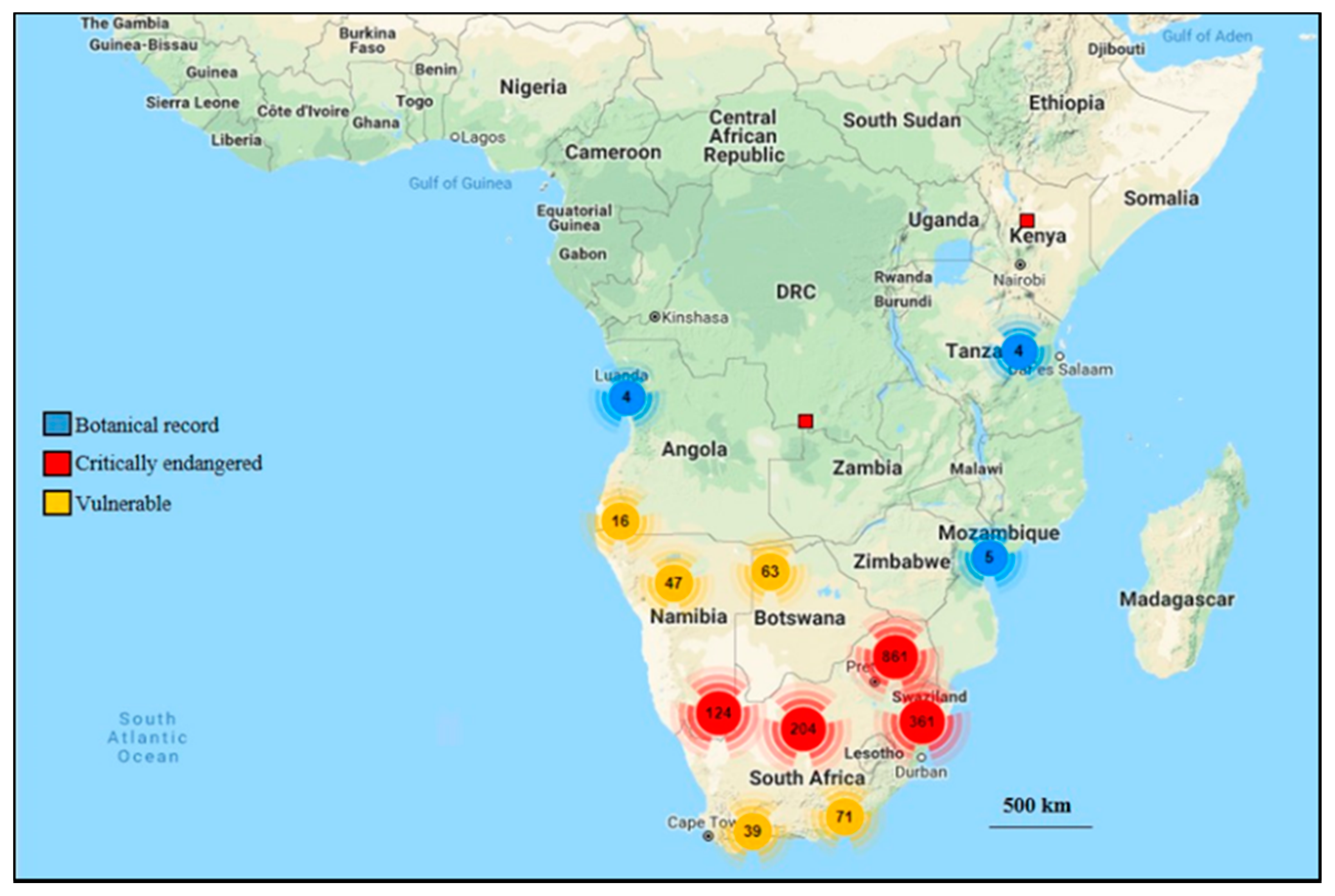
Table 1.
Chemical compounds isolated from the Genus Barleria.
| Plant Species | Plant Part | Chemical Group | Chemical Compounds/Phytoconstituents | Reference |
|---|---|---|---|---|
| B. acanthoides | Whole | Phenolic glycosides | Barlerisides A | [72] |
| Barlerisides B | ||||
| Phenylethanoid glycoside | Verbascoside (acteoside) | |||
| Phenolic acid | p-hydroxycinnamic acid | |||
| Neolignan diglycoside | Barlericin | [73] | ||
| B. cristata | Whole | Phenolic acid | 4-hydroxy-trans-cinnamate | [70] |
| Terpenoid | oleanolic acid | |||
| Bark | Flavonoid | 6-O-α-L-rhamnopyranoside-3,7,3′-O-trimethylated-8-hydroxyquercetin | [74] | |
| 6-O-α-L-rhamnopyranoside quercetagetin | ||||
| 3-O-Methylquercetin | ||||
| Gossypetin 8-methyl ether | ||||
| Quercetagetin | ||||
| Tamarixetin | ||||
| Gossypetin | ||||
| Quercetin | ||||
| Leaves | Phenolic acids | p-Coumaric acid | [75] | |
| Lipid | α -Tocopherol | |||
| Flavonoid | Luteolin | |||
| Flavonoid | 7-O-Methylluteolin | |||
| Iridoid glycosides | Barlerin | |||
| Shanzhiside methyl ester | ||||
| Phenylethanoid glycosides | Desrhamnosyl acteoside | [76] | ||
| Poliumoside | ||||
| Acteoside (verbascoside) | ||||
| B. dinteri | Leaves | Iridoid glycosides | Barlerin | [77] |
| B. lupulina | Aerial | 8-O-acetylipolamiidic acid | [78,79,80,81,82,83,84] | |
| 8-O-acetyl-6-O-(p-methoxy-cis-cinnamoyl)shanzhiside | ||||
| 8-O-acetyl-6-O-(p-methoxy-transcinnamoyl) shanzhiside | ||||
| 6-O-p-methoxy-cis-cinnamoyl-8-O-acetylshanzhiside methyl ester | ||||
| 6-O-p-methoxy-trans-cinnamoyl-8-O-acetylshanzhiside methyl ester | ||||
| 6-O-p-cis-coumaroyl-8-O-acetylshanzhiside methyl ester | ||||
| 6-O-p-trans-coumaroyl-8-O-acetylshanzhiside methyl ester | ||||
| Ipolamiide | ||||
| Ipolamiidoside | ||||
| Shanzhiside | ||||
| Shanzhiside methyl ester | ||||
| 8-O-acetylshanzhiside | ||||
| Barlerin | ||||
| 6-O-acetylshanzhiside methyl ester | ||||
| Acetylbarlerin | ||||
| Mussaenosidic acid | ||||
| Phlorigidoside | ||||
| Iridoid diglucoside | Lupulinoside | |||
| Phenylethanoid glycosides | Forsythoside | |||
| Poliumoside | ||||
| Lignan glucosides | (+)-lyoniresinol 3 α-O- β-glucopyranoside | |||
| Glycoside lipid molecule | 1-octen-3-yl- β -primeveroside | |||
| Glycoside | Benzyl β -primeveroside | |||
| B. noctiflora | Leaves | Phenylethanoid glycoside | Barlerinoside | [85] |
| B. prionitis | Aerial | Terpenoid | Balarenone | [86] |
| Phenylethanoid glycoside | Barlerinoside | [67] | ||
| Phenylethanoid glycoside | Verbascoside | [87] | ||
| Iridoid glycosides | Barlerin | [65,67] | ||
| Acetylbarlerin | ||||
| Shanzhiside methyl ester | [87] | |||
| 6-O-trans-p-coumaroyl-8-O-acetylshanzhiside methyl ester | ||||
| 6-O-cis-coumaroyl-8-O-acetylshanzhiside methyl ester | ||||
| 7-methoxydiderroside | [67] | |||
| Lupulinoside | ||||
| Terpenoid | Pipataline | 87] | ||
| Lupeol | ||||
| Phytosterols | 13,14-seco-stigmasta-5,14-diene-3-β –ol | |||
| Roots | β-sitosterol | [88] | ||
| Aerial | Flavonoid | Apigenin 7-O-β-D-glucoside | [78,86,89,90] | |
| Leaves | 6-hydroxyflavone | [91] | ||
| Scutellarin | ||||
| Aerial | Luteolin-7-O-β-D-glucoside | [92] | ||
| Leaves | Phenolic acid | Melilotic acid | [93] | |
| Syringic acid | [91] | |||
| Vanillic acid | ||||
| p-hydroxybenzoic acid | ||||
| B. strigose Whole | Phenylethanoid glycoside | 4-hydroxyphenylethyl 4-O-β-D-glucopyranosyl-(1→3)-O-α-L-rhamnopyranoside | [94] | |
| Phenylethanoid glycoside | Verbascoside | |||
| Iridoid glycoside | 10-O-trans-coumaroyl-eranthemoside | |||
| Decaffeoylverbascoside | ||||
| Lyoniresinol 3 α -O-β -D-glucoside | ||||
| 7-O-acetyl-8-epi-loganic acid | ||||
| (3R)-1-octen-3-ol-3-O-β-D-xylosyl-(1→6)-β-D-glucoside | ||||
| Phenylethanoid glycosides | Isoverbascoside | |||
| Decaffeoylverbascoside | ||||
| Flavonoid | Apigenin 7-O-α-Lrhamnosyl-(1→6)-O-β-D-glucoside | |||
| B. trispinosa | Aerial | Iridoid glycosides | 6-α-L-rhamnopyranosyl-8-O-acetylshanzihiside methyl ester | [95] |
| Acetyl barlerin | ||||
| Barlerin | ||||
| Shanzhiside methyl ester | ||||
Synonyms: Acteoside = verbascoside.
Table 2.
Antioxidant properties of extracts and compounds isolated from Barleria.
| Plant Species | Plant Part | Extract/Compound | Antioxidant Activity/Models/Assays | Reference |
|---|---|---|---|---|
| B. acanthoides | Whole | Barleriside A, barleriside B | Superoxide scavenging activity, Xanthine oxidase activity | [72] |
| B. albostellata | Leaves, stems | Methanol | DPPH assay; FRAP assay; β-Carotene-linoleic acid model system | [98] |
| B. argillicola | Whole | Methanol | β-Carotene–linoleic acid model system; DPPH assay | [142] |
| B. courtrallica | Leaves | Ethanol | DPPH assay, hydroxyl radical scavenging activity, superoxide radical scavenging activity, ABTS assay and reducing power methods. | [143] |
| B. cristata | Leaves | Ethanol | DPPH assay, Superoxide anion and nitric oxide radical scavenging activity, hydrogen peroxide scavenging activity | [144] |
| Leaves | Ethanol | DPPH assay, ABTS assay, TPTZ assay | [145] | |
| Leaves | Acetone and methanol | DPPH assay; Nitric-oxide Reducing Assay, FRAP | [146] | |
| Leaves | Ethanol | DPPH assay, FRAP | [147] | |
| B. dinteri | Leaves | Acetone and methanol | DPPH assay | [148] |
| B. gibsoni | Leaves | Ethanol | DPPH assay; Nitric oxide radical scavenging activity | [53] |
| B. grandiflora | Leaves | Water and ethanol | FTC method, TBA method | [149] |
| B. greenii | Leaves, stems, roots | Methanol | DPPH assay; FRAP assay; β-Carotene-linoleic acid model system | [98] |
| B. lupulina | Leaves, stems | Methanol | DPPH assay | [42] |
| B. montana | Leaves | Ethanol | DPPH, Reducing power assay, Nitric oxide scavenging activity | [150] |
| Leaves | Methanol | DPPH assay, Hydrogen peroxide method | [151] | |
| B. mysorensis | Leaves | Water | DPPH assay | [152] |
| B. noctiflora | Leaf, roots | Methanol | DPPH assay, Ferrous reducing power, Fe2+ reducing power, Fe2+ chelating activity assay, Nitric oxide scavenging activity, ABTS assay, Superoxide anion scavenging activity, Hydrogen peroxide radical scavenging activity | [153] |
| Whole | Ethanol | DPPH assay | [154] | |
| Aerial | Ethanol and Water | DPPH assay, ABTS assay, Scavenging of hydrogen peroxide (H2O2), Lipid Peroxidation Inhibitory Activity, Hydroxyl radical scavenging activity p-NDA method, Superoxide radical scavenging activity by alkaline DMSO method | [155] | |
| B. prionitis | Leaves, stems, roots | Methanol | DPPH assay; FRAP assay; β-Carotene-linoleic acid model system | [98] |
| Aerial | Ethanol | β carotene bleaching assay, DPPH assay and hydroxyl radical scavenging activity | [140] | |
| Aerial | Shanzhiside methyl ester, 6-O-trans-p-coumaroyl-8-O-acetylshanzhiside methyl ester, barlerin, acetylbarlerin, 7-methoxydiderroside, lupulinoside | DPPH assay | [67] | |
| Whole | Ethyl acetate | DPPH assay | [156] | |
| Leaves, stems | Acetone | FRAP assay, DPPH assay, ABTS Assay | [157] | |
| Leaves, stems | Methanol | DPPH, Reducing power assay | [158] | |
| Bark, leaves | Methanol | DPPH assay | [159] | |
| Leaves | Ethanol | FTC method, TBA method, Scavenging of hydrogen peroxide radicals, DPPH assay | [160] | |
| Whole | Ethanol | DPPH, ABTS Assay, Hydroxyl radical scavenging activity, Reducing power assay, Nitrous oxide Reducing Assay | [69] | |
| Flower | Ethanol and water | DPPH assay | [161] | |
| B. strigosa | Roots | Ethanol and water | ABTS assay, Nitric oxide quenching assay, Ferric reducing assay, DPPH, | [162] |
| Leaves | Methanol | DPPH assay | [163] |
DPPH—(1,1-Diphenyl-2-picrylhydrazyl) free radical-scavenging activity; FRAP—Ferric Reducing Antioxidant Power, ABTS—(2,2′-azino-bis(3-ethylbenzothiazoline-6-sulfonic acid)) free radical-scavenging activity; FTC—Ferric thiocyanate method; TBA—Thiobarbituric acid method; TPTZ—2,4,6-tripyridyl-s-triazine radical scavenging assay; TBA—Thiobarbituric acid method.
Table 3.
Antibacterial activities of extracts from species within Barleria.
| Plant Species | Plant Part | Extract | Antibacterial Activity | Agent Dosage (μg/mL) | Reference |
|---|---|---|---|---|---|
| B. acuminata | Leaves | Ethanol | B. cereus, B. subtilis, E. faecalis, S. aureus, S. epidermidis, E. coli, K. pneumonia, P. mirabilis, S. typhi, S. dysentriae. | 50,000 | [170] |
| B. albostellata | Leaves, stem | Petroleum ether, dichloromethane | B. subtillis, S. aureus, E. coli, K. pneumoniae | 50,000 | [66] |
| B. argillicola | Whole | Methanol | E. coli, P. aeruginosa, S. aureus | 20,000 | [142] |
| B. cristata | Bark | Ethanol | S. aureus, B. subtillis, S. mutans | 10,000 | [74] |
| Leaves | Methanol | K. pneumonia, S. aureus, E. coli, S. paratyphi | 5000 | [144] | |
| Methanol and water | S. pyogenes, E. coli | 300 | [171] | ||
| Petroleum ether, chloroform and water | X. oryzae, B. subtilis, E. coli, P. aeruginosa, P. fluorescences. | - | [172] | ||
| B. dinteri | Leaves | n-hexane, dichloromethane, acetone and methanol | E. coli, E. faecaelis, S. aureus, P. aeruginosa. | 10,000 | [148] |
| B. grandiflora | Aerial | Ethanol | S. aureus; S. mutans | 5000 | [173] |
| B. greenii | Stems, roots | Dichloromethane | B. subtillis, S. aureus, E. coli, K. pneumoniae | 30,000 | [66] |
| B. lupulina | Whole | Methanol | Propionibacterium acnes | - | [64] |
| leaves | Methanol | S. aureus, E. coli; P. aeruginosa, K. pneumoniae, S. typhi | 200,000 | [42] | |
| Leaves, stems | Ethanol | S. aureus, E. coli, P. aeruginosa, K. pneumonia, S. typhi | 20,000 | [174] | |
| Leaves | Methanol | S. aureus, B. pumilus | 1250 | [175] | |
| Leaves | Essential oil | B. pumilus, S. aureus | - | [176] | |
| B. montana | Leaves | Acetone | E. coli; S. typhi; P. aeruginosa; K. pneumoniae; P. vulgaris; B. subtilis; S. pneumoniae; S. aureus; E.coli | 200 | [177] |
| Aerial | Methanol | B. subtilis; B. cereus; B. pumilis; S. aureus; E. coli; P. aeuriginosa; P. vulgaris; S. marceseans | 200,000 | [178] | |
| Leaves | Water, ethanol, methanol, chloroform | E. aerogenes; E. coli; S. pneumoniae; B.subtilis; P. vulgaris | 100 | [179] | |
| B. prionitis | Leaves, stems | Dichloromethane | B. subtilis, S. aureus, E. coli, K. pneumoniae | 50,000 | [66] |
| Bark | Methanol | S. mutants, S. aureus, Pseudomonas sp., Bacillus sp. | 50,000 | [169] | |
| Leaves | Chloroform | S. typhi; B. subtilis; V. cholera; M. luteus; Providencia sp.; L. sporogenus, Citrobacter sp. | 50,000 | [180] | |
| Leaves | Water, petroleum ether, chloroform, acetone | L. rhamnosus | 200,000 | [181] | |
| Leaves | Ethanol | S. typhi; B. subtilis; S. aureus; V. cholera; E. coli | 10,000 | [182] | |
| Leaves, stem | Ethyl acetate | B. pumilus; B. subtilis; S. pyogenes; B. cereus; S. marcescens, C. acidovorans; P. aeruginosa | 100,000 | [183] | |
| Leaves | Methanol | S. mutants; S. aureus; L. acidophilus; Pseudomonas sp. | 10,000 | [184] | |
| Leaves | Petroleum ether, chloroform, water | B. subtilis; E. coli; P. fluorescens; X. oryzae | - | [172] | |
| Leaves | Ethanol | S. aureus; B. subtilis; P. vulgaris; K. pneumonia; E. coli; P. aeruginosa | 10,000 | [185] | |
| Aerial | Ethanol | B. cereus; P. aeruginosa | - | [86] | |
| B. strigosa | Leaves | Butanol | B. subtilis; S. aureus; M. luteus | 2000 | [186] |
Bacillus cereus; Bacillus pumilus; Bacillus subtilis; Bacillus species.; Comomonas acidovorans; Citrobacter species; Enterobacter aerogenes, Enterococcus faecalis; Escherichia coli; Klebsiella pneumoniae; Lactobacillus acidophilus; Lactobacillus rhamnosus; Lactobacillus sporogenus; Micrococcus luteus; Proteus mirabilis; Proteus vulgaris; Pseudomonas aeruginosa; Pseudomonas fluorescences; Pseudomonas sp.; Psuedomonas vulgaris; Salmonella paratyphi; Salmonella typhi; Serratia marceseans; Shigella dysentriae; Staphylococcus aureus; Staphylococcus epidermidis; Streptococcus mutants; Streptococcus pneumoniae; Streptococcus pyogenes; Streptococcus species; Vibrio cholera; Xanthomonas oryzae.
Table 4.
Antifungal activities of extracts from species within Barleria.
| Plant Species | Plant Part | Extract | Antifungal Activity | Agent Dosage (μg/mL) | Reference |
|---|---|---|---|---|---|
| B. albostellata | Leaves and stems | Petroleum ether, dichloromethane | C. albicans | 20,000 | [98] |
| B. cristata | Leaves | Saponin fraction | C. albicans; A. flavous; Penicillium sp.; A. niger; Trichophyton sp. | 1000 | [192] |
| Saponin fraction | A. flavous; A. niger | 5000 | [144] | ||
| Petroleum ether, chloroform, water | A. flavous; C. albicans | - | [172] | ||
| B. grandiflora | Leaves | Water | A. fumigatus | 625 | [193] |
| Aerial | Ethanol | C. albicans | 5000 | [173] | |
| Leaves | Ethanol | C. albicans | 1600 | [194] | |
| B. greenii | Leaves, stems and roots | Dichloromethane | C. albicans | 20,000 | [98] |
| B. montana | Aerial | Methanol | A. niger; R. stolonifera; S. cerevisiae; P. chrysogenum | 200,000 | [178] |
| B. prionitis | Bark | Methanol | S. cerevisiae; C. albicans | 50,000 | [169] |
| Roots and stems | Petroleum ether, dichloromethane | C. albicans | 20,000 | [98] | |
| Leaves, stems and roots | Ethanol | A. fumigatus; C. vaginitis; C. neoformans; C. albicans; B. dermatitidis | 20,000 | [195] | |
| Aerial | Ethanol | C. albicans | 5000 | [173] | |
| Leaves | Ethanol | C. albicans | 1600 | [194] | |
| Aerial | Methanol | C. albicans | 200 | [196] | |
| Methanol | C. albicans, A. niger | 200,000 | [197] |
Aspergillus flavous; Aspergillus fumigatus; Aspergillus niger; Blastomyces dermatitidis; Candida albicans; Candidal vaginitis; Cryptococcus neoformans; Pencillium chrysogenum; Penicillium species; Rhizopus stolonifera; Saccharomyces cerevisiae; Trichophyton species.
Table 5.
Anti-inflammatory activities of extracts, fractions and isolated compounds from species within Barleria.
Table 5.
Anti-inflammatory activities of extracts, fractions and isolated compounds from species within Barleria.
| Plant Species | Plant Part | Extract | Anti-Inflammatory Activity/Assays/Model | Agent Dosage | Reference |
|---|---|---|---|---|---|
| B. albostellata | Leaves and stems | Petroleum ether, dichloromethane, ethanol | COX-1, COX-2 | 0.25 μg/μL | [66] |
| B. cristata | Leaves | Water | CIO in rat paws, prostaglandins inhibitory activity, and acetic acid induced capillary permeability in mice. | 500 mg/kg | [203] |
| Methanol | Inhibited oedema produced by histamine and serotonin in rats. Reduction in the increased peritoneal vascular permeability in mice | 500 mg/kg | [204] | ||
| B. greenii | Stems and roots | Petroleum ether, dichloromethane, ethanol | COX-1, COX-2 | 0.25 μg/μL | [66] |
| B. lupulina | Aerial | Water | Activated the Nrf2 cell defense pathway in human dermal microvascular endothelial cells | - | [205] |
| Aerial | Methanol | Acute and sub-acute inflammation models of albino rats. | 300 mg/kg | [63] | |
| Whole | Methanol, acetone | CIO in rat paws and ethyl phenylpropiolate-induced ear oedema in rats. | 50–200 mg/kg | [206] | |
| B. montana | Leaves | Ethanol | Formalin induced inflammation in male albino wistar rats. | 300 mg/kg | [207] |
| B. prionitis | Leaves, stems and roots | Petroleum ether, dichloromethane, ethanol | COX-1, COX-2 assays | 0.25 μg/μL | [66] |
| Whole | Methanol-aqueous fractions (TAF) | CIO in adrenalectomised rats, activity in acute inflammation induced by carrageenan, histamine and dextran in rats | 100 mg/kg | [202] | |
| Roots | Water fractions | CIO in rat paw model | 400 mg/kg | [208] | |
| Aerial | Shanzhiside methylester, 8-O-acetyl shanzhiside methyl ester, iridoid glycosides, monoterpenoidal fraction | Stimulated rat neutrophils by inhibiting MPO, elastase and MMP-9 enzymes | 10 μg/mL | [209] |
Cyclooxygenase (COX); Carrageenan-induced oedema (CIO); Nuclear factor erythroid 2–related factor 2 (Nrf2); Matrix Metalloproteinase-9 (MMP-9); Myeloperoxidase (MPO).
Table 6.
Anticancer activities of extracts and isolated compounds from species within Barleria.
| Plant Species | Plant Part | Extract/Compounds | Assays/Cell Lines | Agent Dosage | Reference |
|---|---|---|---|---|---|
| B. cristata | Aerial | Isoverbascoside | NQO1 assay, murine hepatoma cell line Hepa-1c1c7 | 3.125 μM | [211] |
| Leaves and bark | Methanol | Brine shrimp lethality assay, brine shrimp cysts | 200 μg/mL | [146] | |
| B. gibsoni | Leaves | Petroleum ether, chloroform | SRB assay, MDA, MB 4355 (Human breast cancer) and Hep G2 (Liver cancer cell line) | 50 µg/mL | [213] |
| B. grandiflora | Leaves | Alcoholic | A-549 (human lung cancer) cells, DLA tumour cells and Vero (African green monkey kidney) normal cells | 300 μg/mL | [212] |
| Ethanol | MTT assay; Human gingival fibroblast cell lines, human dermal fibroblast cell lines | 1000 μg/mL | [173] | ||
| B. lupulina | Leaves | Ethanol | MTT assay; cancerous THP-1 cell lines | 100 μg/mL | [214] |
| Ethanol | MTT assay; HepG2 cells | 1000 μg/mL | [174] | ||
| B. prionitis | Leaves | Ethanol | MTT assay; Human gingival fibroblast cell lines, human dermal fibroblast cell lines, | 1000 μg/mL | [173] |
| Ethanol | SRB assay, breast (MCF-7), colon (DLD-1), lung (A549), breast metastatic (MDMAMB-468), lung metastatic (NCIH358) and colon metastatic (SW620) | 100 μg/mL | [215] | ||
| B. strigosa | Leaves | Butanol | MTT colorimetric assay, Human hepatocellular carcinoma (HepG2), human breast adenocarcinoma (MCF7), human oral epidermoid carcinoma (KB), human colon adenocarcinoma (HT29), murine lymphocytic leukemia (P388), human cervical carcinoma (HeLa) as well as two normal cell lines including African green monkey kidney (Vero) and mouse subcutaneous connective tissue (L929) | 2000 μg/mL | [186] |
MTT—(3-(4, 5-dimethylthiazolyl-2)-2, 5-diphenyltetrazolium bromide); DLA—Dalton’s lymphoma Ascites; NQO1- (NAD(P)H dehydrogenase [quinone] 1); SRB—Sulphorhodamine B.
Table 7.
Antidiabetic activities of extracts and fractions from species within Barleria.
| Plant Species | Plant Part | Extract | Antdiabetic Activity/Assays/Models | Agent Dosage | Reference |
|---|---|---|---|---|---|
| B. bispinosa | Aerial | Methanol | Male Wister rats, Streptozotocin induced diabetic rats | 500 mg/kg | [220] |
| B. cristata | Seeds | Ethanol | Wistar rats, alloxan-induced diabetic rats | 200 mg/kg | [218] |
| Leaves and roots | Ethanol and petroleum ether | Inhibition of alpha-amylase enzyme assay, Inhibition of alpha-glucosidase enzyme assay | - | [147] | |
| B. lupulina | Aerial | Methanol | Male Wister rats, Streptozotocin-diabetic rats | 300 mg/kg | [61] |
| B. montana | Aerial | Methanol | Wistar albino rats; Streptozotocin induced diabetic rats | 400 mg/kg | [221] |
| B. noctiflora | Aerial | Ethyl acetate | Wister rats, Streptozotocin induced type-2 diabetes in rats | 400 mg/kg | [222] |
| Whole | Ethanol | In-vitro anti-diabetic activity was determined by inhibition of α-glucosidase and inhibition of α-amylase studies | 500 μg/mL | [223] | |
| Aerial | Ethyl acetate | Wister rats, Streptozotocin induced diabetic rats | - | [224] | |
| B. prionitis | Leaves and roots | Ethanol | Adult Albino rats, alloxan-induced diabetic rats | 200 mg/kg | [219] |
| Leaves, stems and roots | Alcohol | Albino rats, alloxan-induced hyperglycemic rats | 200 mg/kg | [225] |
Table 8.
Antiulcer activities of extracts and fractions from species within Barleria.
| Plant Species | Plant Part | Extract | Antiulcer Activity/Gastric Cytoprotective Activity/Models | Agent Dosage | Reference |
|---|---|---|---|---|---|
| B. buxifolia | Whole | Methanol | Wistar rats, PL and aspirin induced ulcers | 400 mg/kg | [229] |
| B. gibsoni | Leaves | Ethanol | Wistar rats, PL-induced ulcer models | 500 mg/kg | [53] |
| B. lupulina | Aerial | Methanol | Albino (Wistar) rats, PL ulceration in rats, stress-induced ulceration, drug-induced gastric ulcer in rats, duodenal ulcers in rats. | 200 mg/kg | [62] |
| B. prionitis | Leaves | Methanol | Wistar rats; ethanol induced gastric mucosal lesions, indomethacin induced ulcer models | 500 mg/kg | [52] |
| Ethanol | Male Sprague–Dawley rats and female Swiss albino mice; PL- induced ulcers, aspirin- induced ulcers, CRS-induced ulcers, ethanol-induced ulcer | 200 mg/kg | [228] | ||
| Methanol | Ethanol and Indomethacin Induced ulcer models | 500 mg/kg | [230] | ||
| Chloroform | Rodent experimental models (indomethacin and pylorus ligation) | 250 mg/kg | [52] |
CRS—cold-restraint stress; PL—Pylorus ligated.
Table 9.
Hepatoprotective activities of extracts and fractions from species within Barleria.
| Plant Species | Plant Part | Extract | Hepatoprotective Activity/ASSAYS/Models | Agent Dosage (mg/kg) | Reference |
|---|---|---|---|---|---|
| B. cristata | Leaves | Ethanol | Wistar albino rats, CCl4 induced hepatic damage in rats | 200 | [238] |
| B. cuspidata | Leaves | Methanol | Wistar albino rats, CCl4 induced hepatotoxicity in rats | 400 | [241] |
| B. gibsoni | Aerial | Aqueous alcoholic | Wistar albino rats; inducing agent Paracetamol in Carboxy methyl cellulose | 400 | [242] |
| B. montana | Leaves | Methanol | Wistar albino rats; ethanol-induced rat hepatic injury | 500 | [243] |
| Aerial | Methanol | CCl4 induced hepatotoxicity on rats | 800 | [178] | |
| B. prionitis | Leaves and stems | Fractions from ethanol-aqueous | Charles Foster rats, Swiss albino mice; acute and chronic animal test models, CCl4 toxicity, cetaminophen (APAP) toxicity, D-GalN induced hepatotoxicity | 200 | [240] |
CCl4—Carbon tetrachloride.
Table 10.
Anthelmintic activities of extracts from species within Barleria.
| Plant Species | Plant Part | Extract | Anthelmintic Activity/Assays/Models | Agent Dosage (mg/mL) | Reference |
|---|---|---|---|---|---|
| B. buxifolia | Leaves | Ethanol | IAW P. posthuma | 100 | [258] |
| B. gibsoni | Leaves | Water, ethanol | IAW P. posthuma | 15 | [260] |
| B. prionitis | Whole | Water, ethanol | IAW P. posthuma | 100 | [259] |
| Water, ethanol | IAW P. posthuma | 100 | [261] |
IAW—Indian adult worm; Pheretima posthuma.
Table 11.
Antiarthritic activities of extracts and fractions from species within Barleria.
| Plant Species | Plant Part | Extract | Antiarthritic Activity/Assays/Models | Agent Dosage (mg/kg) | Reference |
|---|---|---|---|---|---|
| B. lupulina | Leaves | Methanol | Albino male mice, female Sprague Dawley rats, formalin-induced arthritis, adjuvant induced arthritis, collagen type II-induced arthritis, monosodium iodoacetate induced osteoarthritis | 600 | [269] |
| B. montana | Leaves | Ethanol | Male Albino Wistar rats, Complete Freund’s in vivo method in induced rats | 400 | [270] |
| Leaves | Ethyl acetate fraction | Sprague Dawley rats, formaldehyde induced arthritis; FCA-induced arthritis rat model | 250 | [268] | |
| B. prionitis | Whole | Methanol | Complete Freund’s induced rat model | 400 | [271] |
FCA—Freund’s complete adjuvant.
Table 12.
Acetylcholinesterase inhibition of extracts and isolated compounds from species within Barleria.
Table 12.
Acetylcholinesterase inhibition of extracts and isolated compounds from species within Barleria.
| Plant Species | Plant Part | Extract/Compound | Inhibition of Acetylcholinesterase/ | Agent Dosage | Reference |
|---|---|---|---|---|---|
| B. albostellata | Leaves, stems and roots | Methanol | Microtitre plate assays based on the colorimetric method; and using the positive control galanthamine | 625 μg/mL | [98] |
| B. greenii | Leaves, stems and roots | Methanol | Microtitre plate assays based on the colorimetric method; and using the positive control galanthamine | 625 μg/mL | [98] |
| B. prionitis | Leaves, stems and roots | Methanol | Microtitre plate assays based on the colorimetric method; and using the positive control galanthamine | 625 μg/mL | [98] |
| Aerial | 8-amino-7-hydroxypipataline | Modified Ellman’s assay, photometric method | - | [86] | |
| 6-O-trans-p-coumaroyl-8-O-actylshanzhiside methyl ester, barlerin, acetylbarlerin, 7-methoydiderroside, lupulinoside | Ellman’s assay | - | [67] |
Table 13.
Biological activity of synthesized nanoparticles from extracts of species of Barleria.
| Plant Species | Plant Part | Extract | Nanoparticles Synthesised | Reported Activity/Phytochemicals Present | Agent Dosage | Reference |
|---|---|---|---|---|---|---|
| B. cristata | Leaves | Water | Ag * | Mosquitocidal potential | 300 μg/mL | [305] |
| Ag | Antibacterial activity against E. coli and S. aureus | - | [306] | |||
| B. longiflora | Leaves | Water | Ag | Antimicrobial activity, inhibition of Enterococcus sp., Streptococcus sp., B. megaterium, P. putida, P. aeruginosa and S. aureus and potential application in photocatalytic dye degradation processes | 10 μg/mL | [307] |
| B. prionitis | Leaves | Water | Ag | Polyphenols, starch, reducing sugars, ascorbic acid and citric acid using GC-MS analysis | - | [308] |
* Silver.
Publisher’s Note: MDPI stays neutral with regard to jurisdictional claims in published maps and institutional affiliations. |
© 2021 by the authors. Licensee MDPI, Basel, Switzerland. This article is an open access article distributed under the terms and conditions of the Creative Commons Attribution (CC BY) license (https://creativecommons.org/licenses/by/4.0/).
Share and Cite
MDPI and ACS Style
Gangaram, S.; Naidoo, Y.; Dewir, Y.H.; El-Hendawy, S. Phytochemicals and Biological Activities of Barleria (Acanthaceae). Plants 2022, 11, 82. https://doi.org/10.3390/plants11010082
AMA Style
Gangaram S, Naidoo Y, Dewir YH, El-Hendawy S. Phytochemicals and Biological Activities of Barleria (Acanthaceae). Plants. 2022; 11(1):82. https://doi.org/10.3390/plants11010082
Chicago/Turabian StyleGangaram, Serisha, Yougasphree Naidoo, Yaser Hassan Dewir, and Salah El-Hendawy. 2022. "Phytochemicals and Biological Activities of Barleria (Acanthaceae)" Plants 11, no. 1: 82. https://doi.org/10.3390/plants11010082
Note that from the first issue of 2016, this journal uses article numbers instead of page numbers. See further details here.