Article

New Aspects of Secretory Structures in Five Alismataceae Species: Laticifers or Ducts?

by
Flávia Maria Leme
1,*,
João Pedro Silvério Pena Bento
1,2,
Vitoria Silva Fabiano
1,2,
Jean David Varilla González
1,2,
Vali Joana Pott
3 and
Rosani do Carmo de Oliveira Arruda
1,2,*
1
Laboratório de Anatomia Vegetal, Instituto de Biociências, Universidade Federal de Mato Grosso do Sul, Cidade Universitária, Caixa Postal 549, Campo Grande 79070-900, MS, Brazil
2
Programa de Pós-Graduação em Biologia Vegetal, Instituto de Biociências, Universidade Federal de Mato Grosso do Sul, Cidade Universitária, Caixa Postal 549, Campo Grande 79070-900, MS, Brazil
3
Herbário CGMS, Instituto de Biociências, Universidade Federal de Mato Grosso do Sul, Cidade Universitária, Caixa Postal 549, Campo Grande 79070-900, MS, Brazil
*
Authors to whom correspondence should be addressed.
Plants 2021, 10(12), 2694; https://doi.org/10.3390/plants10122694
Submission received: 1 July 2021 / Revised: 15 October 2021 / Accepted: 15 October 2021 / Published: 8 December 2021
(This article belongs to the Special Issue Plant Secretory Structures)

Abstract

:
The secretory structures of Alismataceae have been described as secretory ducts, laticifer ducts, laticifer canals or schizogenous ducts. However, these terms are not found in the specialized literature, and ontogenetic analyses for the exact classification of these structures are missing. Accordingly, more studies regarding the secretory structures of Alismataceae are necessary to establish homology in the family or in the order. Thus, the aim of this study was to describe the anatomy, ontogeny, distribution in the organs and exudate composition of the secretory structures present in five Alismataceae species in order to determine whether the family has laticifers or secretory ducts. Samples of leaves, flowers and floral apices were processed for anatomical and histochemical analyses by light microscopy. The analysis indicated the presence of anastomosing secretory ducts in all species, occurring in both leaves and flowers. The exudate contains lipids, alkaloids, proteins and polysaccharides, including mucilage. The secretory duct structure, distribution and exudate composition suggest a defense role against herbivory and in wound sealing. The presence of secretory ducts in all species analyzed indicates a probable synapomorphy for the family.

1. Introduction

Alismataceae is a family consisting of floating to emergent aquatic or marsh herbs with a worldwide distribution [1]. Limnocharitaceae was merged with Alismataceae to form one family comprising 17 genera [1,2,3,4] and ca. 100 species [5]. The family is known for including species with economic uses as food sources [6], ornamentals (aquarium plants) [6,7] and medicinals [6,7], as well as species for beekeeping [1,6]. The largest genera are Echinodorus and Sagittaria, both regularly used as aquarium and pond ornamental plants [1,6]. Leaves of Echinodorus grandiflorus (Cham. and Schltdl.) Micheli and E. macrophyllus (Kunth) Micheli are used for medicinal purposes [7,8,9,10,11,12]. Ethnobotanical investigations and clinical trials indicate that E. grandiflorus leaves have anti-hypertensive, anti-inflammatory, diuretic, and anti-arthritic properties [7,10,11,12,13].
Laticifers have been reported in some families of Alismatales [14], such as Aponogetonaceae [15], Araceae [16,17] and Alismataceae [14]. According to other authors, laticifers have not been recorded in Alismataceae [18,19] and Butomaceae [20], while resin secretory ducts have been described only for Araceae [14].
The secretory structures of Alismataceae have been described as secretory ducts, laticifer ducts or laticifer canals [1,19,20,21,22,23,24], and the secretion is described as latex [23] or milky juice [1]. However, laticifer ducts or laticifer canals are not found in the specialized literature on secretory structures [25,26,27]. In addition, the lack of ontogenetic analyses for the exact classification of these structures does not allow the establishment of homology in the family or order [23].
The secretory ducts or canals are elongated secretory structures lined with an epithelium of live secretory cells delimiting a large intercellular space (lumen) [14,25,26,27,28]. Ontogenetically, they may develop by schizogeny (separation of cells), lysigeny (disintegration of cells) or both (separation and disintegration of cells) [25]. Their development occurs from a group of a few initial meristematic cells that form a rosette in cross-section. These cells undergo divisions in various directions, and the rosette becomes more distinct from the surrounding cells, constituting the future epithelium [14,25,27,29]. The lumen develops in the middle of the epithelium, and further periclinal divisions may occur outside the epithelium, forming a sheath with one or more cell layers. The material secreted by the duct varies between resin, gum or mucilage [14,25,26,29].
Laticifers are a specialized type of secretory structure with an emulsion or suspension of compounds of a varied nature, in which terpenoids predominate, called “latex” [25,26,27,28,30,31]. The latex color may vary according to composition; it may be white (milky) [32,33], yellow [30,34], orange [35], red [36] or colorless [34]. Laticifers consist of one cell with intrusive growth (nonarticulated non-anastomosing type) or a series of connected cells (articulated anastomosing type) that form a uniseriate tube [25,30,34,37]. Articulated anastomosing laticifers or nonarticulated laticifers can be branched or unbranched [25,26,27,30,34,37]. Articulated anastomosing laticifers in a mature phase have terminal walls that disintegrate (multinucleated structure), that can branch and can assume several forms [25,30,34,37].
Laticifers and secretory ducts have been cited in at least 40 and 50 families, respectively, of vascular plants, including phylogenetically unrelated plants such as ferns, gymnosperms, and angiosperms, and they have emerged many times in the course of plant evolution [14,30,38]. Laticifers and secretory ducts have roles in herbivory reduction or resistance in plants. Their products (latex–laticifers; resin, gum or mucilage–secretory ducts) are of great economic importance since they are crucial for the production of pharmaceuticals, enzymes and rubber [28,31,39].
Although there are several anatomical studies on Alismataceae species [19] such as Echinodorus spp. [23,24], Helanthium tenellum [7], Alisma plantago [40], Echinodorus macrophyllus [41] and Sagittaria montevidensis [22], these studies do not provide details regarding the anatomy and ontogeny of the secretory structures called the “laticifer ducts” [1,11,14,22,23,24] or secretory canals [41]. Histochemical studies on the composition of the secretion are also absent. Thus, there is nothing to indicate whether the secreted material has latex or resin characteristics. Therefore, our objective was to study the structure, ontogeny, distribution in the organs and secretion composition of the secretory structures present in Echinodorus grandiflorus, H. tenellum, Hydrocleys nymphoides, Limnocharis flava and Sagittaria rhombifolia in order to determine whether the analyzed species have laticifers or secretory ducts.

2. Results

In E. grandiflorus, H. tenellum, H. nymphoides, L. flava and S. rhombifolia (Figure 1A–F), we found elongated secretory ducts consisting of an intercellular space or lumen lined with one layer of secretory cells, i.e., the epithelium. The lumen, where the secretion is released and stored, is formed by schizogeny during organ development (Figure 2A–F and Figure 3A–H). In the species evaluated, the secretory ducts are extremely narrow, with a diameter ranging from 11.9 µm in H. tenellum to 41.8 µm in H. nymphoides.

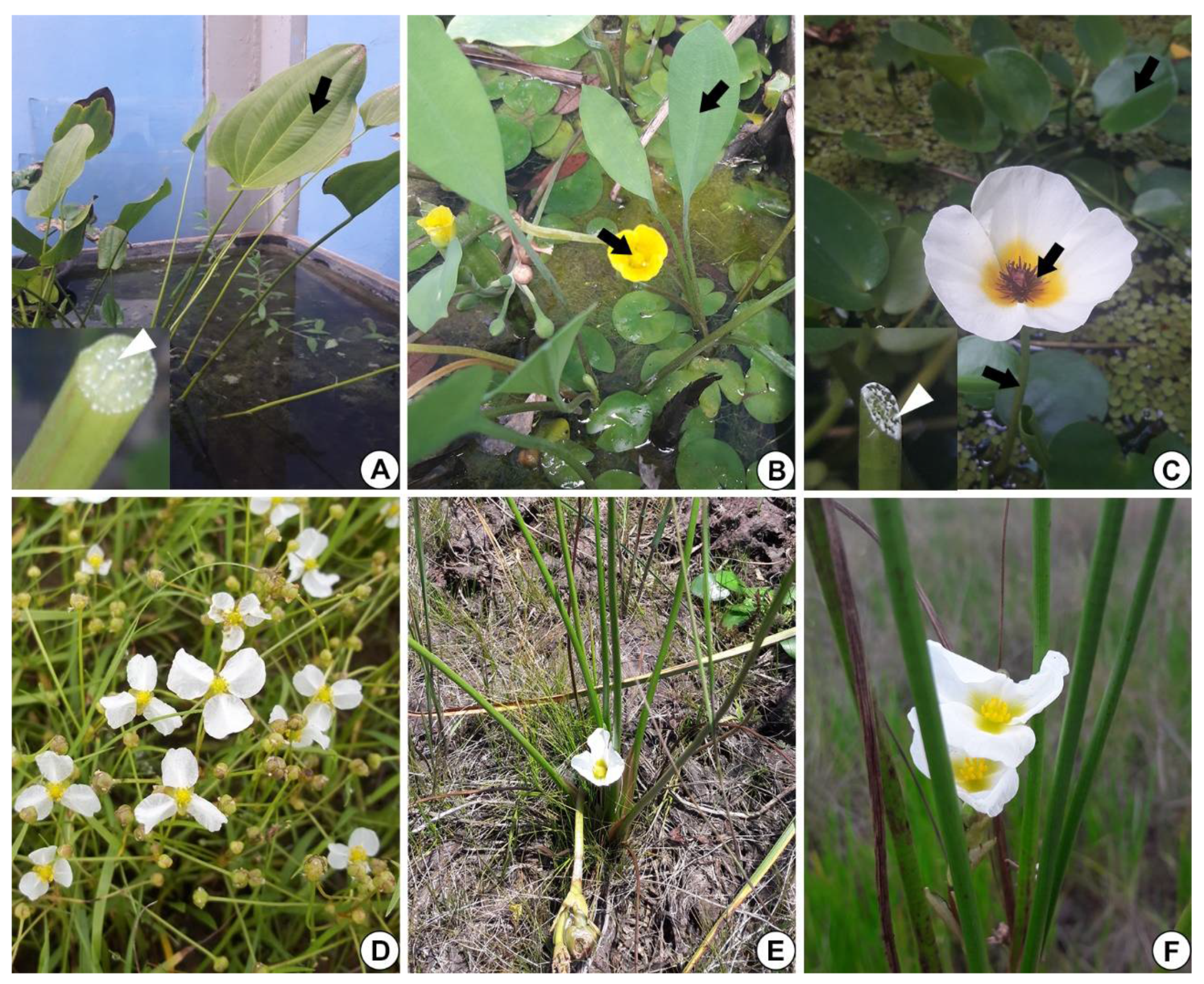
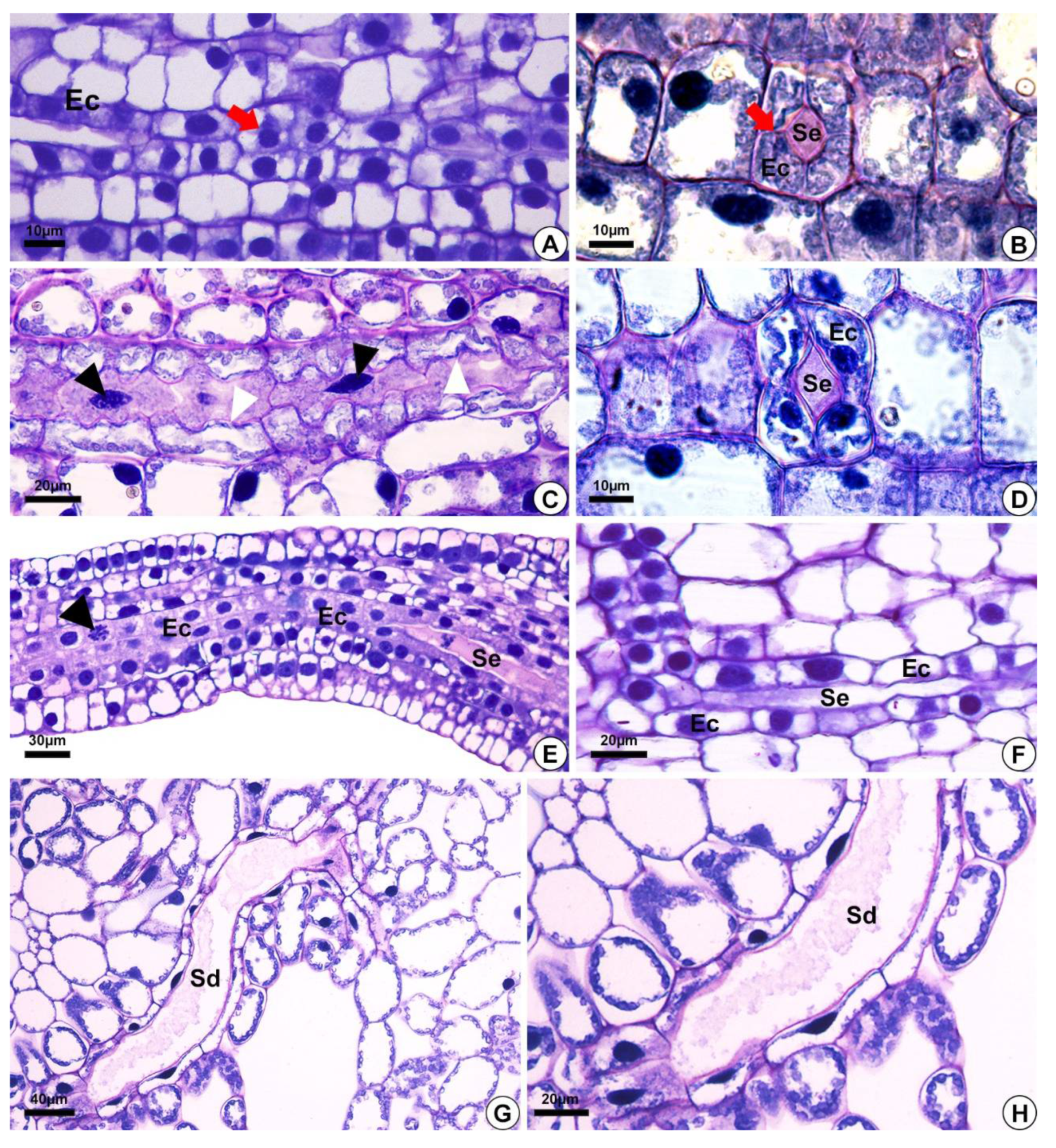
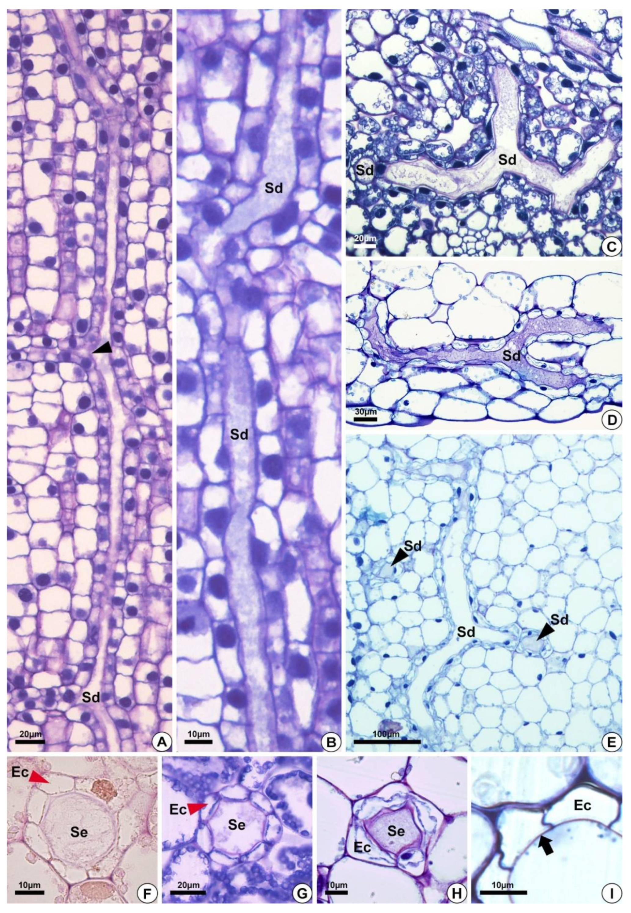
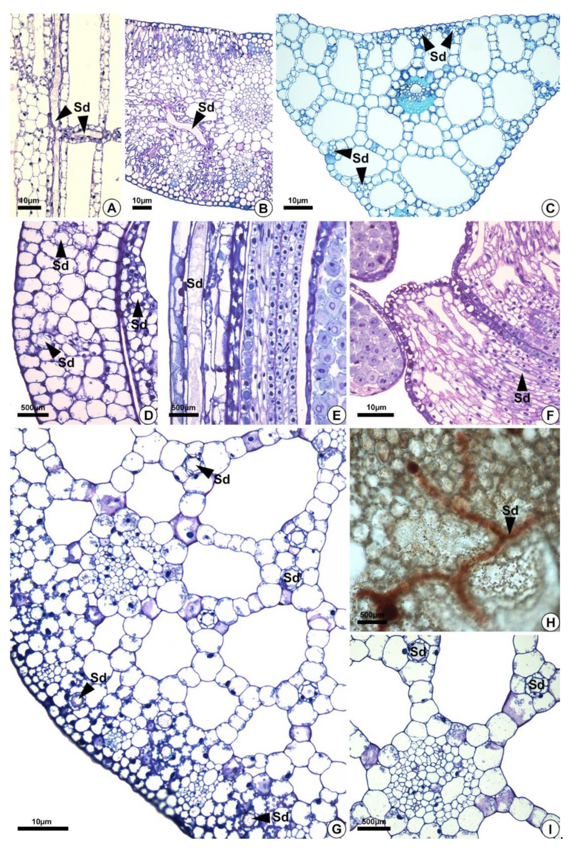
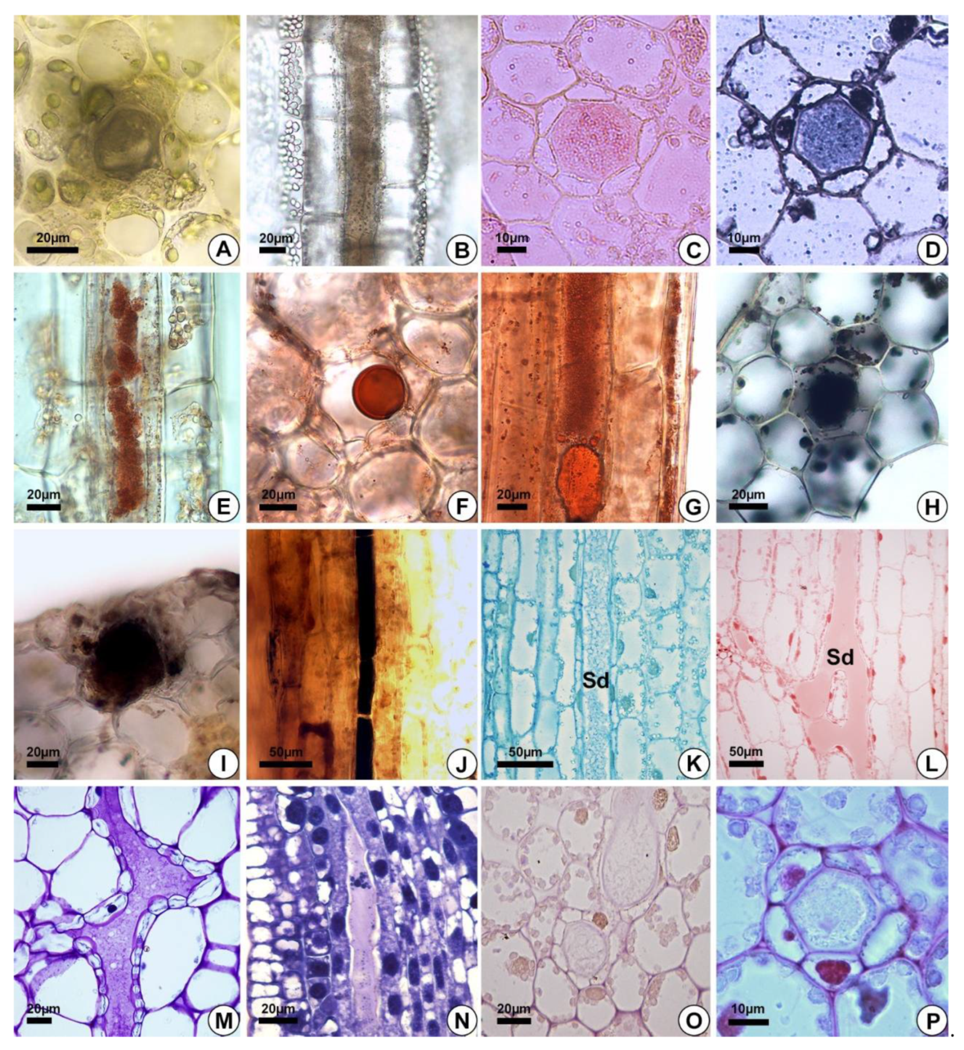
2.1. Origin and Morphology of the Secretory Structures

The secretory ducts of E. grandiflorus, H. tenellum, H. nymphoides, L. flava and S. rhombifolia differentiate early during organ development, being fully visible while other tissues are still meristematic (Figure 2A–E). Thus, the secretory ducts are formed before the complete development of the surrounding tissues. Secretory ducts originate from the ground meristem (Figure 2A–E) by asymmetrical mitotic divisions, followed by the dissolution of the middle lamella between the cells, which gives rise to the lumen in the early stage of development when the epithelium is composed of only four cells (Figure 2B–D). Initially, the cells of the rosette have large nuclei and a dense and uniform cytoplasm. These initial cells are distinguished from the adjacent meristematic cells by asymmetrical cell divisions (Figure 2A,B), dense cytoplasm and fusiform nuclei (Figure 2C). The wall of the epithelial cells becomes progressively undulating in L. flava (Figure 2C); however, in S. rhombifolia, the cell wall is smooth throughout development (Figure 2G,H). The ducts branch through lateral anastomoses with other secretory ducts (Figure 2G). At maturity, most ducts form an interconnected network of canals that extend longitudinally and radially throughout the shoot system (Figure 3A–E). Branched ducts are easily recognized by specific shapes such as a Y-bifurcation pattern (Figure 3C–E).
All species analyzed have ducts with a single-layered epithelium (Figure 3F–H). However, the epithelial cells are morphologically variable in length and width. They are large in H. tenellum and H. nymphoides (Figure 3A,B) but small and narrow in E. grandiflorus, S. rhombifolia and L. flava (Figure 3C–E). In mature secretory ducts, the epithelium is composed of 5 to 8 cells (Figure 3F–H) as seen in cross-section in all species analyzed. Epithelial cells generally have a flattened shape and thin cell walls that project into the lumen, have a slightly dense cytoplasm and large nuclei and contain small plastids. (Figure 3). The cell wall is pectocellulosic, reacting positively with Schiff’s reagent (PAS) (Figure 3F) and staining magenta with toluidine blue (Figure 3H,I).

2.2. Distribution of the Secretory Ducts in the Plant

Secretory ducts are present in the leaves (petiole, blade and midrib) of the five species analyzed and in the flowers of H. nymphoides, L. flava, and S. rhombifolia (Table 1, Figure 4). They occur at higher frequency in H. nymphoides, L. flava, and S. rhombifolia and at lower frequency in H. tenellum and E. grandiflorus. In the five species studied, secretory ducts were found in the petiole (Figure 4A), leaf blade, and midrib (Figure 4B,C). In H. nymphoides, L. flava, and S. rhombifolia flowers, the secretory ducts were distributed in the perianth and stamens (Figure 4D–F). The secretory ducts occur in the subepidermal layer, mesophyll, aerenchyma tissues and diaphragm (Figure 4). In the aerenchyma, they are distributed around the vascular bundles and pass among the aerenchyma spaces (Figure 4G–I).

2.3. Secretion Composition

The secretion in the leaves and petioles of E. grandiflorus, H. tenellum, H. nymphoides, L. flava and S. rhombifolia was milky (Figure 1A,C). Compared to the other species analyzed, H. nymphoides showed the greatest exudation of secretion. The secretion was initially fluid and became thicker after air contact. The histochemical tests were performed on the petiole of the leaves (Figure 5A–K,P) or on floral buds (Figure 5L–O). The results demonstrated that the secretion produced by the secretory ducts was of resin and consisted mainly of lipids (Figure 5C–G), alkaloids (Figure 5H–J), proteins (Figure 5K,L) and polysaccharides (Figure 5M–O) (Table 2). The analysis showed that the secretion was white in fresh material (Figure 1A,C) and dark on the slide (Figure 5A,B). For lipids, the Sudan and the oil red tests were positive in all species of Alismataceae analyzed (Figure 5E–G). We also detected alkaloids (Figure 5H–J), proteins (Figure 5K,L), and polysaccharides (Figure 5M–P) including mucilage (Figure 5M,N,P) in the secretory ducts. Polysaccharides were weakly stained (Figure 5O). No phenolic compounds were found (Table 2).

3. Discussion

Our data support the presence of secretory ducts in E. grandiflorus, H. tenellum, H. nymphoides, L. flava, and S. rhombifolia, as evidenced by anatomy, ontogeny and exudate composition analysis. The presence of resin ducts (sensu lato) in five genera of Alismataceae is a novelty for the family since controversial terms for these secretory structures were reported in previous studies, and secretion was reported as latex [1,14,19,22,23,24,41]. The development of resin ducts begins very early during plant development, differentiating from meristematic tissues in the reproductive and vegetative apices. We observed that resin ducts initiate with asymmetrical cell division, forming four epithelial cells surrounding a lumen. These cells divide again, adding new cells to the epithelium. During development, the secretory ducts anastomose laterally, forming an interconnected network of canals which store the resin composed of varied substances, as typically described for the exudate of some secretory ducts in angiosperms [14,25]. Therefore, our results corroborate the presence of secretory ducts [1,19,22,41] and refute the use of the terms laticifer [14] or laticiferous duct [23,24,42] in E. grandiflorus, H. tenellum, H. nymphoides, L. flava and S. rhombifolia. The confusion of terms used by researchers is due to the difficulty in identifying the real secretory structure present in the members of Alismataceae. Features such as the reduced diameter of the secretory duct, heterogeneous secretion composition, the lack of a sheath surrounding the epithelium, and the white color of the exudate likely caused this misinterpretation. This fact has also occurred in Anacardiaceae, Burseraceae, Clusiaceae, Cactaceae and Calophyllaceae [14,29,43].

3.1. Resin Duct Structure

The anatomy of the internal secretory structures observed in E. grandiflorus, H. tenellum, H. nymphoides, L. flava and S. rhombifolia is consistent with the secretory duct definition: a structure formed by an epithelium of secretory cells that delimits an intercellular space (lumen), where the secretion is stored. In Alismataceae, the ducts produce a complex resin which contains terpenes, phenolic compounds, proteins and polysaccharides, as observed in other families [14,25,26,27]. Notably, the resin ducts of Alismataceae are narrow and lack a sheath (this study), as observed in the ducts of Clusiaceae [29,44]. Sheaths formed by two cell layers surrounding the duct occur in Anacardiaceae [45], Hypericaceae [46], and Araceae [47]. Narrow secretory ducts are not common in angiosperms, but they have also been reported in Philodendron adamantinum Mart. ex Schott (Araceae) [47] and Hypericum perforatum L. (Hypericaceae) [46].
In the present study, epithelial cells have thin walls [44,48], dense cytoplasm mainly during the secretory phase, a fact also observed in Garcinia mangostana L. (Clusiaceae) [49] and Clusia species [29,44], in addition to large vacuoles and many mitochondria, plastids and Golgi apparatus. For Araceae, two types of secretory ducts have been described: one of small diameter in which the secretion accumulates in its lumen and is related to plant defense, with release of the secretion occurring only in the case of injury, and the other of large diameter that releases resin into the external environment and is directly related to the pollination mechanism, having the function to guarantee pollen adherence, specifically present in the adaxial part of the spathe of the inflorescence [47]. The first one is the most common in species of the family and is distributed in roots, stem, leaves and floral organs [47,50]. Although more details are needed to compare Alismataceae ducts to Araceae secretory ducts “type I” [47,50], the structure and diameter show similarities that might be of phylogenetic origin.
The ontogeny of the secretory duct was characterized by asymmetrical mitotic divisions of meristematic cells, followed by the dissolution of the middle lamella between them, resulting in lumen expansion (this study). In Alismataceae species, the lumen is formed in the early stage of duct ontogeny, with secretion already present, and the epithelium has only four cells. In other species, e.g., Clusiaceae, the initial rosette has various cells that will then form the lumen [29,44].

3.2. Secretion Composition and Functions

The resin detected here has a complex composition, containing different chemical classes of substances. Most components are lipophilic, but others have been identified, such as alkaloids, proteins and polysaccharides, including mucilage. Generally, the secretion of secretory ducts may be classified as resin, mucilage or gum [14,25] according to its composition. Mucilage ducts occur in Malvaceae [51] and Calophyllaceae [38]; resin ducts in Araceae [47], Clusiaceae [29] and Anacardiaceae [45]; and gum ducts in Calophyllaceae [38]. However, the complex composition of some resins, which confers white color to the exudate, has caused misinterpretation and has led some researchers to call it “latex”, as in the case of Alismataceae ([19,20], this study), Anacardiaceae [14,52,53] and Clusiaceae [14,44]. Nevertheless, latex is produced only by laticifers [14,25].
The resin produced by ducts is usually related to plant defense against herbivores [28,54,55,56,57]. The presence of terpenes in the resin of Alismataceae indicates that it can provide wound sealing. When the plant is damaged, resin overflows and the terpenes arapidly coagulate when in contact with the air, thus sealing the wounds [55,58]. An anti-herbivory action may also occur because the resin can trap whole insects or their mouthparts [55].
The distribution of resin ducts in the cortex, aerenchyma and surrounding vascular bundles confirms the important defensive and wound healing role of these structures, which are abundantly distributed from peripheral to internal tissues [59,60,61] in E. grandiflorus, H. tenellum, H. nymphoides, L. flava and S. rhombifolia.

3.3. Taxonomic Implications

The families that make up the order Alismatales are characterized by different internal secretory structures such as secretory ducts (resin ducts), laticifers and tanniniferous idioblasts [1,14,19,62]. Secretory ducts are present only in Alismataceae and Araceae, while laticifers are present in Aponogetonaceae, Araceae and Juncaginaceae and tanniniferous idioblasts occur in many families of the order (see Table 3). Of the 14 Alismatales families, only two have secretory ducts and are not closely related according to molecular analysis [63]. Thus, the secretory structures demonstrate an independent origin in the order, as observed in other orders such as Sapindales [14,45].
The presence of secretory ducts in Limnocharitaceae and Alismataceae had led some researchers to suggest that Limnocharitaceae should be included in Alismataceae [18], as later confirmed by molecular analysis [2].
Secretory ducts have been reported for 11 out of 17 genera of Alismataceae [19, this study]. In other genera such as Luronium, the secretory duct was not observed in the Stant study [19], and in the genera Astonia, Albidella, Caldesia, Butomopsis and Burnatia it has not been histologically studied so far. Therefore, the presence of secretory ducts appears to be a putative synapomorphy of Alismataceae. Here, we suggest checking their absence in Luronium [19] by electron microscopy, which can favor a better identification of the secretory structure. Anatomical studies of the other five genera are also essential in order to confirm the synapomorphy of the family.
In taxonomic approaches, Alismataceae secretory ducts are called “puncta pellucida” and “lineae pellucidae” common in Echinodorus, an important diagnostic feature for some species [18] and are apparently the regions where secretory ducts are next to or in contact with the epidermis. Accordingly, the presence and/or distribution of secretory ducts may be an important taxonomic characteristic for species of Alismataceae at the genus and species levels. Therefore, anatomical studies may be of help in the understanding of this plant group and in the elucidation of the convergent relationships between plants and insects from an ecological point of view.

4. Materials and Methods

4.1. Plant Material

Samples of Echinodorus grandiflorus (Cham. and Schltr.) Micheli (43064 CGMS), Helanthium tenellum (Mart. ex Schult. and Schult. f.) Britton (V.J. Pott—n° 8420), Hydrocleys nymphoides (Willd.) Buchenau (V.J. Pott—n°6395), Limnocharis flava (L) Buchenau (V.J. Pott—n° 2561), and Sagittaria rhombifolia Cham. (V.J. Pott—n° 4272), were collected in the aquatic plant aquarium of the Laboratório de Botânica of Instituto de Biociências, Universidade Federal de Mato Grosso do Sul (INBIO/UFMS) and photographed with a cell phone camera of 8 Megapixel (Samsung, Daegu, Korea) (Figure 1). Vouchers were deposited in the CGMS herbarium (INBIO/UFMS). Leaf samples of all species were studied. For H. nymphoides, L. flava and S. rhombifolia, floral scapes and flowers were also analyzed. In addition, floral apices were analyzed for L. flava and S. rhombifolia.

4.2. Histological Analysis

For the histological study (light microscopy—LM), the materials collected were fixed in buffered formalin for 48 h to preserve lipophilic and phenolic substances [66], or in formalin–acetic acid–ethanol (50% FAA) for 48 h to preserve hydrophilic substances [67]. Leaves, flowers and floral apices were dehydrated with an ethanol series (10%, 30%, 50%, 70%, 80%, 90% and 95%), embedded in histological resin using a preinfiltration solution (95% alcohol: pure resin, 1:1) and a pure resin solution (Historesin, Leica Microsystems Inc., Heidelberg, Germany), and cut into longitudinal and transverse 5 µm sections using a rotary microtome (Leica RM 2145, Leica Microsystems Inc., Heidelberg, Germany). The sections were stained with 0.1% toluidine blue in phosphate buffer, pH 6.5 [68,69,70,71], washed under running water, dried in the open air, mounted in Entellan® (Merck KGaA, Darmstadt, Germany) and observed under a Nikon Eclipse Ci light microscope (Tokyo, Japan) with a Motic® Cam Pro 252B digital camera (Beijing, China).

4.3. Histochemical Analysis

Petiole samples were also free-hand sectioned using fresh material, with five fragments and more than 20 sections for each, and the main compounds of the exudate were analyzed using the following reagents: Sudan Black B and Sudan IV for total lipids [72], oil red for rubber [73], Lugol for starch [66,73], ferric chloride for phenolic compounds [66], and Wagner’s reagent for alkaloids [73,74]. Material embedded in historesin was stained with toluidine blue for the detection of phenolic compounds and pectin [67], with periodic acid-Schiff (PAS) for neutral polysaccharides [73,75] and Ruthenium red for acidic mucilage [76], and with Coomassie blue [73,77] and xylidine Ponceau [73,78] for proteins (see [68,70,71]). Photomicrographs were obtained with a Leica DFC 495 digital camera coupled to a Leica DM 5500 B light microscope and a Nikon Eclipse Ci photomicroscope (Tokyo, Japan) with a Motic® (Beijing, China) Cam Pro 252B digital camera.

5. Conclusions

This research demonstrated and confirmed the presence of resin ducts in Echinodorus grandiflorus, Helanthium tenellum, Hydrocleys nymphoides, Limnocharis flava and Sagittaria rhombifolia. The resin ducts are formed by separation of cells (schizogeny) and the epithelium has cells with thin walls, a dense cytoplasm and large nuclei. The ducts are abundantly distributed close to the epidermis and in the aerenchyma. Lipids, proteins and alkaloids were detected in the secretion. The structure, distribution and secretion composition of the secretory ducts suggest their defensive role against herbivory and a protective function in wound sealing. Our novel data reveal a gap of knowledge about ducts in Alismataceae, with further in-depth analysis, including ultrastructural and chemical investigation, being needed to better understand the secretion mechanism of this secretory structure.

Author Contributions

Conceptualization, R.d.C.d.O.A. and F.M.L.; methodology, J.P.S.P.B., V.S.F., J.D.V.G. and V.J.P.; formal analysis, J.P.S.P.B., V.S.F., J.D.V.G. and F.M.L.; investigation, J.P.S.P.B., V.S.F., V.J.P., R.d.C.d.O.A. and F.M.L.; resources, R.d.C.d.O.A. and F.M.L.; data curation, R.d.C.d.O.A. and F.M.L.; writing—original draft preparation, V.S.F., R.d.C.d.O.A. and F.M.L.; writing—review and editing, J.P.S.P.B., V.S.F., R.d.C.d.O.A., V.J.P. and F.M.L.; visualization, R.d.C.d.O.A. and F.M.L.; supervision, R.d.C.d.O.A. and F.M.L. All authors have read and agreed to the published version of the manuscript.

Funding

This research was funded by Fundação Universidade Federal de Mato Grosso do Sul–UFMS/MEC—Brazil and Coordenação de Aperfeiçoamento de Pessoal de Nível Superior—Brasil (CAPES)—Finance Code 001.

Data Availability Statement

Data is contained within the article.

Acknowledgments

The authors, J.P.S.P.B., V.S.F. and J.D.V.G. thank Coordenação de Aperfeiçoamento de Pessoal de Nível Superior–Brasil (CAPES) for fellowships. We thank Arnildo Pott and Elettra Greene for revising the English text; Gisele Catian for providing the Helanthium tenellum image; and Diego Demarco for help with the interpretations of the results.

Conflicts of Interest

The authors declare no conflict of interest.

References

  1. Haynes, R.R.; Les, D.H.; Holm-Nielsen, L.B. Alismataceae. In The Families and Genera of Vascular Plants; Kubitzki, K., Ed.; Springer: Heidelberg, Germany, 1998; Volume 4, pp. 11–18. [Google Scholar]
  2. APG (Angiosperm Phylogeny Group) III. An update of the Angiosperm Phylogeny Group classification for the orders and families of flowering plants: APG III. Bot. J. Linn. Soc. 2009, 161, 105–121. [Google Scholar] [CrossRef] [Green Version]
  3. Chen, L.Y.; Chen, J.M.; Gituru, R.W.; Temam, T.D.; Wang, Q.F. Generic phylogeny and historical biogeography of Alismataceae, inferred from multiple DNA sequences. Mol. Phylogenet. Evol. 2012, 63, 407–416. [Google Scholar] [CrossRef] [PubMed]
  4. The Plant List. Available online: http://www.theplantlist.org/ (accessed on 14 October 2021).
  5. Lehtonen, S. Systematics of the Alismataceae—A morphological evaluation. Aquat. Bot. 2009, 91, 279–290. [Google Scholar] [CrossRef]
  6. Pott, V.J.; Pott, A. Plantas Aquáticas do Pantanal; Embrapa: Brasília, Brazil, 2000; pp. 80–92. [Google Scholar]
  7. Bercu, R. Histoanatomical features of the aquatic plant Helanthium tenellum (Mart.) Britt. (Alismataceae). Ann. West Univ. Timiş. Ser. Biol. 2015, 18, 67–72. [Google Scholar]
  8. ANVISA (Agência Nacional de Vigilância Sanitária). Farmacopéia Brasileira, 6th ed.; 2019; Volume 1. Available online: https://www.gov.br/anvisa/pt-br/assuntos/farmacopeia/farmacopeia-brasileira/arquivos/7989json-file-1 (accessed on 30 January 2021).
  9. Dias, E.G.E.; Valenzuela, V.C.T.; Alves, M.R.; Duarte, M.G.R.; Garcia, E.F. Qualidade e autenticidade de folhas de chapéu-de-couro (Echinodorus grandiflorus) oriundas de fornecedores de São Paulo. Rev. Bras. Plantas Med. 2013, 15, 250–256. [Google Scholar] [CrossRef] [Green Version]
  10. Marques, A.; Provance, D.W., Jr.; Kaplan, M.A.C.; Figueiredo, M.R. Echinodorus grandiflorus: Ethnobotanical, phytochemical and pharmacological overview of a medicinal plant used in Brazil. Food Chem. Toxicol. 2017, 109, 1032–1047. [Google Scholar] [CrossRef]
  11. Oliveira, D.P.; Braga, F.C.; Teixeira, M.M. Medicinal plants and their potential use in the treatment of rheumatic diseases. In Inflammation and Natural Products, 1st ed.; Gopi, S., Amalraj, A., Kunnumakkara, A., Thomas, S., Eds.; Academic Press: Cambridge, MA, USA, 2021; pp. 205–234. [Google Scholar]
  12. Dutra, R.C.; Tavares, C.Z.; Ferraz, S.O.; Sousa, O.V.; Pimenta, D.S. Investigação das atividades analgésica e antiinflamatória do extrato metanólico dos rizomas de Echinodorus grandiflorus. Braz. J. Pharm. 2006, 16, 469–474. [Google Scholar] [CrossRef]
  13. Govindarajalu, E. Further contribution to the anatomy of the alismataceae: Sagittaria guayanensis H.B.K. ssp. lappula (D.Don) bogin. Proc. Indian Acad. Sci. 1967, 65, 142–152. [Google Scholar] [CrossRef]
  14. Prado, E.; Demarco, D. Laticifers and Secretory Ducts: Similarities and Differences. In Ecosystem Services and Global Ecology; Hufnagel, L., Ed.; IntechOpen: London, UK, 2018; pp. 103–123. [Google Scholar]
  15. Gunawardena, A.; Greenwood, J.S.; Dengler, N.G. Cell wall degradation and modification during programmed cell death in lace plant, Aponogeton madagascariensis (Aponogetonaceae). Am. J. Bot. 2007, 94, 1116–1128. [Google Scholar] [CrossRef] [Green Version]
  16. French, J.C. Systematic occurrence of anastomosing laticifers in Araceae. Bot. Gaz. 1988, 149, 71–81. [Google Scholar] [CrossRef]
  17. Keating, R.C. Leaf Anatomical Characters and Their Value in Understanding Morphoclines in the Araceae. Bot. Rev. 2003, 68, 510–523. [Google Scholar] [CrossRef]
  18. Tomlinson, P.B. VII. Helobiae (Alismatidae). In Anatomy of the Monocotyledons; Metcalfe, C.R., Ed.; Clarendon Press: Oxford, UK, 1982; p. 559. [Google Scholar]
  19. Stant, M.Y. Anatomy of the Alismataceae. J. Linn. Soc. 1963, 59, 1–42. [Google Scholar] [CrossRef]
  20. Judd, W.S.; Campbell, C.S.; Kellogg, E.A.; Stevens, P.F.; Donoghue, M.J. Sistemática Vegetal: Um Enfoque Filogenético, 3rd ed.; Artmed: Porto Alegre, Brazil, 2009; p. 632. [Google Scholar]
  21. Dahlgren, R.M.T.; Clifford, H.T.; Yeo, P.F. Evolution within the monocotyledons. In Flowering Plants. Monocotyledon. The Families and Genera of Vascular Plants; Kubitzki, K., Ed.; Springer: Heidelberg, Germany, 1998; Volume 4, pp. 91–106. [Google Scholar]
  22. Colares, M.N.; Delucchi, G.; Nova, M.C.; Vizcaíno, C.E. Anatomia y etnobotanica de las especies medicinales de monocotiledoneas de la estepa pampeana: Alismataceae, Araceae y Arecaceae. Acta Farm. Bonaer. 1997, 16, 137–143. [Google Scholar]
  23. Matias, L.Q.; Soares, A.; Scatena, V.L. Systematic consideration of petiole anatomy of species of Echinodorus Richard (Alismataceae) from north-eastern Brazil. Flora 2007, 202, 395–402. [Google Scholar] [CrossRef]
  24. Matias, L.Q.; Soares, A.; Scatena, V.L. Anatomy of Echinodorus (Alismataceae) scapes from Northeastern Brazil as applied to taxonomy. Edinb. J. Bot. 2008, 65, 11–21. [Google Scholar] [CrossRef] [Green Version]
  25. Fahn, A. Secretory Tissues in Plants; Academic Press: London, UK, 1979. [Google Scholar]
  26. Fahn, A. Plant Anatomy, 4th ed.; Pergamon Press: Oxford, UK, 1990. [Google Scholar]
  27. Evert, R.F.; Eichhorn, S.E. Esau’s Plant Anatomy: Meristems, Cells, and Tissues of the Plant Body: Their Structure, Function, and Development, 3rd ed.; John Wiley & Sons Inc.: Hoboken, NJ, USA, 2006. [Google Scholar]
  28. Pickard, W.F. Research review. Laticifers and secretory ducts: Two other tube systems in plants. New Phytol. 2008, 177, 877–888. [Google Scholar] [CrossRef] [PubMed]
  29. Alencar, A.C.; Tölke, E.D.; Mayer, J.L.S. New perspectives on secretory structures in Clusia (Clusiaceae—Clusiod clade): Production of latex or resins? Botany 2020, 98, 161–172. [Google Scholar] [CrossRef]
  30. Teixeira, S.P.; Marinho, C.R.; Leme, F.M. Structural diversity and distribution of laticifers. In Advances in Botanical Research: Latex, Laticifers and Their Molecular Components: From Functions to Possible Applications; Nawrot, R., Ed.; Academic Press; Elsevier: London, UK, 2020; Volume 93, pp. 27–53. [Google Scholar]
  31. Ramos, M.V.; Freitas, C.D.T.; Morais, F.S.; Prado, E.; Medina, M.C.; Demarco, D. Plant latex and latex-borne defense. In Advances in Botanical Research: Latex, Laticifers and Their Molecular Components: From Functions to Possible Applications; Nawrot, R., Ed.; Academic Press: London, UK, 2020; Volume 93, pp. 1–25. [Google Scholar]
  32. Demarco, D.; Castro, M.M.; Ascensão, L. Two laticifer systems in Sapium haematospermum—New records for Euphorbiaceae. Botany 2013, 91, 545–554. [Google Scholar] [CrossRef]
  33. Demarco, D.; Kinoshita, L.S.; Castro, M.M. Laticíferos articulados anastomosados—Novos registros para Apocynaceae. Rev. Bras. Bot. 2006, 29, 133–144. [Google Scholar] [CrossRef] [Green Version]
  34. Leme, F.M.; Borella, P.H.; Marinho, C.R.; Teixeira, S.P. Expanding the laticifer knowledge in Cannabaceae: Distribution, morphology, origin, and latex composition. Protoplasma 2020, 257, 1183–1199. [Google Scholar] [CrossRef] [PubMed]
  35. Jacob, J.L.; D’Auzac, J.; Prevôt, J.C. The Composition of Natural Latex from Hevea brasiliensis. Clin. Rev. Allergy Immunol. 1993, 11, 225–337. [Google Scholar]
  36. Endress, M.E.; Bruyns, P.V. A Revised Classification of Apocynaceae s.l. Bot. Rev. 2000, 66, 1–56. [Google Scholar] [CrossRef] [Green Version]
  37. Ramos, M.V.; Demarco, D.; Costa-Souza, I.C.; Freitas, C.D.T. Laticifers, latex, and their role in plant defense. Trends Plant Sci. 2019, 24, 553–567. [Google Scholar] [CrossRef] [PubMed]
  38. Costa, E.R.; Tangerina, M.M.; Ferreira, M.J.; Demarco, D. Two Origins, Two Functions: The Discovery of Distinct Secretory Ducts Formed during the Primary and Secondary Growth in Kielmeyera. Plants 2021, 10, 877. [Google Scholar] [CrossRef] [PubMed]
  39. Hagel, J.M.; Yeung, E.C.; Facchini, P.J. Got milk? The secret life of laticifers. Trends Plant Sci. 2008, 13, 631–639. [Google Scholar] [CrossRef]
  40. Bercu, R. Structural aspects of Alisma plantagoAquatica L. (Alismataceae). Ann. West Univ. Timiş. Ser. Biol. 2017, 20, 179–184. [Google Scholar]
  41. Leite, J.P.V.; Pimenta, D.S.; Gomes, R.S.; Dantas-Barros, A.M. Contribuição ao estudo farmacobotânico da Echinodorus macrophyllus (Kunth) Micheli (chapéu-de-couro)—Alismataceae. Rev. Bras. Farmacogn. 2007, 17, 242–248. [Google Scholar] [CrossRef] [Green Version]
  42. Metcalfe, C.R. Recent work on the systematic anatomy of the monocotyledons (with special reference to investigations at the Jodrell Laboratory at Kew). Kew Bull. 1954, 9, 523–532. [Google Scholar] [CrossRef]
  43. Souza, L.R.; Trindade, F.G.; Oliveira, R.A.; Costa, L.C.D.B.; Moreira, V.G.; Da Cunha, M. Histochemical characterization of secretory ducts and essential oil analysis of Protium species (Burseraceae). J. Essent. Oil Res. 2015, 28, 166–171. [Google Scholar] [CrossRef]
  44. Silva, K.M.M.; Luna, B.N.; Joffily, A.; Paiva, S.R.; Barros, C.F. Revealing the development of secretory structures in the leaves of Clusia fluminensis and Clusia lanceolata (Clusiaceae). Flora 2019, 256, 69–78. [Google Scholar] [CrossRef]
  45. Tolke, E.D.; Lacchia, A.P.S.; Lima, E.A.; Demarco, D.; Ascensão, L.; Carmello-Guerreiro, S.M. Secretory ducts in Anacardiaceae revisited: Updated concepts and new findings based on histochemical evidence. S. Afr. J. Bot. 2021, 138, 394–405. [Google Scholar] [CrossRef]
  46. Ciccarelli, D.; Andreucci, A.C.; Pagni, A.M. Translucent glands and secretory canals in Hypericum perforatum L. (Hypericaceae): Morphological, anatomical and histochemical studies during the course of ontogenesis. Ann. Bot. 2001, 88, 637–644. [Google Scholar] [CrossRef] [Green Version]
  47. Gonçalves-Souza, P.; Schlindwein, C.; Paiva, E.A.S. Floral resins of Philodendron adamantinum (Araceae): Secretion, release and synchrony with pollinators. Acta Bot. Bras. 2018, 32, 392–401. [Google Scholar] [CrossRef]
  48. Pessoa, M.J.G.; Pireda, S.; Simioni, P.; Bautz, N.; Da Cunha, M. Structural and histochemical attributes of secretory ducts and cavities in leaves of four species of Calophyllaceae J. Agardh in Amazonian savannas. Plant Biol. 2021, 23, 1128–1140. [Google Scholar] [CrossRef]
  49. Dorly, D.; Tjitrosemito, S.; Poerwanto, R.; Juliarni, J. Secretory Duct Structure and Phytochemistry Compounds of Yellow Latex in Mangosteen Fruit. HAYATI J. Biosci. 2008, 15, 99–104. [Google Scholar] [CrossRef] [Green Version]
  50. French, J.C. Systematic survey of resin canals in roots of Araceae. Bot. Gaz. 1987, 148, 360–371. [Google Scholar] [CrossRef]
  51. Garcia, T.B.; Costa, E.R.; Kikuchi, T.Y.S.; Aguiar-Dias, A.C.A.; Demarco, D. Coalescent cavities: A novel process of secretory duct formation in Theobroma L. (Malvaceae). In Plant Ontogeny: Studies, Analyses and Evolutionary Implications; Demarco, D., Ed.; Nova: New York, NY, USA, 2020; pp. 1–20. [Google Scholar]
  52. Venning, F.D. The ontogeny of the laticiferous canals in the Anacardiaceae. Am. J. Bot. 1948, 35, 637–644. [Google Scholar] [CrossRef]
  53. Luo, D.; Xu, Y.Y. Preliminary study on the blocked laticiferous canals in the bark of Rhus verniciflua Stokes. Sci. Sin. 1984, 20, 418–422. [Google Scholar]
  54. Gleason, F.; Chollet, R. Plant Biochemistry, 1st ed.; Jones & Bartlett Learning: Sudbury, MA, USA, 2012. [Google Scholar]
  55. Esau, K. Plant Anatomy; John Wiley and Sons Inc.: New York, NY, USA, 1965. [Google Scholar]
  56. Agrawal, A.A.; Konno, K. Latex: A model for understanding mechanisms, ecology, and evolution of plant defense against herbivory. Annu. Rev. Ecol. Evol. Syst. 2009, 40, 311–331. [Google Scholar] [CrossRef] [Green Version]
  57. Cusimano, N.; Bogner, J.; Mayo, S.J.; Boyce, P.C.; Wong, S.Y.; Hesse, M.; Hetterscheid, W.L.A.; Keating, R.C.; French, J.C. Relationships within the Araceae: Comparison of morphological patterns with molecular phylogenies. Am. J. Bot. 2011, 98, 654–668. [Google Scholar] [CrossRef] [PubMed]
  58. Farrell, B.D.; Dussourd, D.E.; Mitter, C. Escalation of plant defense: Do latex and resin canals spur plant diversification? Am. Nat. 1991, 138, 881–900. [Google Scholar] [CrossRef]
  59. Ekchaweng, K.; Evangelisti, E.; Schornack, S.; Tian, M.; Churngchow, N. The plant defense and pathogen counter defense mediated by Hevea brasiliensis serine protease HbSPA and Phytophthora palmivora extracellular protease inhibitor PpEPI10. PLoS ONE 2017, 12, e0175795. [Google Scholar] [CrossRef]
  60. Amorin, A.; Borba, H.R.; Carauta, J.P.P.; Lopes, D.; Kaplan, M.A.C. Anthelmintic activity of the latex of Ficus species. J. Ethnopharmacol. 1999, 64, 255–258. [Google Scholar] [CrossRef]
  61. Lewinsohn, T.M. The geographical distribution of plant latex. Chemoecology 1991, 2, 64–68. [Google Scholar] [CrossRef]
  62. Kuo, J.; McComb, A.J. Posidoniaceae. In Flowering Plants·Monocotyledons. The Families and Genera of Vascular Plants; Kubitzki, K., Ed.; Springer: Heidelberg, Germany, 1998; pp. 404–408. [Google Scholar]
  63. Li, X.; Zhou, Z. Phylogenetic studies of the core Alismatales inferred from morphology and rbcL sequences. Prog. Nat. Sci. 2009, 19, 931–945. [Google Scholar] [CrossRef]
  64. Van Bruggen, H.W.E. Aponogetonaceae. In Flowering Plants·Monocotyledons. The Families and Genera of Vascular Plants; Kubitzki, K., Ed.; Springer: Heidelberg, Germany, 1998; pp. 21–25. [Google Scholar]
  65. CooK, C.D.K. Hydrocharitaceae. In Flowering Plants·Monocotyledons. The Families and Genera of Vascular Plants; Kubitzki, K., Ed.; Springer: Heidelberg, Germany, 1998; Volume 4, pp. 234–248. [Google Scholar]
  66. Lillie, R.D. Histopathologic Technic and Practical Histochemistry; McGraw-Hill Book Company: New York, NY, USA, 1965. [Google Scholar]
  67. Johansen, D.A. Plant Microtechnique; McGraw-Hill Book Company: New York, NY, USA, 1940. [Google Scholar]
  68. Marinho, C.R.; Teixeira, S.P. Novel reports of laticifers in Moraceae and Urticaceae: Revisiting synapomorphies. Plant Syst. Evol. 2019, 305, 13–31. [Google Scholar] [CrossRef]
  69. O’Brien, T.P.; Feder, N.; McCully, M.E. Polychromatic staining of plant cell walls by toluidine blue O. Protoplasma 1964, 59, 367–373. [Google Scholar] [CrossRef]
  70. Canaveze, Y.; Machado, S.R. The occurrence of intrusive growth associated with articulated laticifers in Tabernaemontana catharinensis A. DC., a new record for Apocynaceae. Int. J. Plant Sci. 2016, 177, 458–467. [Google Scholar] [CrossRef] [Green Version]
  71. Canaveze, Y.; Mastroberti, A.A.; Mariath, J.E.; Machado, S.R. Cytological differentiation and cell wall involvement in the growth mechanisms of articulated laticifers in Tabernaemontana catharinensis A. DC. (Apocynaceae). Protoplasma 2019, 256, 131–146. [Google Scholar] [CrossRef] [Green Version]
  72. Pearse, A.G.E. Histochemistry: Theoretical and Applied, 4th ed.; C. Livingstone: Edinburgh, UK, 1985; Volume 2. [Google Scholar]
  73. Ventrella, M.C.; Almeida, A.L.; Nery, L.A.; Coelho, V.P.M. Métodos Histoquímicos Aplicados às Sementes; Editora UFV: Viçosa, Brazil, 2013. [Google Scholar]
  74. Furr, M.; Mahlberg, P.G. Histochemical analyses of laticifers and glandular trichomes in Cannabis sativa. J. Nat. Prod. 1981, 44, 153–159. [Google Scholar] [CrossRef]
  75. Jensen, W.E. Botanical Histochemistry: Principles and Practice; W. H. Freeman: San Francisco, CA, USA, 1962. [Google Scholar]
  76. Gregory, M.; Baas, P. A survey of mucilage cells in vegetative organs of the dicotyledons. Isr. J. Bot. 1989, 38, 125–174. [Google Scholar]
  77. Fisher, D.B. Protein staining of ribboned epon sections for light microscopy. Histochemie 1968, 16, 92–96. [Google Scholar] [CrossRef] [PubMed]
  78. Vidal, B.C. Dichroism in collagen bundles stained with xylidine-Ponceau 2R. Ann. Histochim. 1970, 15, 289–296. [Google Scholar]
Figure 1. Alismataceae species analyzed. (A) Echinodorus grandiflorus leaf (arrow). Inset: detail of the petiole with white secretion (white arrowhead). (B) Limnocharis flava showing leaves and flowers (arrows). (C) Hydrocleys nymphoides: leaf, petiole and flower (arrows). Inset: note on the left side the petiole with white secretion (white arrowhead). (D) Helanthium tenellum (Image D: Giseli Catian). (E,F) Sagittaria rhombifolia.
Plants 10 02694 g001
Figure 2. Light micrographs after staining with toluidine blue, depicting the development of secretory ducts in Helanthium tenellum, Hydrocleys nymphoides, Limnocharis flava and Sagittaria rhombifolia. (A,F) Hydrocleys nymphoides. (BD) Limnocharis flava. (E) Helanthium tenellum. (G,H) Sagittaria rhombifolia. Longitudinal (A,C,E,F) and cross-section (B,D) of the floral meristem. (A,B) Secretory ducts originated from the ground meristem by asymmetrical mitotic divisions (red arrow) (A). The initial epithelial cells start to pull away (schizogeny) and give rise to the lumen (B). (C) Secretory duct development. Note epithelial cells with fusiform nuclei (black arrowheads), and an undulating cell wall (white arrowheads). (DF) Epithelial cells with large nuclei and dense cytoplasm. (E) Epithelial cells in division (arrowhead), with an increase in the number of secretory cells around the lumen. (G,H) Mature secretory ducts. (G) Branching of secretory ducts by anastomosis. (H) Note the presence of secretion. Sd = secretory ducts. Ec = epithelial cells. Se = secretion.
Plants 10 02694 g002
Figure 3. Structure of the secretory ducts of Hydrocleys nymphoides (A,B), Sagittaria rhombifolia (C,F,G), Limnocharis flava (D,E,H), and Echinodorus grandiflorus (I). (AE) Longitudinal sections. (FI) Cross-sections showing secretory ducts in the aerenchyma tissue of the petiole. All preparations were stained with toluidine blue, except for the one shown in (F), which was stained with Schiff reagent (PAS). (A) Secretory ducts with anastomoses in different directions (black arrowhead). (B) Elongated secretory ducts. (CE) Anastomosing secretory ducts “Y”-shaped. (D) Epithelial cells surrounding the lumen. (E) Secretory ducts in longitudinal (Sd) and cross-sections (black arrowheads–Sd). (FH) Cross-section showing secretory ducts with a layer of epithelium (red arrowheads) composed of variable epithelial cell numbers. (I) Pectocellulosic cell wall of epithelial cells (arrows). Sd = secretory ducts. Ec = epithelial cells. Se = secretion.
Plants 10 02694 g003
Figure 4. Distribution of secretory ducts in the petiole, leaf blade, and perianth of Sagittaria rhombifolia and Helanthium tenellum. (A,B,DI) Sagittaria rhombifolia. (C) Helanthium tenellum. All preparations were stained with toluidine blue, except for (H) which was stained with oil red reagent that reacted positively in orange for lipids. (A) Longitudinal section of the petiole. Note the secretory ducts distributed vertically and longitudinally in the aerenchyma (arrowheads). (B,C) Secretory ducts in the leaf blade and the midrib of a cross-section. (DF) Longitudinal section of the floral organs. (D,E) Secretory ducts in the perianth (arrowheads). (F) Secretory ducts in the filament (arrowheads). (GI) Cross-sections. (G) Cross-section of a petiole. Note the secretory ducts in the cortex and the aerenchyma (arrowheads). (H) Secretory ducts in the diaphragm showing lipids in orange. (I) Secretory ducts in the aerenchyma around the vascular bundle. Sd = secretory ducts.
Plants 10 02694 g004
Figure 5. Histochemical analyses of the secretion. (A,F,G,L,P) Hydrocleys nymphoides. (BE,K,O) Sagittaria rhombifolia. (J,N) Helanthium tenellum. (I,M) Limnocharis flava. (H) Echinodorus grandiflorus. (A,B) Secretory ducts without staining. (CG) Positive reaction of the secretion for total lipids (Sudan IV (C), Sudan black (D) and oil red (EG)). (HJ) Alkaloids (Wagner’s reagent). (K,L) Proteins (Coomassie blue (K) and xylidine Ponceau (L)). (M,N) Secretion with acidic substances (toluidine blue). (O) Neutral polysaccharides (PAS). (P) Positive reaction of the secretion for acidic mucilage (Ruthenium red). Sd = secretory ducts.
Plants 10 02694 g005
Table 1. Distribution of secretory ducts in the vegetative and floral organs of Echinodorus grandiflorus, Helanthium tenellum, Hydrocleys nymphoides, Limnocharis flava and Sagittaria rhombifolia. Symbols: (+) presence; (−) absence; (NA) not analyzed.
OrganEchinodorus grandiflorusHelanthium tenellumHydrocleys nymphoidesLimnocharis flavaSagittaria rhombifolia
LeafPetiole+++++
Blade+++++
FlowerFloral scapeNANA+++
Floral organsNANA+++
Table 2. Histochemical data obtained for the secretory duct secretion of Echinodorus grandiflorus, Helanthium tenellum, Hydrocleys nymphoides, Limnocharis flava and Sagittaria rhombifolia. Symbols: (+) presence; (−) absence.
ReagentTarget CompoundColorEchinodorus grandiflorusHelanthium tenellumHydrocleys nymphoidesLimnocharis flavaSagittaria rhombifolia
Schiff (PAS)Neutral polysaccharidespink++++
LugolStarch grainsblue
Sudan Black BTotal lipidsblack+++++
Sudan IVTotal lipidslight orange+++++
Oil RedLipidsorange+++++
Comassie BlueProteinblue+++++
Wagner’s reagentAlkaloidsblue-black to reddish+++++
Toluidine BluePhenolic compoundsgreen
Ferric chloridePhenolic compoundsbrownish
Table 3. Survey of secretory structures in Alismatales based on the specialized literature.
Family/SpeciesLaticifersSecretory CanalSecretory StructureReferences
Alismataceae
Echinodorus grandiflorus, Helanthium tenellum, Hydrocleys nymphoides, Limnocharis flava, Sagittaria rhombifolia
AbsentPresentSecretory ducts anastomosedThis study
AlismataceaePresentAbsentNot specified[14]
Alismataceae
(Limnocharitaceae)
Not specifiedNot specifiedSecretory ducts (“laticifers”) [1,18,19]
AlismataceaeNot specifiedNot specifiedSecretory ducts or latex canal [42]
Echinodorus macrophyllusNot specifiedPresentSecretory canal[41]
E. glandulosus, E. lanceolatus, E. palaefolius, E. paniculatus, E. pubescens, E. subalatus subsp. subalatus.Not specifiedNot specifiedLaticiferous ducts [24]
Sagittaria guayanensis ssp. lappulaNot specifiedNot specifiedLaticifers or laticifer canal (articulated) [11]
Sagittaria montevidensisNot specifiedNot specifiedSchizogenous duct [22]
Sagittaria acutifolia, Alisma plantago, Baldellia ranunculoides, Damasonium alisma, Echinodorus, Liminophyton obtusifolium, Ranalisma humile, Wisneria schweinfurthiiNot specifiedNot specifiedSecretory ducts[19]
Sagittarialatifolia, Luronium natans and WisneriaNot specifiedNot specifiedTannin cells[19]
AponogetonaceaePresentAbsent [14]
PresentAbsent [15]
PresentAbsentArticulated laticifers and tannin cells[64]
AraceaePresentPresentNot specified[14,17]
PresentPresentNot specified[16,50]
ButomaceaeAbsentAbsentNot specified[18]
CymodoceaceaeAbsentAbsentTannin cells[18]
HydrocharitaceaeAbsentAbsentAbsence of schizogenous secretory ducts[19,65]
Juncaginaceae
Triglochin
PresentAbsentNot specified[1,20]
MaundiaceaeAbsentAbsentNot specified[1]
PosidoniaceaeAbsentAbsentTannin cells[62]
RuppiaceaeAbsentAbsentTannin cells[1]
ScheuchzeriaceaeAbsentAbsentTannin cells [18]
Publisher’s Note: MDPI stays neutral with regard to jurisdictional claims in published maps and institutional affiliations.

