Article

Characterization of Four Complete Mitogenomes of Monolepta Species and Their Related Phylogenetic Implications

1
Anhui Provincial Key Laboratory of the Conservation and Exploitation of Biological Resources, College of Life Sciences, Anhui Normal University, Wuhu 241000, China
2
Department of Entomology, China Agricultural University, Beijing 100193, China
3
Guangdong Key Laboratory of Animal Conservation and Resource Utilization, Guangdong Public Laboratory of Wild Animal Conservation and Utilization, Guangdong Institute of Applied Biological Resources, Guangzhou 510260, China
*
Authors to whom correspondence should be addressed.
Insects 2024, 15(1), 50; https://doi.org/10.3390/insects15010050
Submission received: 6 December 2023 / Revised: 5 January 2024 / Accepted: 8 January 2024 / Published: 11 January 2024
(This article belongs to the Section Insect Systematics, Phylogeny and Evolution)

Abstract

:

Simple Summary

Monolepta is one of the largest groups in the subfamily Galerucinae, which has considerable ecological and economic significance. However, the lack of mitogenomic data for Monolepta limits our understanding of the taxonomy and phylogeny of this genus. Here, the completed mitogenomes of four Monolepta species were obtained using high-throughput sequencing technology. We compared the main features of newly sequenced mitogenomes, as well as the rate of evolution, base compositions, and relative synonymous codon usage (RSCU) of the mitogenomes among Monolepta species. Furthermore, combined with all available mitochondrial genomes and ND1 data, the relationships of the section “Monoletites” at the suprageneric and species levels were explored. The section “Monoleptites” was proved to be a monophyletic group, while Monolepta was a non-monophyletic group. This study supported that the characteristic of “antennal segment 2 equals 3” of the true “Monolepta” evolved multiple times in several subgroups. This study will provide the basal data for further study of the taxonomy and phylogeny of Galerucinae.

Abstract

Monolepta is one of the diverse genera in the subfamily Galerucinae, including 708 species and 6 sub-species worldwide. To explore the information on the mitogenome characteristics and phylogeny of the section “Monoleptites”, especially the genus Monolepta, we obtained the newly completed mitochondrial genomes (mitogenomes) of four Monolepta species using high-throughput sequencing technology. The lengths of these four new mitochondrial genomes are 16,672 bp, 16,965 bp, 16,012 bp, and 15,866 bp in size, respectively. All four mitochondrial genomes include 22 transfer RNA genes (tRNAs), 13 protein-coding genes (PCGs), 2 ribosomal RNA genes (rRNAs), and one control region, which is consistent with other Coleoptera. The results of the nonsynonymous with synonymous substitution rates showed that ND6 had the highest evolution rate, while COI displayed the lowest evolution rate. The substitution saturation of three datasets (13 PCGs_codon1, 13 PCGs_codon2, 13 PCGs_codon3) showed that there was no saturation across all datasets. Phylogenetic analyses based on three datasets (ND1, 15 genes of mitogenomes, and 13 PCGs_AA) were carried out using maximum likelihood (ML) and Bayesian inference (BI) methods. The results showed that mitogenomes had a greater capacity to resolve the main clades than the ND1 gene at the suprageneric and species levels. The section “Monoleptites” was proven to be a monophyletic group, while Monolepta was a non-monophyletic group. Based on ND1 data, the newly sequenced species whose antennal segment 2 was shorter than 3 were split into several clades, while, based on the mitogenomic dataset, the four newly sequenced species had close relationships with Paleosepharia. The species whose antennal segment 2 was as long as 3 were split into two clades, which indicated that the characteristic of “antennal segment 2 as long as 3” of the true “Monolepta” evolved multiple times in several subgroups. Therefore, to explore the relationships among the true Monolepta, the most important thing is to perform a thorough revision of Monolepta and related genera in the future.

Graphical Abstract

1. Introduction

The genus Monolepta (Chevrolat, 1837) [1] is one of the largest genera of leaf beetles that belongs to the section “Monoleptites” in the subfamily Galerucinae (Coleoptera: Chrysomelidae), including 708 species and 6 sub-species worldwide [2]. In the Oriental region, 342 species were distributed, occupying almost half the species in this genus. In China, 73 species have been described, only 2 species distributing in the Palaearctic region, and 71 species in the Oriental region [3]. Both their larvae and adults are phytophagous, with most larvae living in soil and feeding on plant roots, and adults feeding on plant stems and leaves. Several well-known agricultural pest species belonging to this genus (e.g., M. hieroglyphica and M. signata) cause serious losses of some crops, such as soybean, corn, and rice, as well as some vegetables, around the world [4].
The section “Monoleptites”, established by Chapuis (1875), includes 36 genera in the tribe Luperini [5,6]. This section is separated from other sections of the tribe Luperini because it harbors a first hind tarsal segment that is distinctly longer than the remainder combined [7,8]. Monolepta is the biggest genus of the section “Monoleptites”, which is very complicated. There are two types (type I and type II) of antennae in Monolepta: in type I, segment 2 is equal to segment 3; in type II, segment 3 is longer than segment 2. Recently, much review work has been conducted by Wagner. Wagner re-described the type species Monolepta bioculata (Faricius, 1781) and re-checked many generic characteristics of Monolepta. Monolepta was considered to have closed anterior coxae cavities, but after the examination of the type species, it is known to have open rather than closed cavities [9]. In addition, there are no obvious concavities on the pronotum, and antennal segments 2 and 3 are equal in length. So, the “true” Monolepta belongs to antennae type II. Due to ‘unstable’ morphological characteristics and no description of male aedeagus, many species no longer belong to this genus. For example, eleven genera from Afrotropical and Oriental regions were established from the original. Monolepta is based on the characteristics of an anterior coxae, the ratio of its antennal length, and the convex on the pronotum and aedeagus [10,11,12,13,14,15,16,17,18]. As a result, 180 species are recorded from the African region. In China, research on Monolepta mainly focuses on its taxonomy and phylogeny. Yang et al. [3] provided a comprehensive catalog and species key of Monolepta. After 2015, several species were reviewed and new species were described: four species from Taiwan were transferred to genus Paleosepharia, Laboissière, 1936 [19], including M. formosana Chûjô, 1935 [20]; M. amiana Chûjô, 1962 [21]; M. yasumatsui Kimoto, 1969 [22]; and M. nantouensis Kimoto, 1996 [23]. M. sublata (Gressitt & Kimoto, 1963) [24] was designated as the type species of genus Chinochya Lee, 2020 [25]; M. tsoui Lee, 2009 [26] was transferred to the new genus Tsouchya Lee, 2020 [25]; five new species were described [27]; and M. hieroglyphica and M. quadriguttata became synonyms of M. signata [28].
The phylogenetic relationship of Monolepta at the species level has been explored by many researchers. Bolz and Wagner [29] first explored the phylogenetic relationship of “Monoleptites” including 16 species from Monolepta, using 20 external morphological characters and 14 male and female external genitalia features. The result showed that Monolepta was polyphyletic, with its four species standing out from most Monolepta species, that is, M. versicolora was closed to a clade of (Galerudolphia + Barombiella), and M. duplicata, M. didyma, and M. thomsoni were closed to Candezea centromaculata. Stapel et al. [8] explored the phylogenetic status of Afrotropical galerucines, including 14 species from Monolepta, based on morphological and molecular data (ND1 and ITS2), and the results supported that Monolepta was polyphyletic and, additionally, indicated that an elongated metatarsus has evolved multiple times in Galerucinae. Nie et al. [30] used mitochondrial genomes, including from five species of “Monoleptites” which showed Monolepta were paraphyletic, too.
The mitochondrial genome was a very powerful marker to explore the phylogeny of the Coleoptera in different ranks [31,32,33,34,35,36,37]. However, only six complete mitogenomes of Monolepta have been released by the NCBI database (https://www.ncbi.nlm.nih.gov/, accessed on 1 January 2024). The lack of available data severely limits comprehension of the classification and phylogenetic relationship of the genus Monolepta. In this study, four complete mitochondrial genomes were newly obtained. Firstly, we compared the main features, evolutionary rate, base compositions, and relative synonymous codon usage (RSCU) of whole mitochondrial genomes among Monolepta species. Then, we combined this with all available mitochondrial genomes and ND1 data to reconstruct the phylogenetic relationship of the section “Monoletites” at the suprageneric and species levels.

2. Materials and Methods

2.1. Taxon Sampling and DNA Extraction

The four adult samples were collected from different locations in China and preserved in absolute ethanol at −20 °C before DNA extraction. Genomic DNA was extracted from the head and prothorax of each specimen with a DNeasy Blood and Tissue kit (QIAGEN, Beijing, China) and eluted in 150 μL TE buffer, then kept at −80 °C until used. All newly sequenced species were identified by Professor Xing-Ke Yang and Dr. Qi-Long Lei. The voucher samples of the four taxa were kept at the Anhui Provincial Key Laboratory of the Conservation and Exploitation of Biological Resources, College of Life Sciences, Anhui Normal University.

2.2. Sequencing and Assembly

The genomic DNA was used to sequence the mitochondrial genomes using high-throughput sequencing on the Illumina Novo 6000 platform at Berry Genomics Corporation (Beijing, China) and libraries with 150 bp paired-end sequencing and 350 bp insert size sequencing. The principle of library preparation for sequencing was prepared using one sample with one library. The software Trimmomatic v.0.36 was used to trim the adapters [38]. Then, prinseq was used to remove low-quality and short reads [39]. Getorganelle v.1.7.7.0 was used for de novo assembly with high-quality reads under k-mer sizes of 21, 45, 65, 85, 105 and t-value 15 [40]. The gene annotations, checking for circularization, and extracting the individual protein-coding genes were performed in Geneious Prime 2020.2.4 [41]. We also used CGView Server (http://cgview.ca, accessed on 1 October 2023) to draw a map of the mitogenomes [42]. The formulas to calculate AT-skew and GC-skew were AT-skew = [A% − T%] / [A% + T%] and GC-skew = [G% − C%] / [G% + C%] [43]. The codon usage and relative synonymous codon usage of 13 PCGs were calculated by Phylosuite [44,45]. Calculating the rate of nonsynonymous (Ka) to synonymous (Ks) substitutions of 13 PCGs was performed by DnaSP 6.0 (Barcelona, Spain) [46]. Base compositions of the mitochondrial genome were analyzed in MEGA v.11 [47]. MITOS Web Server (http://mitos2.bioinf.uni-leipzig.de/index.py, accessed on 1 November 2023) was used to forecast the secondary structures and identify the anticodons of the tRNAs from the mitochondrial genomes [48]. The test of substitution saturation, with three datasets (13 PCGs_codon1, 13 PCGs_codon2, 13 PCGs_codon3), was performed by DAMGE v.7 with the GTR model [49].

2.3. Phylogenetic Analyses

The phylogenetic analyses of Monolepta species were carried out based on three types of datasets: (1) ND1 gene from 34 taxa, using Exosoma sp. as an outgroup (accession number: AY116139) (Table S1); (2) 15 genes (13 PCGs and 2 rRNAs) from all available sixteen species of “Monoleptites” using two Oides species as outgroups (accession number: MF946622, MF960098) (Table 1); (3) 13 PCG amino acids (13 PCGs_AA) from 18 species, which is the same as in (2). Nucleotide sequences of 13 PCGs were aligned with TransAlign [50]. Two rRNAs and thirteen PCGs_AA were aligned with MUSCLE v.3.8.31 [51]. Using Gblocks 0.91, we selected conserved blocks from multiple alignments to filter the gaps and ambiguous sites in sequences under default parameters [52]. The aligned genes were concatenated by SequenceMatrix v.1.9 [53]. Under Bayesian inference (BI), we used Phylobayes MPI v1.5a under the CAT-GTR model for all searches to perform phylogenetic inferences [54]. Two parallel and independent tree searches were performed until the discrepancies were lower than 0.1 (maxdiff less than 0.1). A consensus tree was computed using the remaining trees from two runs after the initial 25% trees were discarded as burn-in. IQ-TREE v.2 was used to reconstruct the phylogenetic tree under maximum likelihood (ML) optimization [55]. The MFP-MERGE model was used for the bootstrapping phase and node support in all ML analyses was calculated by using 1000 SH-aLRT replicates [56] and 1000 UFBoot2 bootstraps (-B 1000, -alrt 1000), respectively [57].

3. Results

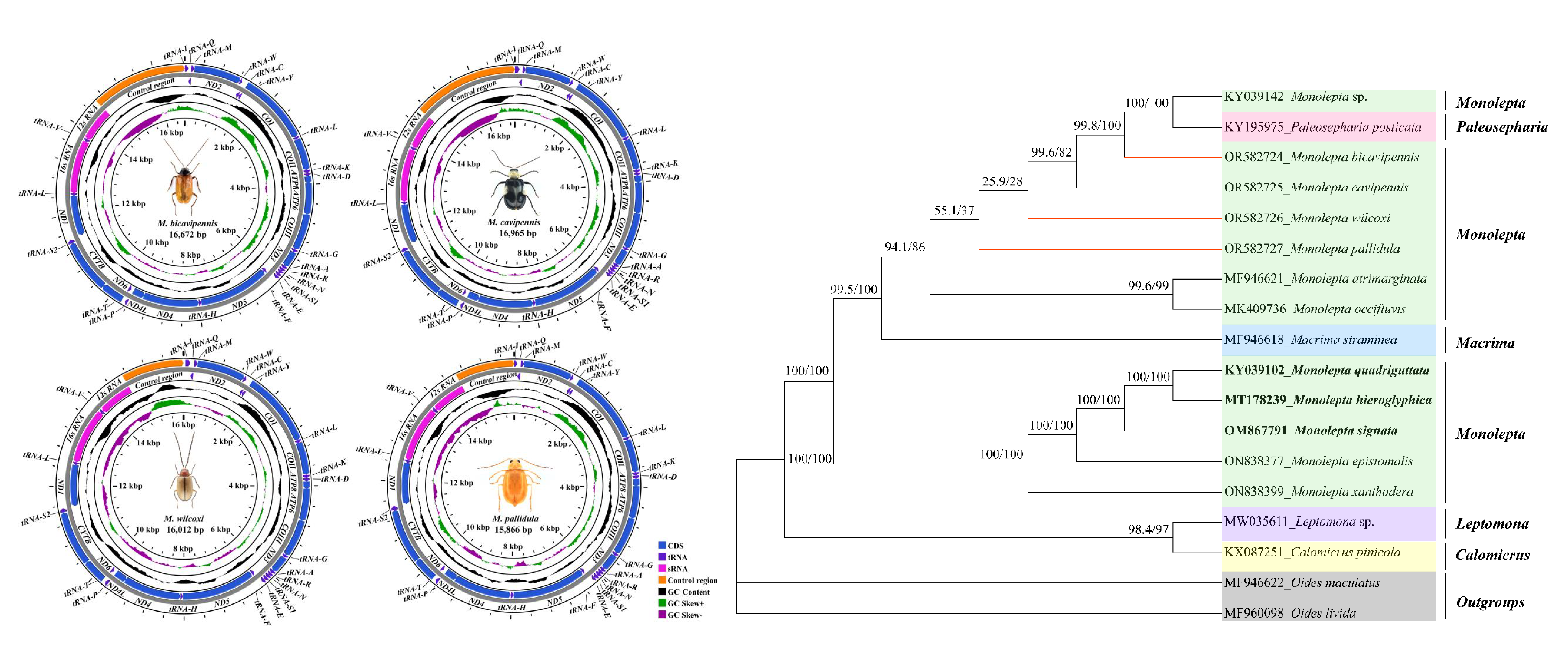
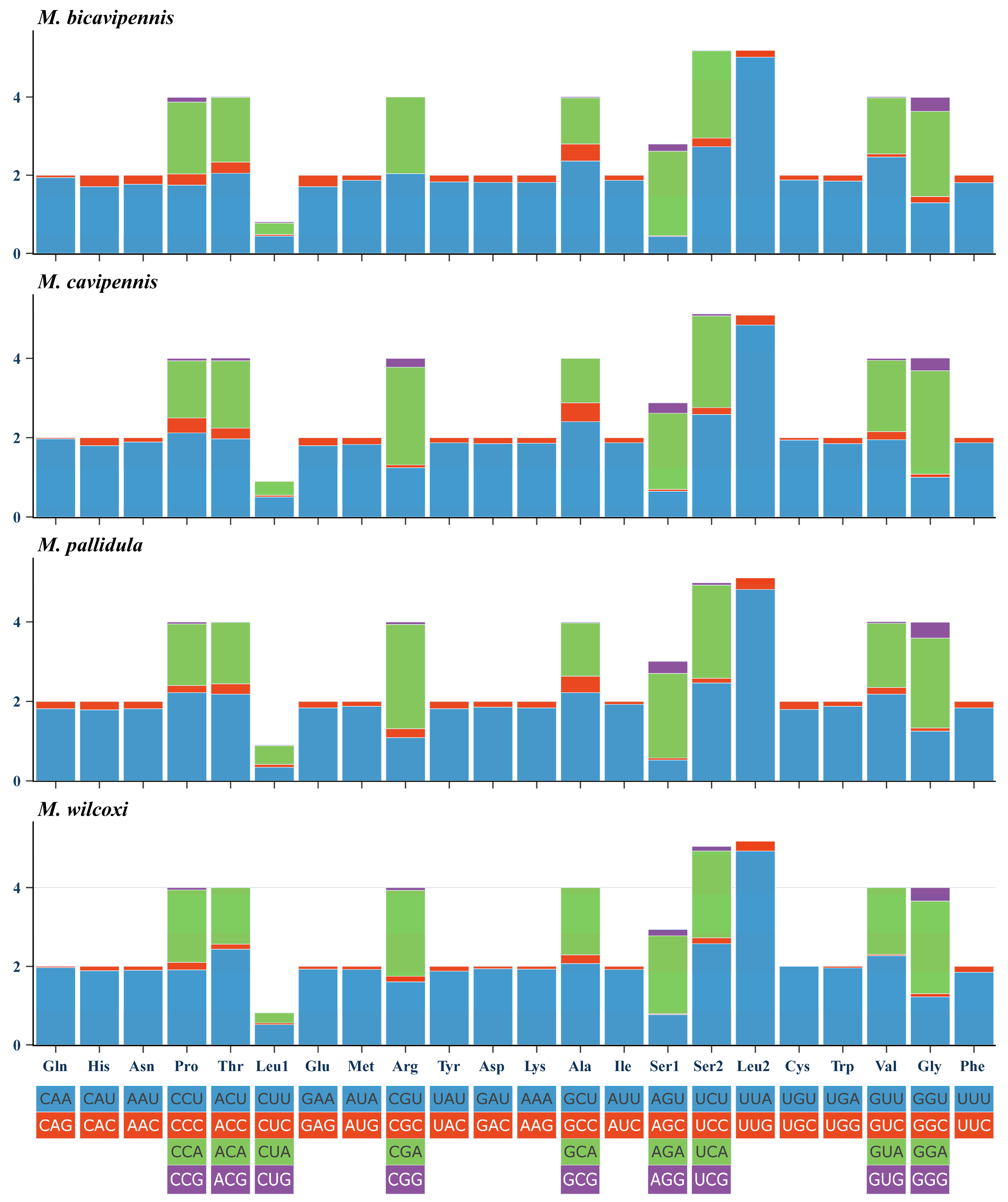
3.1. Sequence Data, Mitogenomic Organization, and Composition in Monolepta

Raw reads (about 15 Gb) were obtained for each sample using high-throughput sequencing technology. A total of four newly sequenced complete mitogenomes of Monolepta were obtained in this study, which were M. bicavipennis Chen, 1942 [60]; M. cavipennis (Baly, 1878) [61]; M. pallidula (Baly, 1874) [62]; and M. wilcoxi Gressitt & Kimoto, 1965 [63]. All newly sequenced mitochondrial genomes were submitted to GenBank, with accession numbers OR582724-OR582727 (Table 1). The four newly obtained sequences were circled and ranged from 15,866 bp (M. pallidula) to 16,965 bp (M. wilcoxi) in length, with significant variation in the size of the species mainly occurring in the control regions. All newly sequenced mitogenomes contained 37 genes (13 PCGs, 22 tRNAs, and 2 rRNAs) and a large non-coding region (control region), which is usually present in most insect mitochondrial genomes (Figure 1). Overall, the mitogenomic structure and nucleotide composition of these four species exhibited typical features of the family Chrysomelidae. Among these 37 genes, 9 genes from 13 PCGs and 14 genes from tRNAs were transcribed on the majority strand (J-strand), with the remaining genes oriented on the minority strand (N-strand). The four new mitogenomes only had a few overlaps between their detected genes, and the organization was very compact (Table S2). A UUU anticodon in tRNA-Lys, unique to Chrysomeloidea, was derived in the four newly sequenced mitogenomes of Monolepta, which is consistent with previous research [30,35,37].
The AT content of the newly sequenced mitogenomes exhibited a high degree of similarity in nucleotide composition (Table 2). All new mitogenomes had a significant bias of the total nucleotide composition toward A and T: 79.1% in M. bicavipennis; 79.3% in M. cavipennis; 79.8% in M. wilcoxi; and 78.8% in M. pallidula (Table 2). The skew metrics of four mitogenomes showed that AT-skew was positive and GC-skew was negative in PCGs, rRNAs, tRNAs, and control regions. The skew analysis indicated that the obvious bias was toward the use of A and C in the whole genomes (Table 2).
The lengths of 13 PCGs were not significantly different, ranging from 11,118 bp to 11,121 bp. For the four new mitogenomes, the details for start and stop codons of protein-coding genes can be seen in Table S2. Except for the ND1 gene starting with TTG, the rest of the PCGs started with ATN, all stop codons of 13 PCGs were TAA/TAG or just one single T. There is a very high similarity between the relative synonymous codon usage (RSCU) of the four sequenced mitogenomes and that of other previously determined beetles, and shows a codon usage bias (Figure 2): A and U were more frequently used than G and C. UUA, UCU, CGA, and GGA were the most frequently used codons.
The location and characteristics of the two rRNA genes are similar to those of previously studied beetles. The 16S rRNA gene is located between tRNA-Leu (TAG) and tRNA-Val. The 12S rRNA gene is located between tRNA-Val and the control region. The arrangement of the tRNA genes of the four new sequenced species is very conserved. The secondary structure of all 22 tRNAs is folded into the typical cloverleaf structure, except tRNA-Ser1 (AGN). Compared with the typical cloverleaf structure, tRNA-Ser1 (AGN) lacks the DHU-stem, with several unmatched base pairs in the anticodon stem (Figure 3). More information on the four newly observed mitochondrial structures can be seen in the Supplementary Data (Table S2).
The average ratio of nonsynonymous (Ka) to synonymous (Ks) substitution could be used to estimate non-neutral changes relative to neutral changes and the degree of the selective pressure of a PCG [64]. In this study, the Ka/Ks substitution ratios of 13 PCGs were less than one, and ranged from 0.09672 (COI) to 0.54603 (ND6) (Figure 4). The results demonstrated that all PCGs were under purifying selection. The evolution rate of 13 PCGs was as follows: ND6 > ATP8 > ND4L > ND2 > ND5 > ND3 > ND1 > ATP6 > ND4 > CYTB > COIII > COII > COI. Among them, COI showed the lowest evolution rate, while ND6 and ATP8 exhibited a faster evolutionary rate and greater diversity than other PCGs.
Substitution saturation testing reduces the amount of phylogenetic information contained in sequences and affects phylogenetic analyses involving deep branches. The substitution saturation of three datasets (13 PCGs_codon1, 13 PCGs_codon2, 13 PCGs_codon3) was assessed using DAMBE v.7. All the analyzed results showed a lower ISS value (simple index of substitution saturation) than ISS.c value (critical ISS value) (p < 0.05), which indicated all four datasets were not saturated (Figure 5). All those data types are feasible to use in phylogenetic analyses.

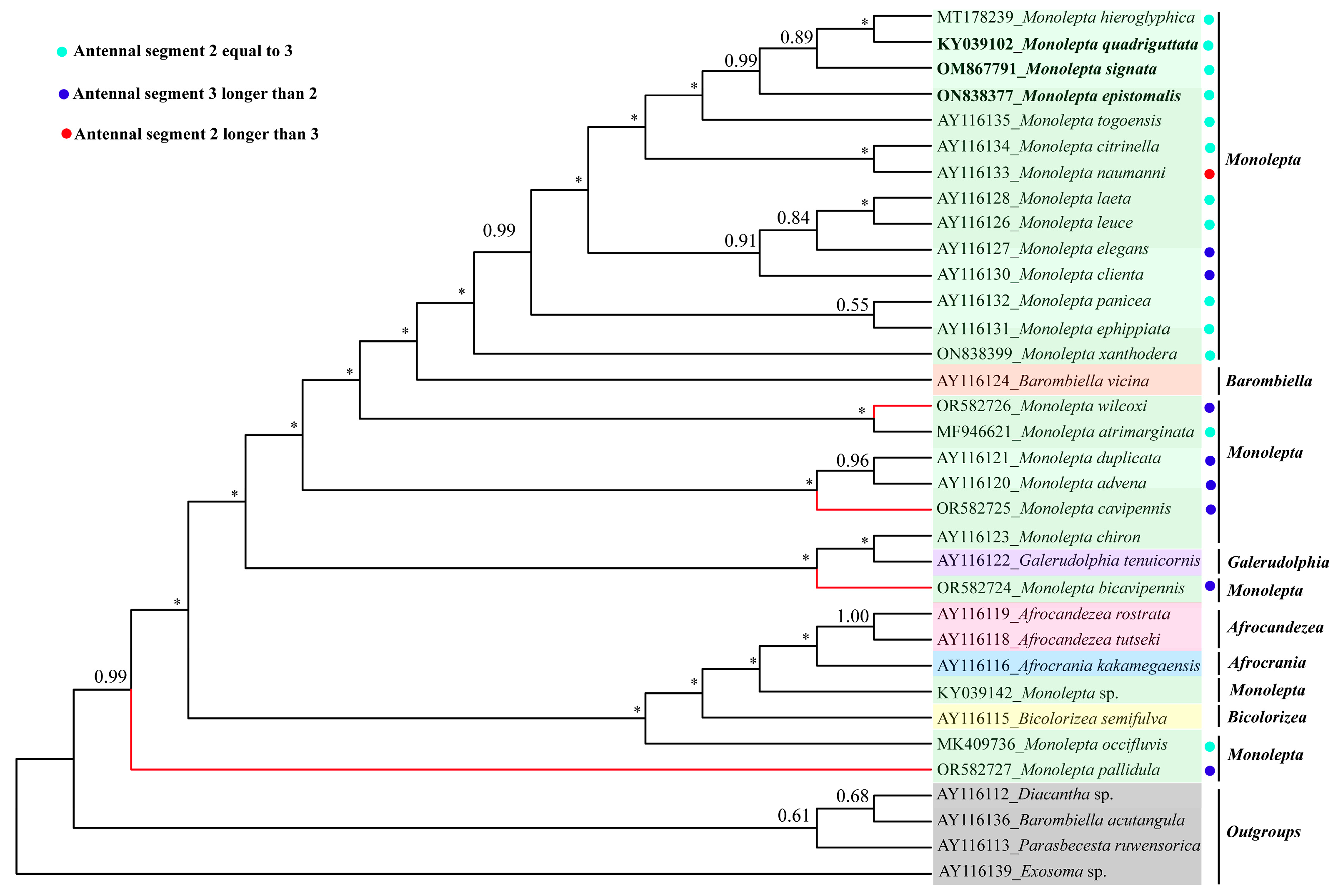
3.2. The Phylogeny of Monolepta

To verify the phylogenetic position of M. bicavipennis, M. cavipennis, M. wilcoxi, and M. pallidula within Monolepta, we used three datasets including 15 genes, 13 PCGs_AA, and ND1 to reconstruct the phylogenetic relationships using IQ-TREE and Phylobayes methods.
The ND1 dataset, including 34 taxa, was mainly pulled from the previous report by Stapel et al. [8], which was used to reconstruct the phylogenetic trees using the above two methods. The results showed that the topologies of IQ-TREE and Phylobayes tree were similar (Figure 6 and Figure S1). Monolepta was polyphyletic, with Afrocandezea, Afrocrania, Barombiella, and Pseudocrania emerging inside the Phylobayes tree. The newly sequenced Chinese species were divided into several distantly related branches. M. wilcoxi and M. atrimarginata were sister groups with low bootstrap value support; M. cavipennis and (M. advena + M. duplicata) and M. bicavipennis and (Galerudolphia tenuicornis + M. chiron) were sister groups, respectively; and M. pallidula was separated from all Monolepta species. Additionally, M. quadriguttata, M. hieroglyphica, and M. signata formed a clade with 89% bootstrap values, which is consistent with the results of Ge et al. [28].
For the 15 genes dataset (13 PCGs + 2 rRNAs) and 13 PCGs_AA, the 18 taxa were mainly pulled from NCBI and newly sequenced mitogenomes. The topologies of trees in Phylobayes and IQ-TREE were similar, except for the position of Macrima straminea, M. atrimarginata, and M. occifluvis (Figure 7 and Figures S2–S4). All Monolepta species were divided into two clades based on two datasets using different tree building methods (Figure 7 and Figures S1, S2 and S4), except for M. occifluvis and M. atrimarginata, which separated from all Monolepta species in the Phylobayes tree (Figure S3). The results showed that “Monoleptites” was a monophyletic group, while Monolepta was polyphyletic, splitting into several distant branches nested within Paleosepharia and Macrima. The four newly sequenced species had close relationships with Paleosepharia. M. quadriguttata, M. hieroglyphica, and M. signata formed one branch with a 100% bootstrap value and were proposed to be one species by Ge et al. [28] which was sister to M. epistomalis with the support of a 100% bootstrap value. The species of Monolepta with equal lengths were divided into two clades.

4. Discussion and Conclusions

In this study, the complete mitogenomes of four species of Monolepta, M. bicavipennis, M. cavipennis, M. wilcoxi, and M. pallidula, were sequenced successfully. All newly sequenced mitogenomes had similar structural characteristics and nucleotide compositions to previously published Chrysomelidae data. By combining the available mitogenomes of “Monoleptites” (18 taxa), their base compositions were calculated herein, and the results showed that all four mitogenomes were obviously biased towards A and T, which were similar to other beetles. The results of the ratio of nonsynonymous to synonymous substitution indicated that ND6 had the highest evolution rate, while COI displayed the lowest evolution rate, which is different from other leaf beetles. Hebert et al. [65] argued that a COI-based DNA barcoding identification system could be developed for all animals, that is, COI divergences can serve as an effective tool in species recognition; in this recognition system, intraspecific divergences are rarely greater than 2% and most are less than 1%. In the previous study, the COI (766 bp extracted according to primers of LCO and HCO) intraspecific divergences of M. hieroglyphica, M. quadriguttata, and M. signata were shown to be 1.3%, while those of ND1 were less than 1% (0.03%), and M. hieroglyphica and M. quadriguttata were confirmed to be synonyms of M. signata [28]. However, the divergences of the related, near-allied species of M. signata and M. wilcoxi was 12% for COI-barcode and 17% for ND6. Taking the highest interspecific evolution rate and lower intraspecific divergences of ND6, we propose that ND6 may be enabling the discrimination of closely allied species in Monolepta as an effective DNA marker.
Before constructing the phylogenetic tree, we tested the substitution saturation of 13 PCGs_codon1, 13 PCGs_codon2, and 13 PCGs_codon3. The results indicated all those data types were feasible to use to construct phylogenetic relationships. The phylogenetic relationship of the section “Monoleptites” was reconstructed based on three different datasets (15 genes, 13 PCGs_AA and ND1) using two different methods (IQ-TREE and Phylobayes). The topology of the phylogenetic tree based on ND1 was very similar to the topology built by Stapel et al. [8], based on ND1 and ITS2, which indicated that Monolepta was a non-monophyletic group. The Chinese distributed species of Monolepta were split into several clades and grouped with the African Monolepta species. Barombiella, Afrocrania, and Afrocandezea are restricted to African genera, which created one clear clade nested within M. occifluvis and Monolepta sp. with weak support. The phylogenetic inference based on the mitogenome showed a neater topology and higher node-supported value than that based on ND1 data. The mitogenomic data showed greater power to resolve most expected main clades than the ND1 gene at the suprageneric and species levels; this is mainly due to the larger number of variable characters, whereas each site also contains more information on average than ND1. The phylogenetic analyses based on mitogenomic data revealed that Monolepta was a non-monophyletic group. All the newly sequenced species whose antennal segment 2 was shorter than 3 had near relationships with Paleosepharia, and also had near relationships with Macrima. Monolepta was a very complicated group. Wilcox [66] was especially aware of the many inconsistent allocations of species to Monolepta. He commented that this genus needed to be revised and many species should be transferred to other genera. Lee [67] stated that some species of the genus Monolepta could be members of Paleosepharia and transferred four Monolepta species to Paleosepharia. In addition, the current study showed that the species whose antennal segments 2 and 3 are equal were divided into two clades. Wagner [9] re-described the type species of Monolepta and stated that the true “Monolepta” are those with a second and third antennomere of the same length. However, the current study supported that the characteristic of “antennal segment 2 as long as 3” of the true “Monolepta” evolved multiple times in several subgroups. So, to better understand the status of Monolepta and the suprageneric phylogenetic relationships at the species level, the revision of Monolepta’s relationship with related genera should be conducted first. Then, more taxon sampling and more molecular markers will be required in the future.

Supplementary Materials

The following supporting information can be downloaded at: https://www.mdpi.com/article/10.3390/insects15010050/s1. Figure S1. Phylogenetic tree reconstructed with IQ-TREE method based on ND1 gene (34 species) under MFP + MERGE model. Newly sequenced species in this study are highlighted in red color. The numbers above nodes in the tree are the SH-aLRT (left) and bootstrap values (right). Tips of synonymous species are highlighted in black color. Different colored backgrounds represent the different genera or outgroups. Figure S2. Phylogenetic tree reconstructed with IQ-TREE method based on amino acids of 13 PCGs-AA (18 species) under MFP + MERGE model. Newly sequenced species in this study are highlighted in red color. The numbers above nodes in the tree are the SH-aLRT (left) and bootstrap values (right). Tips of synonymous species are highlighted in black color. Different colored backgrounds represent the different genera. Figure S3. Phylogenetic tree reconstructed using Bayesian inference method based on 15 genes (13 PCGs + 2 rRNAs) (18 species) under CAT-GTR model. Newly sequenced species in this study are highlighted in red color. The numbers above nodes are Bayesian posterior probabilities. Tips of synonymous species are highlighted in black color. Asterisk indicates that the bootstrap value of the node is lower than 0.50. Different colored backgrounds represent the different genera. Figure S4. Phylogenetic tree reconstructed with IQ-TREE method based on 15 genes (13 PCGs + 2 rRNAs) (18 species) under MFP + MERGE model. Newly sequenced species in this study are highlighted in red color. The numbers above nodes in the tree are the SH-aLRT (left) and bootstrap values (right). Tips of synonymous species are highlighted in black color. Different colored backgrounds represent the different genera. Table S1. Samples and register gene information (ND1 data). Table S2. Organization of four newly sequenced mitogenomes of Monolepta.

Author Contributions

Conceptualization, J.-S.H., X.-K.Y. and R.-E.N.; Methodology, R.-E.N.; Software, I.Z., X.J. and R.-R.G.; Formal analysis, I.Z., R.-R.G. and C.-Y.S.; Resources, J.-S.H. and R.-E.N.; Data curation., I.Z., X.J., Q.-L.L. and R.-R.G.; Writing—original draft, Q.-L.L., R.-R.G., X.-K.Y., J.-S.H. and R.-E.N.; Writing—review and editing, Q.-L.L., I.Z., X.J., R.-R.G. and R.-E.N.; Visualization, C.-Y.S.; Supervision, J.-S.H. and R.-E.N.; Funding acquisition, R.-E.N. All authors have read and agreed to the published version of the manuscript.

Funding

This research was funded by grants from the Anhui Provincial University Outstanding Youth Program (2022AH020021), the National Science Foundation of China (Nos. 32170443), partly by the Science & Technology Fundamental Resources Investigation Program (2022FY202100).

Data Availability Statement

The following information was supplied regarding the availability of DNA sequences: the new mitogenomes are deposited in GenBank of NCBI and the accession numbers are OR582724-OR582727.

Acknowledgments

We express our sincere gratitude to Ming Bai and Yu-Xia Yang for collecting some samples.

Conflicts of Interest

The authors declare no conflicts of interest.

References

  1. Chevrolat, L.A.A. Monolepta. In Catalogues Des Coléopterès De La Collection De M. LeComte Dejean, Troisième éd.; revue, corrigée et augmentée, livraison, Dejean, P.F.M.A., Ed.; Méquignon-Marvis Père et Fils: Paris, France, 1837; Volume 5, pp. 385–503. [Google Scholar]
  2. Nie, R.E.; Bezděk, J.; Yang, X.K. How many genera and species of Galerucinaes. str. do we know? Updated statistics (Coleoptera, Chrysomelidae). Zookeys 2017, 720, 91–102. [Google Scholar] [CrossRef] [PubMed]
  3. Yang, X.K. Genus Monolepta Chevrolat, 1837. In Chinese Leaf Beetles; Yang, X.K., Ge, S.Q., Nie, R.E., Ruan, Y.Y., Li, W.Z., Eds.; Science Press: Beijing, China, 2015; pp. 261–274. [Google Scholar]
  4. Zhang, C.; Yuan, Z.H.; Wang, Z.Y.; He, K.L.; Bai, S.X. Population dynamics of Monolepta hieroglyphica (Motschulsky) in cornfields. J. Appl. Entomol. 2014, 51, 668–675. [Google Scholar]
  5. Chapuis, F. Famille des Phytophages. In Histoire Naturelle Des Insects; Lacordaire, T., Chapuis, F., Eds.; Genera des Coleopteres: Paris, France, 1875; Volume 11, pp. 1–420. [Google Scholar]
  6. Wilcox, J.A. Chrysomelidae: Galerucinae: Luperini: Luperina. In Coleopterorum Catalogus Supplementa. Pars 78 (3); Wilcox, J., Ed.; BIBLIO: Gravenhage, The Netherlands, 1973; pp. 433–664. [Google Scholar]
  7. Bolz, H.; Wagner, T. Neobarombiella, a diverse, newly described genus of Afrotropical Galerucinae (Coleoptera, Chrysomelidae). Zootaxa 2012, 3463, 1–112. [Google Scholar] [CrossRef]
  8. Stapel, H.; Misof, B.; Wagner, T. A molecular and morphological phylogenetic analysis of Afrotropical Monolepta species and related Galerucinae (Coleoptera: Chrysomelidae). Arthropod Syst. Phylogeny 2008, 66, 3–17. [Google Scholar] [CrossRef]
  9. Wagner, T. Monolepta Chevrolat, 1837, the most speciose galerucine taxon: Redescription of the type species Monolepta bioculata (Fabricius, 1781) and key to related genera from Africa (Chrysomelidae, Coleoptera). J. Nat. Hist. 2007, 41, 81–100. [Google Scholar] [CrossRef]
  10. Wagner, T. Revision of afrotropical Monolepta species (Coleoptera, Chrysomelidae, Galerucinae). Part I: Species with red and black coloured elytra, pronotum and head, with description of new species. Entomol. Z. 2000, 110, 226–237. [Google Scholar]
  11. Wagner, T. Revision of Afrotropical Monolepta Chevrolat, 1837 (Coleoptera, Chrysomelidae, Galerucinae). Part II: Species with red elytra, pronotum and elytra, with descriptions of new species. Bonn. Zool. Beitr. 2001, 50, 49–65. [Google Scholar]
  12. Wagner, T. Revision of Afrotropical Monolepta species (Coleoptera, Chrysomelidae, Galerucinae). Part III: Species with red elytra and yellow prothorax, including description of new species. Dtsch. Entomol. Z. 2002, 49, 27–45. [Google Scholar] [CrossRef]
  13. Steiner, I.; Wagner, T. Afronaumannia gen. nov., a new monophyletic group of leaf beetles from Africa (Coleoptera: Chrysomelidae, Galerucinae). Entomol. Z. 2005, 115, 15–24. [Google Scholar]
  14. Wagner, T. Description of Monoleptoides gen.nov. from the Afrotropical Region, including the revision of nine species (Coleoptera: Chrysomelidae: Galerucinae). Bonn Zool. Bull. 2011, 60, 169–199. [Google Scholar]
  15. Wagner, T.; Bieneck, S. Galerucine type material described by Victor Motschu in 1858 and 1866 from the Zoological Museum Moscow (Coleoptera: Chrysomelidae, Galerucinae). Entomol. Z. 2012, 122, 205–216. [Google Scholar]
  16. Hazmi, I.R.; Wagner, T. Revision of Neolepta Jacoby, 1884 and related Galerucines from the Oriental region, including descriptions of two new genera (Coleoptera: Chrysomelidae: Galerucinae). Raffles Bull. Zool. 2013, 61, 73–95. [Google Scholar]
  17. Heunemann, L.O.; Dalstein, V.; Schulze, M.; Wagner, T. Bicolorizea gen. nov. from tropical Africa (Coleoptera: Chrysomelidae, Galerucinae). Mitt. Mus. Naturkunde Berl. Dtsch. Entomol. Z. 2015, 125, 235–246. [Google Scholar]
  18. Wagner, T. Doeberllepta gen. nov., a new monotypic genus for a widely distributed galerucine species from Africa (Coleoptera: Chrysomelidae). Entomol. Blätter 2017, 113, 245–253. [Google Scholar]
  19. Laboissière, V. Observations sur les Galerucini asiatiques principalement du Tonkin et du Yunnan et descriptions de nouveaux genres et espèces (5e partie). Ann. Soc. Entomol. Fr. 1936, 105, 239–261. [Google Scholar] [CrossRef]
  20. Chûjô, M.H. Sauter’s. Formosa-Ausbeute: Subfamily Galerucinae (Coleoptera: Chrysomelidae). Arb. Morph. Taxon. Ent. Berlin-Dahlem 1935, 2, 160–174. [Google Scholar]
  21. Chûjô, M. A taxonomic study on the Chrysomelidae (Insecta: Coleoptera) from Formosa XI. Subfamily Galerucinae. Philipp. J. Sci. 1962, 91, 1–239. [Google Scholar]
  22. Kimoto, S. Notes on the Chryosmelidae from Taiwan, China. II. Esakia 1969, 7, 1–68. [Google Scholar] [CrossRef] [PubMed]
  23. Kimoto, S. Notes on the Chrysomelidae from Taiwan, China, XIII. Entomol. Rev. Jpn. 1996, 51, 27–51. [Google Scholar]
  24. Gressitt, J.L.; Kimoto, S. The Chrysomelidae (Coleopt.) of China and Korea. Part II. Pac. Insects Monogr. 1963, 1B, 301–1026. [Google Scholar]
  25. Lee, C.F. Revision of Taiwanese species of Atrachya Chevrolat, 1836 (Coleoptera, Chrysomelidae, Galerucinae): Descriptions of three new genera, two new species, and designations of three new synonyms. Zookeys 2020, 940, 117–159. [Google Scholar] [CrossRef] [PubMed]
  26. Lee, C.F. A taxonomic revision of Monolepta pallidula species group in Taiwan (Coleoptera: Chrysomelidae: Galerucinae). Zootaxa 2009, 2170, 15–27. [Google Scholar] [CrossRef]
  27. Lei, Q.L.; Xu, S.Y.; Yang, X.K.; Nie, R.E. Five new species of the leaf-beetle genus Monolepta Chevrolat (Coleoptera, Chrysomelidae, Galerucinae) from China. Zookeys 2021, 1056, 35–57. [Google Scholar] [CrossRef] [PubMed]
  28. Ge, Y.C.; Shi, C.M.; Bai, M.; Cao, Z.Y.; Cao, L.P.; Wang, Z.Y.; Dong, J.A.; Wang, Y.Y. Molecular data confirm Monolepta hieroglyphica (Motschulsky, 1858) and M. quadriguttata (Motschulsky, 1860) being synonyms of M. signata (Oliver, 1808). Insect Syst. Evol. 2023, 54, 402–412. [Google Scholar] [CrossRef]
  29. Bolz, H.; Wagner, T. Revision of Galerudolphia Hincks, 1949 (Coleoptera: Chrysomelidae, Galerucinae). Insect Syst. Evol. 2004, 35, 361–400. [Google Scholar] [CrossRef]
  30. Nie, R.E.; Breeschoten, T.; Timmermans, M.J.T.N.; Nadein, K.; Xue, H.J.; Bai, M.; Huang, Y.; Yang, X.K.; Vogler, A.P. The phylogeny of Galerucinae (Coleoptera: Chrysomelidae) and the performance of mitochondrial genomes in phylogenetic inference compared to nuclear rRNA genes. Cladistics 2018, 34, 113–130. [Google Scholar] [CrossRef] [PubMed]
  31. Sheffield, N.C.; Song, H.; Cameron, S.L.; Whiting, M.F. A comparative analysis of mitochondrial genomes in Coleoptera (Arthropoda: Insecta) and genome descriptions of six new beetles. Mol. Biol. Evol. 2008, 25, 2499–2509. [Google Scholar] [CrossRef]
  32. Sheffield, N.C.; Song, H.; Cameron, S.L.; Whiting, M.F. Nonstationary evolution and compositional heterogeneity in beetle mitochondrial phylogenomics. Syst. Biol. 2009, 58, 381–394. [Google Scholar] [CrossRef]
  33. Pons, J.; Ribera, I.; Bertranpetit, J.; Balke, M. Nucleotide substitution rates for the full set of mitochondrial protein-coding genes in Coleoptera. Mol. Phylogenet. Evol. 2010, 56, 796–807. [Google Scholar] [CrossRef]
  34. Yuan, M.L.; Zhang, Q.L.; Zhang, L.; Guo, Z.L.; Liu, Y.J.; Shen, Y.Y.; Shao, R. High-level phylogeny of the Coleoptera inferred with mitochondrial genome sequences. Mol. Phylogenet. Evol. 2016, 104, 99–111. [Google Scholar] [CrossRef] [PubMed]
  35. Timmermans, M.J.T.N.; Barton, C.; Haran, J.; Ahrens, D.; Culverwell, C.L.; Ollikainen, A.; Dodsworth, S.; Foster, P.G.; Bocak, L.; Vogler, A.P. Family-level sampling of mitochondrial genomes in Coleoptera: Compositional heterogeneity and phylogenetics. Genome Biol. Evol. 2016, 8, 161–175. [Google Scholar] [CrossRef] [PubMed]
  36. Nie, R.E.; Vogler, A.P.; Yang, X.K.; Lin, M.Y. Higher-level phylogeny of longhorn beetles (Coleoptera: Chrysomeloidea) inferred from mitochondrial genomes. Syst. Entomol. 2021, 46, 56–70. [Google Scholar] [CrossRef]
  37. Nie, R.E.; Andújar, C.; Gómez-Rodríguez, C.; Bai, M.; Xue, H.J.; Tang, M.; Yang, C.T.; Tang, P.; Yang, X.K.; Vogler, A.P. The phylogeny of leaf beetles (Chrysomelidae) inferred from mitochondrial genomes. Syst. Entomol. 2020, 45, 188–204. [Google Scholar] [CrossRef]
  38. Bolger, A.M.; Lohse, M.; Usadel, B. Trimmomatic: A flexible trimmer for Illumina sequence data. Bioinformatics 2014, 30, 2114–2120. [Google Scholar] [CrossRef] [PubMed]
  39. Schmieder, R.; Edwards, R. Quality control and preprocessing of metagenomic datasets. Bioinformatics 2011, 27, 863–864. [Google Scholar] [CrossRef]
  40. Jin, J.J.; Yu, W.B.; Yang, J.B.; Song, Y.; dePamphilis, C.W.; Yi, T.S.; Li, D.Z. GetOrganelle: A fast and versatile toolkit for accurate de novo assembly of organelle genomes. Genome Biol. 2020, 21, 241. [Google Scholar] [CrossRef]
  41. Kearse, M.; Moir, R.; Wilson, A.; Stones-Havas, S.; Cheung, M.; Sturrock, S.; Buxton, S.; Cooper, A.; Markowitz, S.; Duran, C.; et al. Geneious Basic: An integrated and extendable desktop software platform for the organization and analysis of sequence data. Bioinformatics 2012, 28, 1647–1649. [Google Scholar] [CrossRef]
  42. Grant, J.R.; Stothard, P. The CGView Server: A comparative genomics tool for circular genomes. Nucleic Acids Res. 2008, 36, W181–W184. [Google Scholar] [CrossRef]
  43. Perna, N.T.; Kocher, T.D. Patterns of nucleotide composition at fourfold degenerate sites of animal mitochondrial genomes. J. Mol. Evol. 1995, 41, 353–358. [Google Scholar] [CrossRef]
  44. Zhang, D.; Gao, F.; Jakovlić, I.; Zou, H.; Zhang, J.; Li, W.X.; Wang, G.T. PhyloSuite: An integrated and scalable desktop platform for streamlined molecular sequence data management and evolutionary phylogenetics studies. Mol. Ecol. Resour. 2020, 20, 348–355. [Google Scholar] [CrossRef]
  45. Xiang, C.Y.; Gao, F.L.; Jakovlić, L.; Lei, H.P.; Hu, Y.; Zhang, H.; Zou, H.; Wang, G.T.; Zhang, D. Using PhyloSuite for molecular phylogeny and tree-based analyses. iMeta 2023, 2, e87. [Google Scholar] [CrossRef]
  46. Rozas, J.; Ferrer-Mata, A.; Sánchez-DelBarrio, J.C.; Guirao-Rico, S.; Librado, P.; Ramos-Onsins, S.E.; Sánchez-Gracia, A. DnaSP 6: DNA sequence polymorphism analysis of large data sets. Mol. Biol. Evol. 2017, 34, 3299–3302. [Google Scholar] [CrossRef] [PubMed]
  47. Tamura, K.; Stecher, G.; Kumar, S. MEGA11 Molecular evolutionary genetics analysis version 11. Mol. Biol. Evol. 2021, 38, 3022–3027. [Google Scholar] [CrossRef] [PubMed]
  48. Bernt, M.; Donath, A.; Jühling, F.; Externbrink, F.; Florentz, C.; Fritzsch, G.; Pütz, J.; Middendorf, M.; Stadler, P.F. MITOS: Improved de novo metazoan mitochondrial genome annotation. Mol. Phylogenet. Evol. 2013, 69, 313–319. [Google Scholar] [CrossRef] [PubMed]
  49. Xia, X.H. DAMBE7: New and Improved Tools for Data Analysis in Molecular Biology and Evolution. Mol. Biol. Evol. 2018, 35, 1550–1552. [Google Scholar] [CrossRef]
  50. Bininda-Emonds, O.R. transAlign: Using amino acids to facilitate the multiple alignment of protein-coding DNA sequences. BMC Bioinform. 2005, 6, 156. [Google Scholar] [CrossRef] [PubMed]
  51. Edgar, R.C. MUSCLE: Multiple sequence alignment with high accuracy and high throughput. Nucleic Acids Res. 2004, 32, 1792–1797. [Google Scholar] [CrossRef]
  52. Castresana, J. Selection of conserved blocks from multiple alignments for their use in phylogenetic analysis. Mol. Biol. Evol. 2000, 17, 540–552. [Google Scholar] [CrossRef]
  53. Patrick, P.S.; Hammersley, J.; Loizou, L.; Kettunen, M.I.; Rodrigues, T.B.; Hu, D.E.; Tee, S.S.; Hesketh, R.; Lyons, S.K.; Soloviev, D.; et al. Dual-modality gene reporter for in vivo imaging. Proc. Natl. Acad. Sci. USA 2014, 111, 415–420. [Google Scholar] [CrossRef]
  54. Lartillot, N.; Rodrigue, N.; Stubbs, D.; Richer, J. Phylobayes MPI: Phylogenetic reconstruction with infinite mixtures of profiles in a parallel environment. Syst. Biol. 2013, 62, 611–615. [Google Scholar] [CrossRef]
  55. Minh, B.Q.; Schmidt, H.A.; Chernomor, O.; Schrempf, D.; Woodhams, M.D.; von Haeseler, A.; Lanfear, R. IQ-TREE 2: New models and efficient methods for phylogenetic inference in the genomic era. Mol. Biol. Evol. 2020, 37, 1530–1534. [Google Scholar] [CrossRef] [PubMed]
  56. Guindon, S.; Dufayard, J.F.; Lefort, V.; Anisimova, M.; Hordijk, W.; Gascuel, O. New algorithms and methods to estimate maximum-likelihood phylogenies: Assessing the performance of PhyML 3.0. Syst. Biol. 2010, 59, 307–321. [Google Scholar] [CrossRef] [PubMed]
  57. Hoang, D.T.; Chernomor, O.; von Haeseler, A.; Minh, B.Q.; Vinh, L.S. UFBoot2: Improving the ultrafast bootstrap approximation. Mol. Biol. Evol. 2018, 35, 518–522. [Google Scholar] [CrossRef] [PubMed]
  58. Jiang, Z.L.; Wang, D.M.; Wang, B.F.; Song, X.Y. Genome survey and mitochondrial genome analysis of Monolepta hieroglyphica. J. Environ. Entomol. 2019, 41, 1287–1296. [Google Scholar]
  59. Song, N.; Yin, X.M.; Zhao, X.C.; Chen, J.H.; Yin, J. Reconstruction of mitogenomes by NGS and phylogenetic implications for leaf beetles. Mitochondrial DNA Part A 2018, 29, 1041–1050. [Google Scholar] [CrossRef] [PubMed]
  60. Chen, S.X. Galerucinae nouveaux de la faune Chinoise. Notes D’entomologie Chin. 1942, 9, 9–67. [Google Scholar]
  61. Baly, J.S. Descriptions of the phytophagous Coleoptera collected by the late Dr. F. Stoliczka during Forsyth’s expedition to Kashgar in 1873–1874. Cistula Entomol. 1878, 2, 369–383. [Google Scholar]
  62. Baly, J.S. Catalogue of the phytophagous Coleoptera of Japan, with descriptions of the species new to science. Trans. Ent. Soc. Lond. 1874, 22, 161–217. [Google Scholar] [CrossRef]
  63. Gressitt, J.L.; Kimoto, S. Second supplement to ‘The Chrysomelidae (Coleopt.) of China and Korea. Pac. Insects 1965, 7, 799–806. [Google Scholar]
  64. Hurst, L.D. The Ka/Ks ratio: Diagnosing the form of sequence evolution. Trends Genet. 2002, 18, 486–487. [Google Scholar] [CrossRef]
  65. Hebert, P.D.; Ratnasingham, S.; deWaard, J.R. Barcoding animal life: Cytochrome c oxidase subunit I divergences among closely related species. Proc. Biol. Sci. 2003, 270 (Suppl. 1), S96–S99. [Google Scholar] [CrossRef] [PubMed]
  66. Wilcox, J.A. Coleopterum Catalogus Supplementa (Chrysomelidae: Galerucinae, Oidini, Galerucini, Metacyclini, Sermylini), 2nd ed.; BIBLIO: Gravenhage, The Netherlands, 1971; pp. 1–770. [Google Scholar]
  67. Lee, C.F. The genus Paleosepharia Laboissiere, 1936 in Taiwan: Review and nomenclatural changes (Coleoptera, Chrysomelidae, Galerucinae). Zookeys 2018, 744, 19–41. [Google Scholar] [CrossRef] [PubMed]
Figure 1. Circle maps of the four complete mitochondrial genomes of Monolepta species, with different colors to distinguish different genes.
Insects 15 00050 g001
Figure 2. Relative synonymous codon usage (RSCU) of the four new mitogenomes.
Insects 15 00050 g002
Figure 3. The secondary structure of tRNA-Ser1 (AGN) in the four newly determined mitogenomes, and the predicted secondary structure of tRNA-Val in the M. bicavipennis mitogenome. The pink circle represents a mismatched base, and the orange square represents a matched base.
Insects 15 00050 g003
Figure 4. Non-synonymous (Ka) to synonymous (Ks) substitution rates of 13 PCGs among four sequenced species.
Insects 15 00050 g004
Figure 5. The chart of substitution saturation for the three different mitogenomes’ datasets. The plots show uncorrected pairwise divergences in transitions (s) (blue) to transversions (v) (orange) compared with divergences calculated by GTR model.
Insects 15 00050 g005
Figure 6. Phylogenetic tree reconstructed by Bayesian inference method based on ND1 gene (34 species) under CAT-GTR model. Newly sequenced species in this study are highlighted in red color. The numbers above nodes are Bayesian posterior probabilities. Tips of synonymous species are highlighted in black color. Asterisk indicates that the bootstrap value of the node is lower than 0.50. Different colored backgrounds represent the different genera or outgroups.
Insects 15 00050 g006
Figure 7. Phylogenetic tree reconstructed by Bayesian inference method based on 13 PCGs-AA (18 species) under CAT-GTR model. Newly sequenced species in this study are highlighted in red color. The numbers above nodes are Bayesian posterior probabilities. Tips of synonymous species are highlighted in black color. Asterisk indicates that the bootstrap value of the node is lower than 0.50. Different colored backgrounds represent the different genera.
Insects 15 00050 g007
Table 1. The information of 18 examined species with NCBI accession numbers and their references.
SubfamilyTribeSpeciesLength (bp)Acc. No.References
GalerucinaeLuperiniCalomicrus pinicola15,436KX087251unpublished
GalerucinaeLuperiniLeptomona sp.16,697MW035611unpublished
GalerucinaeLuperiniMacrima straminea15,567MF946618[30]
GalerucinaeLuperiniMonolepta atrimarginata15,143MF946621[30]
GalerucinaeLuperiniMonolepta bicavipennis16,672OR582724this study
GalerucinaeLuperiniMonolepta cavipennis16,965OR582725this study
GalerucinaeLuperiniMonolepta epistomalis15,161ON838377unpublished
GalerucinaeLuperini* Monolepta hieroglyphica16,299MT178239[58]
GalerucinaeLuperiniMonolepta occifluvis15,998MK409736unpublished
GalerucinaeLuperini* Monolepta quadriguttata16,130KY039102[59]
GalerucinaeLuperiniMonolepta pallidula15,866OR582727this study
GalerucinaeLuperini* Monolepta signata16,329OM867791unpublished
GalerucinaeLuperiniMonolepta sp.15,792KY039142[59]
GalerucinaeLuperiniMonolepta wilcoxi16,012OR582726this study
GalerucinaeLuperiniMonolepta xanthodera14,782ON838399unpublished
GalerucinaeLuperiniPaleosepharia posticata15,729KY195975unpublished
GalerucinaeOidiniOides livida16,127MF960098[30]
GalerucinaeOidiniOides maculatus15,089MF946622[30]
Note: * The synonymous species of M. quadriguttata, M. signata, and M. hieroglyphica were not combined because it is better to keep the original names they had when they were submitted.
Table 2. Base composition of the four mitogenomes.
SpeciesWhole MitogenomeProtein-Coding
Genes
12S rRNA Genes16S rRNA GenesControl Region
A + T%A + T%AT-SkewGC-SkewA + T%A + T%A + T%
M. bicavipennis79.177.6 −0.1450.00381.283.083.4
M. cavipennis79.377.6−0.1420.01781.782.083.1
M. pallidula78.877.3−0.1430.01580.783.085.9
M. wilcoxi79.878.4 −0.1430.01883.383.182.5
Disclaimer/Publisher’s Note: The statements, opinions and data contained in all publications are solely those of the individual author(s) and contributor(s) and not of MDPI and/or the editor(s). MDPI and/or the editor(s) disclaim responsibility for any injury to people or property resulting from any ideas, methods, instructions or products referred to in the content.

