Article

Antioxidant and Anticancer Potential of Bioactive Compounds from Rhinacanthus nasutus Cell Suspension Culture

by
Pattralak Songserm
1,2,
Poramaporn Klanrit
3,
Poramate Klanrit
4,5,
Jutarop Phetcharaburanin
4,5,
Pornthap Thanonkeo
2,6,
Jirawan Apiraksakorn
2,6,
Khamphee Phomphrai
7 and
Preekamol Klanrit
2,5,6,*
1
Graduate School, Khon Kaen University, Khon Kaen 40002, Thailand
2
Department of Biotechnology, Faculty of Technology, Khon Kaen University, Khon Kaen 40002, Thailand
3
Research Group of Chronic Inflammatory Oral Diseases and Systemic Diseases Associated with Oral Health, Department of Oral Biomedical Sciences, Faculty of Dentistry, Khon Kaen University, Khon Kaen 40002, Thailand
4
Department of Biochemistry, Faculty of Medicine, Khon Kaen University, Khon Kaen 40002, Thailand
5
Cholangiocarcinoma Research Institute, Faculty of Medicine, Khon Kaen University, Khon Kaen 40002, Thailand
6
Fermentation Research Center for Value Added Agricultural Products (FerVAAP), Khon Kaen University, Khon Kaen 40002, Thailand
7
School of Molecular Science and Engineering, Vidyasirimedhi Institute of Science and Technology, Rayong 21210, Thailand
*
Author to whom correspondence should be addressed.
Plants 2022, 11(15), 1994; https://doi.org/10.3390/plants11151994
Submission received: 26 June 2022 / Revised: 22 July 2022 / Accepted: 26 July 2022 / Published: 30 July 2022
(This article belongs to the Section Plant Genetics, Genomics and Biotechnology)

Abstract

:
The potential benefits of natural plant extracts have received attention in recent years, encouraging the development of natural products that effectively treat various diseases. This is the first report on establishing callus and cell suspension cultures of Rhinacanthus nasutus (L.) Kurz. A yellow friable callus was successfully induced from in vitro leaf explants on Murashige and Skoog medium supplemented with 1 mg/L 2,4-dichlorophenoxyacetic acid and 1 mg/L 1-naphthalene acetic acid. A selected friable callus line was used to establish the cell suspension culture with the same medium. The antioxidant assays showed that the leaf- and ethanolic-suspension-cultured cell (SCC) extracts exhibited high antioxidant potential. In addition, the in vitro cytotoxicity revealed by the MTT assay demonstrated potent antiproliferative effects against the oral cancer cell lines ORL-48 and ORL-136 in a dose-dependent manner. Several groups of compounds, including terpenoids, phenolics, flavonoids, quinones, and stilbenes, were identified by UHPLC–QToF–MS, with the same compounds detected in leaf and SCC extracts, including austroinulin, lucidenic acid, esculetin, embelin, and quercetin 3-(2″-p-hydroxybenzoyl-4″-p-coumarylrhamnoside). The present study suggests the value of further investigations for phytochemical production using R. nasutus cell suspension culture.

1. Introduction

Rhinacanthus nasutus (L.) Kurz (R. nasutus), also known as snake jasmine or Thong Pan Chang (in Thai), is a high-value medicinal and versatile plant that belongs to the Acanthaceae family. It is widely cultivated in tropical and subtropical areas, including India, China, and Thailand [1]. Different parts of R. nasutus, including the leaves and roots, have also been used in traditional medicine for the treatment of various diseases and disorders, including skin diseases such as eczema, herpes, inflammation, hepatitis, diabetes, and hypertension, as well as bacterial, fungal, or viral infections [2,3,4]. R. nasutus has been shown to contain significant levels of essential bioactive compounds; for example, phenolic compounds, including phenolic acids, stilbenes, lignans, and flavonoids, as well as other groups of compounds, including steroids, terpenoids, and quinones, which have been isolated from and characterized in the leaves and roots [4,5,6,7]. These bioactive compounds offer several attractive biological properties and therapeutic potential, such as antibacterial, antifungal, antiviral, antioxidant, neuroprotective, and anticancer effects, making R. nasutus the subject of research in several fields [7,8,9,10,11,12].
Most of the relevant research about R. nasutus has mainly focused on extracting bioactive compounds from plant parts, identifying the bioactive compounds, and investigating their potential biological properties. However, the main drawback of using plant parts from nature or traditional cultivation is the potential for many diseases and pathogens, decreasing their survival and leading to contamination. Moreover, the accumulation levels of bioactive compounds vary depending on the geography and environmental influences. Thus, plant tissue and cell cultures are alternative techniques used to circumvent these problems. Compared with traditional growth, plant tissue cultures offer several advantages, including the rapid multiplication of plant species in a short period, the production of pathogen-free plants, and the continuous production of commercially important bioactive compounds using a callus or cell suspension culture.
Plant callus and cell suspension cultures exhibit superior secondary metabolite production over an extended subculture period compared to extraction from natural plant leaves [13]. The induction of leaf-derived callus and cell suspension cultures is an alternative technique for inducing the same group of bioactive compounds present in the leaf. The callus induction is the first step in the establishment of cell suspension cultures, in which friable callus fragments are placed into suitable sterile liquid media [14]. The types and concentrations of plant growth regulators (PGRs) used in the system are essential parameters for successful callus induction. The fine cell suspension culture provides an excellent opportunity to investigate the properties and potentialities of plant cells. Moreover, the cell suspension culture offers a valuable platform for producing high-value secondary metabolites and other invaluable bioactive compounds of commercial interest [15,16].
Several studies have demonstrated the in vitro production of secondary metabolites from many plants for various purposes. For example, the roots of Panax ginseng are used for the production of ginsenoside and hypocotyls of Celosia argentea are used as explants for the production of betalains as color compounds [17,18]. To date, many studies on the micropropagation of R. nasutus to generate new whole plants using various explant techniques have been published [19,20]. However, only a few studies have reported on the leaf-derived callus induction of R. nasutus without further establishment for bioactive compound production [21,22,23]. For callus induction, it was found that using Murashige and Skoog (MS) medium supplemented with different concentrations of PGRs, such as 5 mg/L 6-benzylaminopurine (BAP) and 2.5 mg/L 2,4-dichlorophenoxyacetic acid (2,4-D) [21], 0.1 mg/L thidiazuron (TDZ) and 0.5 mg/L 2,4-D [24], and 4 mg/L kinetin (KT) and 0.5 mg/L indole-3-butyric acid (IBA) [23] or 2 mg/L 2,4-D alone, was successful in generating different types of callus [21,22]. However, the callus texture was compact and was not suitable for establishing a cell suspension culture system. Moreover, the study of bioactive compound production using suspension culture and the potential of the suspension-cultured cell (SCC) extract from R. nasutus have not been investigated.
The aims of this work were to establish callus and cell suspension cultures of R. nasutus and to investigate the antioxidant and anticancer potentials of the extracts. In this study, leaf explants were used for callus induction with different PGRs at various concentrations. Several parameters were determined, including the percentages of callus induction, callus appearance, and the growth profile of the cell suspension culture. In addition, the metabolite profiles of leaf and SCC extracts of R. nasutus were explored using ultra high performance liquid chromatography–quadrupole time-of-flight mass spectrometry (UHPLC–QToF –MS).

2. Results

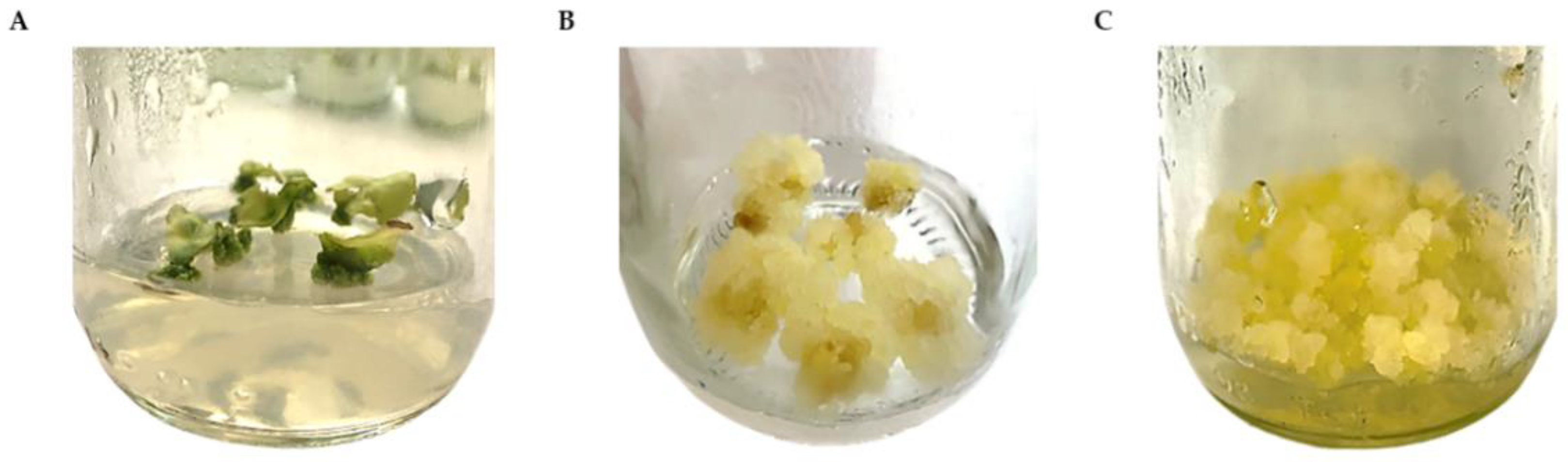
2.1. Callus Induction and Proliferation

R. nasutus is a small shrub with a height of 50–200 cm. The leaves are simple, opposite, and ovate to oblong. The flowers contain two upper lips and three lower lips (Figure 1). After sterile R. nasutus plants were established from shoot tips, the young leaf segments developed from 6-week-old shoots were used as explants for the callus induction experiment. The leaf explants were cultured on MS medium supplemented with different concentrations of 2,4-D and 1-naphthalene acetic acid (NAA) (Table 1).
The callus was initiated from the cut edges of leaf explants, and the callus formation was initiated within 3 weeks in almost all treatments (Figure 2A). The combination of 2,4-D (0.5–2.0 mg/L) and NAA (1.0 and 2.0 mg/L) was found to be effective for callus induction. The maximum callogenesis response of 100% was observed in the presence of 2,4-D (0.5, 1, 1.5, and 2 mg/L) and 1.0 mg/L NAA. In addition, the induced callus was yellowish and friable in texture (Figure 2B). The supplementation of the medium with 2 mg/L NAA resulted in lower percentages of callus induction (<35%), and using either 2,4-D or NAA alone did not promote new callus growth (Table 1). No callus developed on the explants placed on the MS medium without PGRs, as expected. After five weeks, the callus derived from leaf explants was subcultured for proliferation on fresh MS medium supplemented with 1.0 mg/L 2,4-D and 1.0 mg/L NAA (callus induction medium, CIM) every two weeks for twenty-one subcultures. The yellow friable callus proliferated well and appeared as bright yellow and soft (Figure 2C).

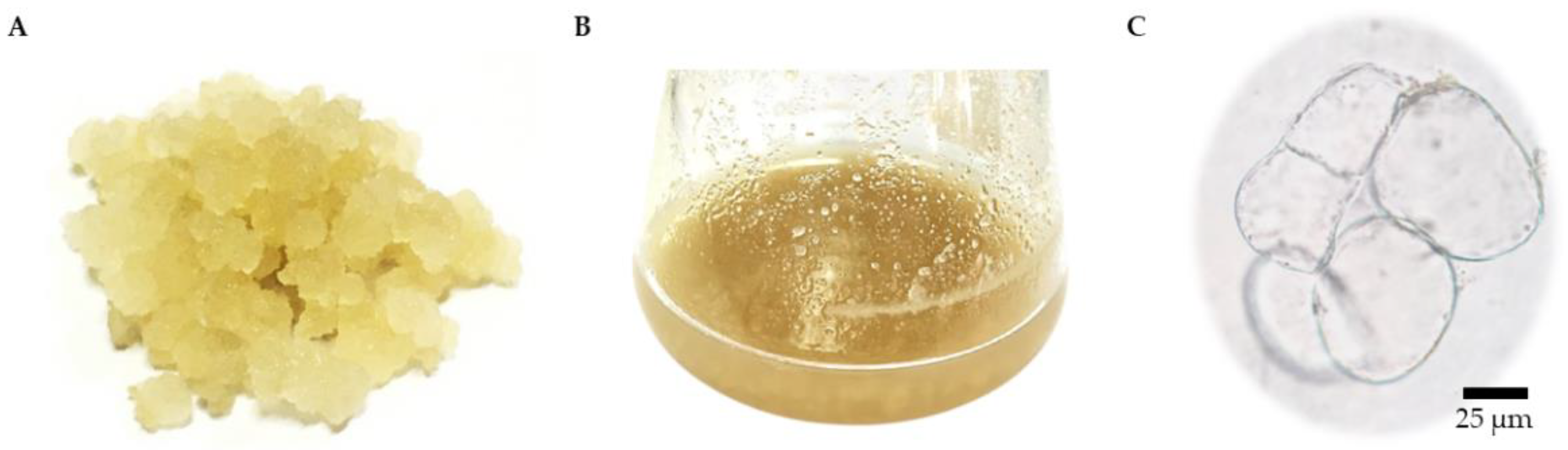
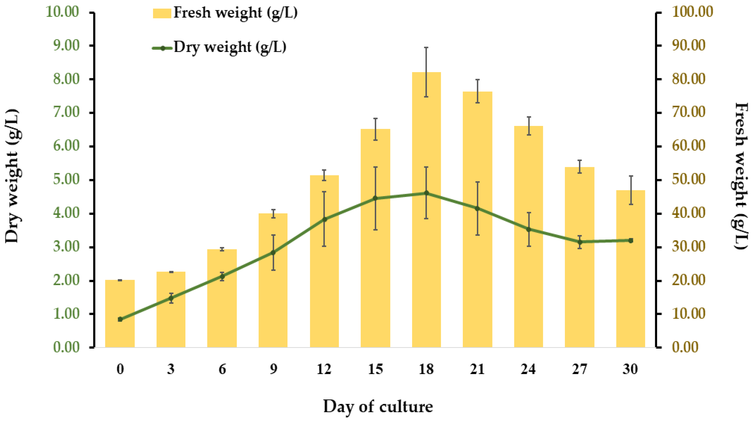
2.2. Establishment of Cell Suspension Culture

A fast-growing cell line producing a yellow friable callus was used to initiate a cell suspension culture (Figure 3A). SCCs were grown in liquid CIM, i.e., MS medium supplemented with 1 mg/L 2,4-D and 1 mg/L NAA (Figure 3B). Homogeneous cell masses were observed under a bright-field microscope with Evans blue staining, showing oval and spherical cells with thin walls (Figure 3C). The growth pattern of the R. nasutus suspension culture was typical of plant cell cultures, with a fresh weight (FW) pattern similar to the dry weight (DW) pattern. The cells continued to grow until they reached maximum biomass accumulation on day 18, when FW and DW values of 82.14 g/L and 4.62 g/L, respectively, were observed (Figure 4). After this, the biomass decreased.
To determine the most appropriate time point to collect SCCs and further test for their antioxidant and anticancer potential, the total phenolic content (TPC) was used as a marker to ensure that the obtained fast-growing cell line actually produced bioactive compounds. The SCCs were collected at three time points, namely days 9, 15, and 18, and were tested for TPC values. The SCCs showed the highest TPC on day 15, with a value of 14.67 ± 0.37 mg GAE/g DW (Figure S1). The SCCs observed on day 9 and day 15 were viable, as demonstrated by the callus color and texture, as well as the images viewed under a bright-field microscope with Evans blue staining. However, the SCCs observed on day 18 were mostly nonviable. Based on these results, the cell line culture for 15 days was used to prepare the crude extracts of the SCCs and to further investigate their antioxidant and anticancer properties compared to those of the leaf extract.

2.3. Total Phenolic and Total Flavonoid Contents

The leaf and SCC samples were extracted with 80% (v/v) ethanol using the maceration extraction method at 65 °C for 6 h. The TPC and TFC values obtained from the extracts were analyzed using the Folin–Ciocalteu (FC) and aluminum chloride colorimetric assays, respectively. The TPC and TFC values of the ethanolic leaf extract were significantly higher than those of the SCCs. The leaf extract showed a TPC of 45.27 ± 1.34 mg GAE/g DW and a TFC of 8.67 ± 0.10 mg QE/g DW, whereas the SCC extract showed TPC and TFC values of 14.67 ± 0.09 mg GAE/g DW and 0.54 ± 0.06 mg QE/g DW, respectively.

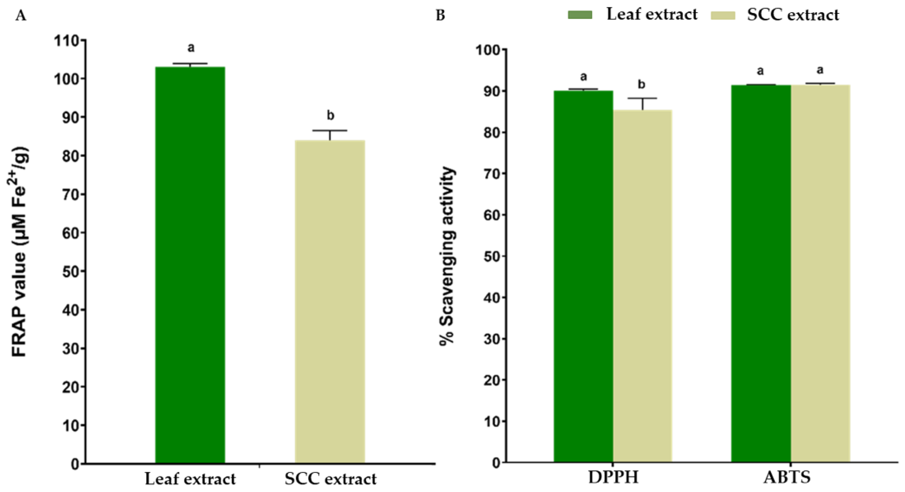
2.4. Antioxidant Properties

The leaf and SCC extracts were tested for their antioxidant capacity using three methods, namely the ferric-reducing antioxidant power (FRAP), 2,2-diphenyl-1-picrylhydrazyl (DPPH), and 2,2′-azino-bis (3-ethylbenzothiazoline-6-sulfonic acid) (ABTS) assays (Figure 5). The FRAP value of the ethanolic leaf extract (103.05 ± 0.89 µM Fe2+/g DW) was higher than that of the SCC extract (84.01 ± 2.55 µM Fe2+/g DW) (Figure 5A). The DPPH scavenging activity of the leaf extract was 86.78%, which was higher than that of the SCC extract (83.29%). For the ABTS assay, the leaf and SCC extracts showed strong antioxidant capacity levels of 91.40% and 91.25%, respectively (Figure 5B).

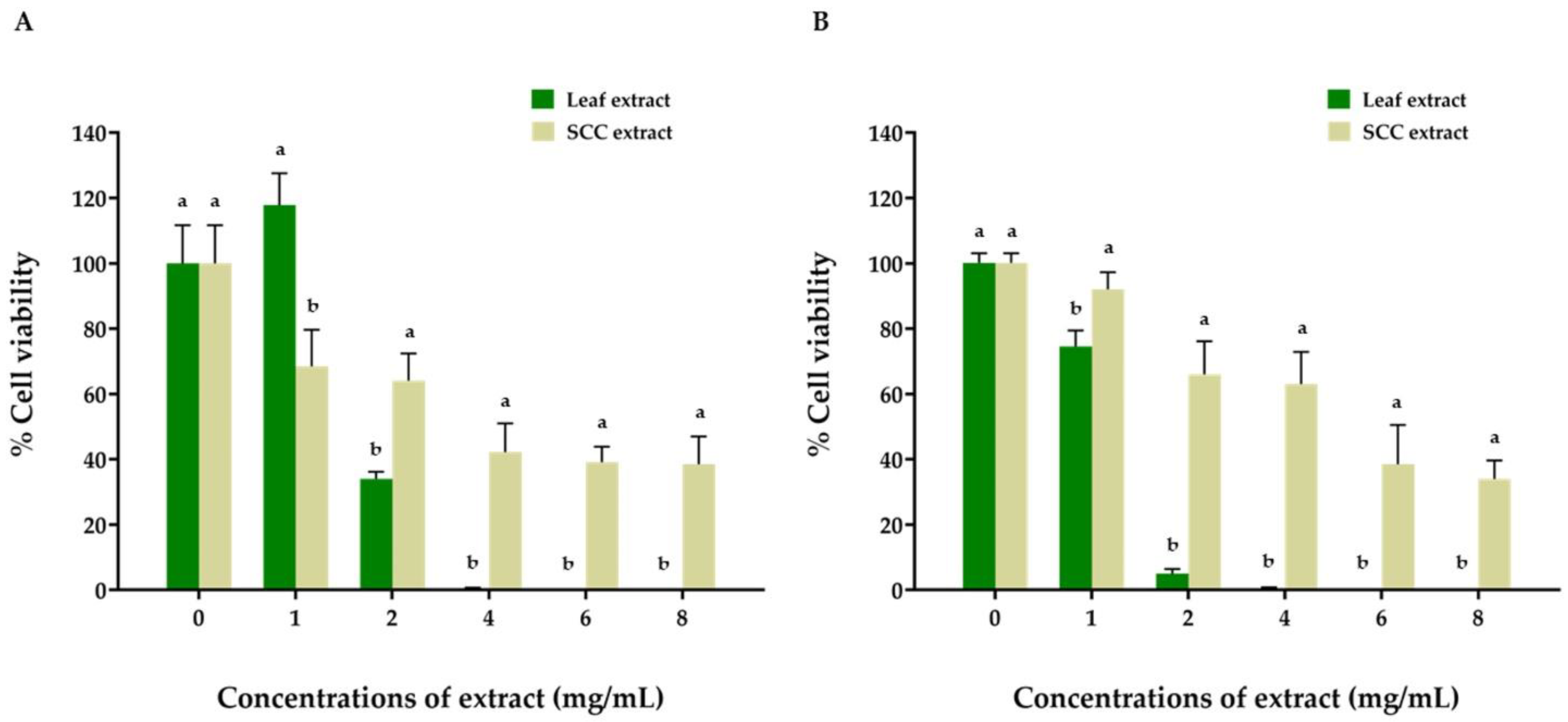
2.5. Anticancer Activity

The cell viability of the two ORL-136 and ORL-48 lines was assessed using the MTT assay following 48 h of treatment with the leaf and SCC extracts at concentrations of 1, 2, 4, 6, and 8 mg/mL. The results showed that both cell lines responded to the cytotoxic effects of both extracts in a dose-dependent manner based on the percentages of cancer cell viability (Figure 6). The leaf extract was shown to have a greater inhibitory effect on the growth of the ORL-48 (with an IC50 value of 2.35 mg/mL) and ORL-136 (with an IC50 of 1.24 mg/mL) cell lines than the SCC extract. In particular, after treatment with 4 mg/mL leaf extract, the results showed the significantly inhibited growth of the two cell lines, with a cell viability rate of 0%. In contrast, the SCC extract showed inhibitory effects on ORL-48 and ORL-136 cell growth at the highest concentration of 8 mg/mL, with cell viability levels of 38% and 34% and IC50 values of 4.38 mg/mL and 5.31 mg/mL, respectively.
The morphological analysis of the ORL-48 and ORL-136 cell lines, compared with the control group (no added extract), showed that the normal cancer cells were squamous and highly densely packed. No cancer cells survived in either cell line after treatment with 4 mg/mL leaf extract (Figure S2). Under treatment with the SCC extract at the highest dose of 8 mg/mL, some ORL-48 cancer cells survived; however, the shapes of the cells were abnormal, changing to spheroid with loose contact and more spaces between cells (Figure S3). The levels of cell death gradually increased in a dose-dependent manner. Similar results were observed for the ORL-136 cell line under the same treatment conditions.

2.6. Analysis of Metabolites in the Ethanolic Leaf and SCC Extracts by UHPLC–QToF–MS

The metabolite compositions of the leaf and SCC extracts were investigated using UHPLC–QToF–MS analysis in both positive and negative ionization modes and verified with public databases. Several groups of compounds were identified in the leaf and SCC extracts (Tables S1 and S2), belonging to 32 different classes (Table S3). High percentages of the secondary metabolites detected in both extracts were terpenoids or phenolic compounds, including phenolic acids, flavonoids, quinones, and stilbenes. Terpenoid compounds such as austroinulin, inulicin, lucidenic acid, and corosolic acid; phenolic compounds such as (E)-2-glucosyl-3,4′,5 trihydroxy stilbene, ferulic acid, trans-cinnamic acid, 4-hydroxycoumarin, and esculetin; quinones, including embelin, isoplumbagin, and 1,3,5,8-tetra hydroxy-6-methoxy-2-methyl anthraquinone; and flavonoids, including rutin and quercetin 3-(2″-p-hydroxybenzoyl-4″-p-coumarylrhamnoside), were observed in the ethanolic leaf extract (Table 2). The same groups of compounds were also detected in the SCC extract, with differences in compound types (Table 3). Interestingly, five compounds were observed in both the leaf and SCC extracts, namely, austroinulin, lucidenic acid, esculetin, embelin, and quercetin 3-(2″-p-hydroxybenzoyl-4″-p-coumarylrhamnoside) (Figure S4). The compounds present only in the SCC extract were camelledionol, pterostilbene glycinate, n-feruloyltyramine, 1,4-naphthoquinone, and hesperetin.

3. Discussion

Plant tissue and cell cultures, especially callus and cell suspension cultures, offer a promising platform for the production of high-value bioactive compounds from plants to be used for medical and pharmaceutical purposes [25,26,27]. PGRs, particularly auxins and cytokinins, play an important role in the growth and development of plant cells, and they have been widely used to induce callus formation in many plant species [27,28,29]. Calluses, unorganized, undifferentiated, and growing masses of cells formed by plant cell division, can be obtained from various types of explants placed on a solid medium containing either auxin alone or auxin together with cytokinin [30]. In the present investigation, a callus formation response of 100% using leaf explants of R. nasutus was achieved by using auxin alone (0.5–2 mg/L 2,4-D and 1 mg/L NAA), which was supplemented in MS medium. The leaf explant cultured on MS medium without any PGRs failed to produce a callus, as expected. Other studies have reported the use of only 2,4-D (2–5 mg/L) to induce a callus in R. nasutus [21,22]. However, our study showed no callus response when using 2,4-D alone at a concentration of 1 or 2 mg/L. This contradictory result could be attributed to the differences in the explant types used in the experiments, the different genetic backgrounds of the plants, and environmental influences [31]. In the case of using the combination of auxin and cytokinin for callus induction in R. nasutus, a few studies have demonstrated the use of 2,4-D together with either BAP or TDZ to induce callus formation. One study found that MS medium containing 0.1 mg/L 2,4-D and 0.5 mg/L TDZ was optimal for a high degree of callus induction (96%) [24], while another study demonstrated a callus induction frequency of 100% when culturing leaf explants on MS medium supplemented with 2.5 mg/L 2,4-D and 5 mg/L BAP [21]. In addition, other types of auxin and cytokinin, i.e., 0.5 mg/L IBA and 4 mg/L KT, respectively, were used to induce calluses, showing a callus induction rate of 98.8% [23].
Table 2. A list of metabolites in R. nasutus leaf extract.
Proposed
Compound
Class *Subclass *Retention Time (min)Product Ions (m/z)MassMolecular
Formula
Remarks
Terpenoids
AustroinulinPrenol lipidsDiterpenoids43.58345.24322.25C20 H34 O3Found in Stevia/anti-inflammatory [32]
InulicinPrenol lipidsTerpene lactones26.71307.16308.16C17 H24 O5May suppress angiogenesis and lung cancer cell growth [33]
Lucidenic acidPrenol lipidsSesquiterpenoids41.91481.26458.27C27 H38 O6Lucidenic acid A, B, N caused cell cycle arrest in G1 phase/inhibited PMA-induced HCC invasion and apoptosis in human leukaemia cells HL-60 [34,35,36]
Corosolic acidPrenol lipidsTriterpenoids43.30495.35472.36C30 H48 O4Inhibits human colorectal cancer cells [37]/inhibits liver cancer cell growth under high-glucose conditions [38]
Ganoderic acidPrenol lipidsTriterpenoids40.42555.29532.31C30 H44 O8Inhibits cell proliferation, viability, and ROS and mRNA expression of TNF [39]
Punctaporin BPrenol lipids-41.32251.17252.17C15 H24 O3Found in Carolina cucumber and Vernonia cinerea (L.) Less [40]
Phenolics
(E)-2-glucosyl-3,4′,5 trihydroxy stilbeneStilbenesStilbene glycosides27.74413.12390.13C20 H22 O8Complementary and alternative medicine for cancer therapy [41]
Ferulic acidCinnamic acids and derivativesHydroxycinnamic acids and derivatives16.07193.05194.06C10 H10 O4Able to suppress tumors in breast cancer, cervical carcinoma cells, prostate cancer cells, and pancreatic cancer cells [42]
Trans-cinnamic acidCinnamic acids and derivativesCinnamic acids33.20149.06148.05C9 H8 O2Some inhibitory activity against enzymes from the human liver and the human cholangiocarcinoma cell line [43]
4-hydroxy coumarinCoumarins and derivativesHydroxycoumarins28.54163.04162.03C9 H6 O3Inhibits cell proliferation in the gastric carcinoma cell line [44]
EsculetinCoumarins and derivativesHydroxycoumarins13.12177.02178.03C9 H6 O4Inhibits migration and invasion of laryngeal cancer [45]
Quinones
EmbelinPrenol lipidsQuinone and hydroquinone lipids27.41293.18294.19C17 H26 O4Inhibits TNF-α and cancer cell metastasis [46,47,48]
IsoplumbaginNaphthalenesNaphthoquinones23.39187.04188.05C11 H8 O3Suppresses oral, lung, prostate, and cervical cancer cells [49]
1,3,5,8-tetra hydroxy -6-methoxy-2-methyl anthraquinoneOthersAnthraquinones28.85334.09316.06C16 H12 O7Inhibits cancer progression by kinases, topoisomerases, telomerases, matrix metallo proteinases, and G-quadruplexes [50]
Flavonoids
2′,4′-dihydroxy-7-methoxy-8-prenylflavanFlavonoidsFlavans26.69363.16340.17C21 H24 O4Also found in Cassia fistula anticancer [51]
RutinFlavonoidsFlavonoid glycosides14.92609.15610.15C27 H30 O16Anticancer effects on human cervical cancer cells [52]
Vitexin 4′-O-galactosideFlavonoidsFlavonoid glycosides15.52593.15594.16C27 H30 O15Many flavonoids have been isolated from and identified in flowers of the genus Iris [53]
Quercetin 3-(2′′-p-hydroxybenzoyl-4′′-p-coumaryl rhamnoside)FlavonoidsFlavones43.83715.17714.16C37 H30 O15Quercetin suppresses cyclooxygenase-2 (COX-2) expression in human breast cancer cells [54]
* Class and subclass were assigned according to the Human Metabolome Database (HMDB) (https://hmdb.ca) (accessed on 9 May 2022).
Table 3. A list of metabolites in R. nasutus SCC extract.
Proposed
Compound
Class *Subclass *Retention Time (min)Product Ions (m/z)MassMolecular
Formula
Remarks
Terpenoids
AustroinulinPrenol lipidsDiterpenoids43.10345.24322.25C20 H34 O3Found in Stevia/anti-inflammatory [32]
Lucidenic acidPrenol lipidsSesquiterpenoids38.46463.31462.30C27 H42 O6Lucidenic acid A, B, N caused cell cycle arrest in G1 phase/inhibited PMA-induced HCC invasion and apoptosis in human leukaemia cells HL-60 [34,35,36]
CamelledionolPrenol lipidsTriterpenoids41.48441.34440.33C29 H44 O3Inhibits the A549, LLC, HL-60, and MCF-7 cancer cell lines [55]
Phenolics
Pterostilbene glycinateStilbenes-18.38336.12313.13C18 H19 N O4Effective in treat ment of melanoma and has anticancer activity [56]
EsculetinCoumarins and derivativesHydroxycoumarins13.08177.02178.03C9 H6 O4Inhibits migration and invasion of laryngeal cancer [45]
N-feruloyltyramineCinnamic acids and derivativesHydroxycinnamic acids and derivatives19.03312.12313.13C18 H19 N O4Significantly fights against the oxidative damage induced by H2O2 and inhibits HepG2 cells [57]
Quinones
EmbelinPrenol lipidsQuinone and hydroquinone lipids27.50293.18294.19C17 H26 O4Inhibits TNF-α and cancer cell metastasis [46,47,48]
1,4-naphtho quinoneNaphthalenesNaphthoquinones14.04159.04158.04C10 H6 O2Its derivatives demonstrate good anticancer, antioxidant, antimicrobial, and anti-inflammatory effects [58,59]
Flavonoids
HesperetinFlavonoidso-methylated flavonoids20.359301.07302.08C16 H14 O6A drug used for lowering cholesterol and treating multiple cancers [60]
Quercetin 3-(2″-p-hydroxybenzoyl
-4″-p-coumaryl rhamnoside)
FlavonoidsFlavones43.83715.17714.16C37 H30 O15Quercetin suppresses cyclooxygenase-2 (COX-2) expression in human breast cancer cells [54]
* Class and subclass were assigned according to the Human Metabolome Database (HMDB) (https://hmdb.ca) (accessed on 9 May 2022).
Although a high callus induction frequency was reported in all previously mentioned studies, only green or greenish-yellow compact calluses were produced, suggesting that the auxins and cytokinins used were not suitable for friable callus induction and proliferation in R. nasutus. The presence of green and compact calluses could be explained by the high concentration of cytokinin, which possibly promotes shoot and chlorophyll formation [61]. In contrast, our study showed for the first time that using two types of auxin (2,4-D and NAA) could effectively induce a bright yellow callus with a soft texture, possibly due to the presence of 2,4-D, which has been proposed to inhibit chlorophyll synthesis [62]. Additionally, NAA and subculturing for several cycles, as shown in our study, might play an important role in promoting friable callus formation, making it an attractive technique for establishing suspension cultures [13,63]. Similar results in other plants were reported by Anjusha et al. [31], who showed yellow friable callus induction on a medium containing only auxin (1 mg/L 2,4-D) from Gynochthodes umbellata, a medicinal plant belonging to family Rubiaceae, and from Asteracantha longifolia Nees, a medicinal plant belonging to the Acanthaceae family, which produced a friable white callus in the presence of auxin alone (2 mg/L 2,4-D) [64].
This research was the first attempt to establish a suspension culture of R. nasutus. The friable callus induced on the CIM containing 1 mg/L NAA and 1 mg/L 2,4-D was selected due to its fast-growing and bright-yellow color characteristics. Then, it was subcultured every two weeks for 21 subculture cycles until it was ready to initiate a cell suspension culture. The growth pattern of the R. nasutus suspension culture presented here was typical of the plant cell culture [65]. Slow growth was observed at the beginning of the cycle, then increases in both FW and DW were observed, potentially due to cell division and an increase in the water content of cells [66]. The highest bioactive compound accumulation was observed on day 15 of the culture, which was in agreement with the data from other authors [18,65]. The population of SCCs shrank after day 18, suggesting that cell death predominated during this period [67]. Based on the results from previous experiments, it was confirmed that the selected cell line was suitable for establishing a suspension culture and further investigating its antioxidant and anticancer properties compared to those of the leaf.
The current study revealed a higher TPC in the leaf extract of R. nasutus than reported in other studies based on the same method of extraction and solvent [5,68,69,70]. Similarly, the TFC obtained in the present study was comparable to those reported by other investigators [68,69,70]. Therefore, the solvent and extraction system are appropriate for the extraction of phenolic acids and flavonoids from R. nasutus. However, the TPC and TFC values of the SCC extract shown in the current study were relatively low compared to those of the leaf extract. Due to the presence of undifferentiated cells in calluses, it is possible that calluses can produce and accumulate lower levels of bioactive compounds than leaves, which are composed of fully differentiated cells. Notably, the suspension culture system is an effective system in which the bioactive compound accumulation can be substantially enhanced using elicitors, as observed in hairy root cultures of R. nasutus and other plants [28,71]. Since there are no reports comparing leaf and SCC extracts from R. nasutus, our study provides initial information on the established suspension culture system.
Much attention has been given to the antioxidant as well as other therapeutic properties of many medicinal herbs, including R. nasutus. The high antioxidant capacity of the leaf extract could be attributable to the high TPC observed in the present study, since the majority of the phenolic compounds have the ability to scavenge free radicals in many plant species, including R. nasutus [69,72,73]. Although flavonoids were detected in smaller amounts in the extract, evidence of many flavonoids contributing to the antioxidant activity was also reported [69]. Interestingly, although a lower TPC and TFC were observed in the SCC extract, the SCC extract exhibited a strong antioxidant potential equivalent to that of the leaf extract, as demonstrated by high scavenging activities of greater than 80% in the DPPH and ABTS assays as well as the high FRAP value. Thus, it is possible that other groups of phytochemicals, including carotenoids, alkaloids, terpenoids or quinones, present in the SCC extract contributed to the high antioxidant capacity, as demonstrated in other studies [4,5,74]. It should be noted that higher ABTS values may be attributed to the ABTS•+ (radical) stability over a wide pH range, the high sensitivity test, and the fast radical–antioxidant reaction. Moreover, ABTS•+ is reactive toward most antioxidants, supporting the evaluation of a wide range of antioxidants, including hydrophilic and lipophilic compounds [75,76] The observed antioxidant capacity levels of the R. nasutus leaf extract were in good agreement with the results from other investigators [69,72,77]. In summary, the SCC extract exhibited a high antioxidant potential equivalent to that of the leaf extract, suggesting the possibility of using the SCC extract in further applications.
In addition to the antioxidant capacity of the plant extract that is of particular interest, the potential for the plant extract to serve as an alternative or combined treatment for cancer is also worth exploring. Various other studies investigated the use of R. nasutus leaf and root extracts for cancer treatment. For the root extract, the previous studies showed effective inhibition of tumor cell growth in several cancer cell lines, including human cervical carcinoma (HeLa), human epithelial carcinoma (KB), human epidermoid carcinoma (Hep-2), human promyelocytic leukemia (HL-60), human uterus carcinoma (SiHa), human breast cancer (MCF-7), human colorectal adenocarcinoma (C-32, HT-29, Caco-2), human lung carcinoma (LLC, NCI-H187, A-549), mouse leukemia cell (P-388), and mouse colon adenocarcinoma (Colon-26) [8,78,79,80]. Additionally, the antiproliferative activity and cytotoxicity against HeLa cells, human prostate carcinoma cells (PC-3), human bladder carcinoma cells (T24), and multidrug-resistant CEM/ADR5000 cells were observed when applying the leaf extract [78,81,82].
This study provides the first evidence of the anticancer potential of R. nasutus leaf and SCC extracts against the oral cancer cell lines ORL-48 (oral cavity-gingiva) and ORL-136 (oral cavity-tongue). Based on the cancer cell viability and morphology, the leaf and SCC extracts could inhibit cancer cell proliferation, although the cytotoxic inhibitory effect of the SCC extract was lower than that of the leaf extract. Additionally, the SCC extract, which can be produced continuously in the laboratory, will also be beneficial in bioactive compound production for the treatment of oral cancer and other types of human cancer.
Considering the metabolites identified in the leaf and SCC extracts, the results obtained from this study are consistent with other reports of the presence of terpenoid, phenolic, flavonoid, and quinone compounds in R. nasutus leaves, conferring significant free radical scavenging activity and cancer-inhibitory effects [41,49,50,56,58,59,83,84]. Similar metabolite profiles were observed between the leaf and SCC extracts, with the leaf extract potentially showing greater metabolite accumulation. This information is not surprising, since the leaves at the mature stage normally accumulate several plant metabolites. In contrast, an undifferentiated callus may accumulate fewer types and smaller amounts of metabolites, depending on the selected cell line. However, some compounds, including camelledionol, pterostilbene glycinate, n-feruloyltyramine, 1,4-naphthoquinone, and hesperetin, were observed only in the SCC extract. The differences in metabolite accumulation in natural plant parts and those in the SCC might be attributed to various internal and external factors. In particular, callus induction and proliferation are under genetic control, so these factors might trigger differential gene expression, leading to different enzymes being involved in the biosynthesis of different compounds. In addition, external factors, including major and minor elements, hormones present in the soil and culture media, as well as the growing conditions (light, humidity, and temperature), are possible influences affecting the differences in metabolite production between the plant and the SCC [30].
Austroinulin isolated from S. rebaudiana has anti-inflammatory effects on nitric oxide (NO) production [32]. In addition, the ethanolic leaf extract of S. rebaudiana possessed antioxidant potential for use as a natural antioxidant agent [85]. Some terpenoid extracts of G. lucidum contain lucidenic acids with high antioxidant activity [86,87], and the impact of lucidenic acids (A and N) on cell growth inhibits PMA-induced invasion in hepatocellular carcinoma [34]. Esculetins, a class of coumarins and derivatives, and embelin, a naturally occurring benzoquinone obtained from the Embelia ribes Burm plant, possess a wide range of medicinal properties, including antioxidant, anticancer, anti-inflammatory, antibacterial, antifungal, and antiviral properties [45,46,47,48]. Additionally, the free radical scavenging reactions and antioxidant activity of embelin have been studied. Other studies in R. nasutus and Citrus lumia have reported that quercetin 3-(2″-p-hydroxy benzoyl-4″-p-coumarylrhamnoside) has antioxidant and anticancer potential [5,54]. Other compounds from R. nasutus are worth exploring in more detail, such as lignans, which significantly inhibited neuraminidase activity, suggesting their antiviral potential [7].

4. Materials and Methods
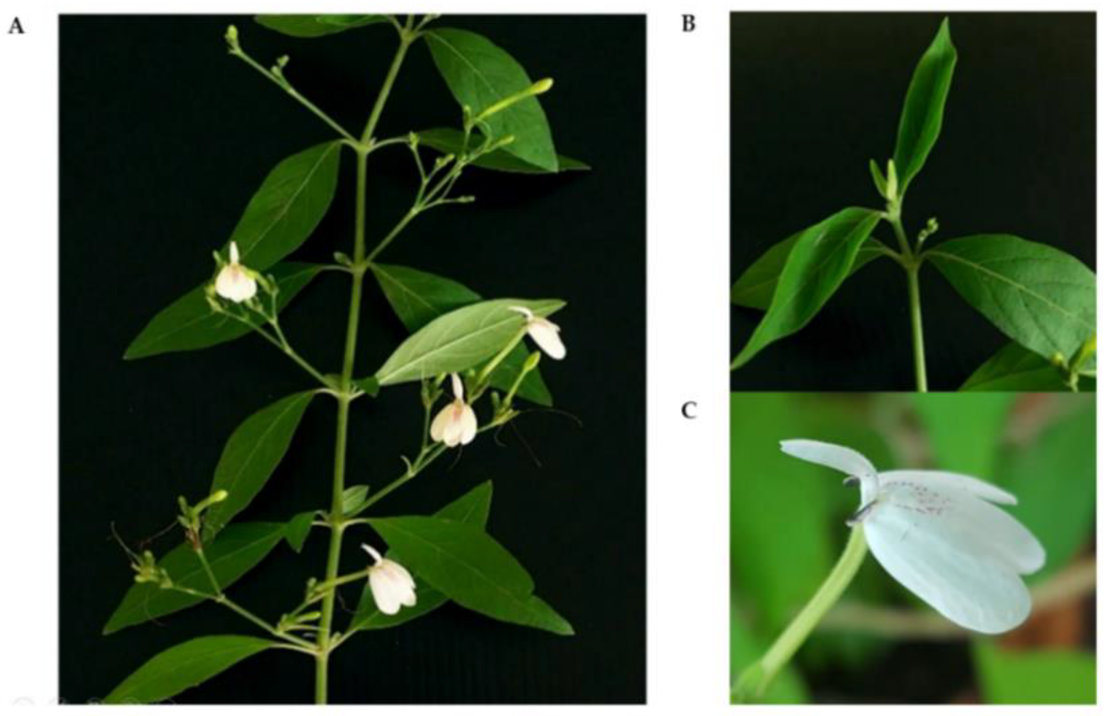
4.1. Plant Material

R. nasutus plants were grown and samples were collected in Khon Kaen Province, Thailand. The plants were identified at the Department of Biology, Faculty of Science, Khon Kaen University. The voucher specimen was deposited in the herbarium of the Department of Biology, Faculty of Science, Khon Kaen University (herbarium specimen number: KKU25555).

4.2. Explant Preparation

To prepare sterile plants for subsequent callus induction experiments, shoot tips of R. nasutus were used as explants. The shoot tips were washed with water with a few drops of dishwashing liquid for 5 min; after this, they were washed thoroughly under running tap water for 15 min. Inside a laminar flow cabinet (ESCO, Singapore), the shoot tips of R. nasutus plants (2.5 cm in length) were washed thoroughly with dishwashing liquid and running tap water and then dipped in 95% and 70% (v/v) ethanol for 1 and 4 min, respectively. After being washed three times in sterile distilled water, the shoot tips (5 mm in length) were treated with 20% and 10% (v/v) sodium hypochlorite (polysorbate 20 added) for 5 and 10 min, respectively. After five rinses with sterile distilled water, the surface-sterilized shoot tips were cultured on MS solid medium [88] supplemented with 3% (w/v) sucrose and 0.8% (w/v) PhytagelTM at pH 5.7. The cultures were kept at 25 ± 2 °C under a light intensity of 2000 lux for 16/8 h light/dark.

4.3. Callus Induction, Cell Line Selection and Proliferation

Leaf segments, with dimensions of approximately 0.8 cm by 0.8 cm, developed from sterilized 6-week-old shoots were used as explants for callus induction. The CIM contained MS salts and vitamins with 3% (w/v) sucrose and 0.2% (w/v) PhytagelTM supplemented with different concentrations and combinations of PGRs, i.e., 2,4-D (0.5, 1.0, 1.5, and 2.0 mg/L) and NAA (1.0 and 2.0 mg/L). The cultures were kept at 25 ± 2 °C with a light intensity of 2000 lux under a 16 h light/8 h dark photoperiod regime. The callus induction frequency (%), color, and texture, as visually assessed, were evaluated after 3 weeks of culture. The callus induction experiment was performed following a factorial design for each combination. Five leaf explants were placed into a jar for each treatment as one replicate, with three replicates per treatment. All experiments were repeated three times independently. After five weeks, the rapidly growing callus cell line was selected and subcultured on fresh CIM every two weeks.

4.4. Establishment of Cell Suspension Cultures and Growth Measurements

The fast-growing friable callus cell line with a creamy color was selected to establish a cell suspension culture. A preliminary test for the TPC was also performed. The growth of the friable callus was measured by sacrificial sampling. Two grams (FW) of a two-week-old friable callus was cultured in 250-mL Erlenmeyer flasks containing 100 mL of liquid CIM supplemented with 1 mg/L 2,4-D and 1 mg/L NAA. The cell suspensions were maintained at 110 rpm on a rotary shaker at 25 ± 2 °C, 2000 lux, and 16/8 h light/dark. To determine the cell suspension growth profile, the samples were harvested at 3-day intervals for a period of 30 days. All determinations were carried out in triplicate. The callus was separated from the medium by vacuum filtration, and the FW and DW were measured. Bright-field microscopy (Olympus, Tokyo, Japan) was used to examine the callus morphology. Evans blue staining was used to observe the cell viability [67].

4.5. Preparation of Extracts

Leaf and SCC samples of R. nasutus were dried at 45 °C for 24 h and then ground into a fine powder. One gram of each sample powder was subjected to maceration extraction using 50 mL of 80% (v/v) ethanol at 65 °C for 6 h. The supernatant was collected and then filtered through filter paper. The solvent was evaporated (EYELA N-1100, Tokyo, Japan) and further lyophilized (Christ, Osterode, Germany). The obtained dried powder was stored at −20 °C until further use in subsequent experiments. For the analysis of plant metabolites by UHPLC–QToF–MS), dried samples (15% w/v) were used following the maceration extraction procedure previously mentioned.

4.6. Determination of the TPC and TFC

The TPC values of leaf and SCC extracts were estimated using the FC method, following the method described by Folin and Ciocalteu [89] with slight modifications. Briefly, twenty microliters (20 μL) of each extract (10 mg/mL) was mixed with 60 μL of distilled water and 20 μL of FC reagent (1 N). After 5 min, 100 μL of 8% (w/v) sodium carbonate (Na2CO3) was added and the final volume was adjusted to 300 μL using distilled water. The reaction mixture was incubated for 30 min at 25 °C in the dark. The absorbance at 765 nm was measured using a microplate reader (Thermo Scientific Varioskan® Flash, Waltham, MA, USA). Gallic acid was used as a reference, and the results are presented as milligrams of gallic acid equivalents per gram of dry weight (mg GAE/g DW).
For TFC determination, the aluminum chloride (AlCl3) colorimetric assay as described by Zongo et al. with slight modifications was employed [90]. In brief, 100 μL each of the leaf and SCC extracts (2 mg/mL) was mixed with 100 μL of 2% AlCl3. The mixed reactions were incubated for 1 h at 25 °C in the dark before the absorbance of the mixture was assayed at 420 nm. Quercetin was chosen as the standard, and the TFC was expressed as quercetin equivalents in milligrams per gram of dry sample (mg QE/g DW).

4.7. Determination of Antioxidant Capacity

Three methods, namely the FRAP assay (OxiSelect™ Ferric-Reducing Antioxidant Power Assay Kit, Cell Biolabs, San Diego, CA, USA), ABTS assay (OxiSelect™ Trolox Equivalent Antioxidant Capacity Assay Kit, Cell Biolabs) and DPPH assay (Sigma, Ronkonkoma, NY, USA), were used to evaluate the total antioxidant capacity of leaf and SCC extracts. The experiments were carried out using 96-well plates, and the absorbance at specific wavelengths was measured using a microplate reader (Thermo Scientific Varioskan® Flash, Waltham, MA, USA) according to the methodology described by Benzie et al. (1996), Smeriglio et al. (2017), and Shalaby and Shanab (2013) with some modifications.
The method used by Benzie et al. [91] was adapted to determine the antioxidant capacity using the FRAP assay with slight modifications. Briefly, 100 μL of each extract (2 mg/mL) was mixed with 100 μL of FRAP reagent and incubated for 10 min at 25 °C, then the absorbance was read at 540 nm. Iron (II) oxide (Fe2+) served as the standard (0–500 μM), and the FRAP values are expressed as μM Fe2+/g DW.
For the ABTS assay with Trolox as the reference compound, the ABTS•+ radical solution was generated by mixing 4.3 mM (w/v in water) potassium persulfate (K2S2O8) and 1.8 mM ABTS solution at a ratio of 1:5 (v/v). The mixtures were incubated for 1 h at 25 °C in the dark. Before reacting with the samples, the ABTS•+ radical solution was diluted with distilled water to obtain an absorbance of 0.7 ± 0.02 at 734 nm. Then, ten microliters of each extract (2 mg/mL) was added to 200 μL of ABTS•+ solution, the mixture was incubated in the dark for 6 min, and the absorbance at 734 nm was recorded [92].
The DPPH assay was performed according to Shalaby and Shanab [93] with some modifications. Ten milligrams of leaf and SCC extracts was dissolved in 1 mL of distilled water. Then, 100 µL of each sample was reacted with 100 µL of DPPH radical (0.1 mM in methanol). The mixtures were shaken vigorously and then kept for 30 min at 25 °C. All determinations were conducted with three replicates using Trolox as the standard equivalent (0 to 500 mg/L). The absorbance at 515 nm was measured, and the antioxidant capacity of the extracts was calculated as follows.
The antioxidant capacity of the extracts was calculated using ABTS and DPPH assays. The percent inhibition of radicals was calculated as the % scavenging activity using the following equation:
% Scavenging activity = [(A control − A sample) × 100]/A control
where A control is the absorbance of the ABTS or DPPH radical and A sample is the absorbance of the ABTS or DPPH radical and sample.

4.8. Determination of Anticancer Activity

The effects of leaf and SCC extracts on oral cancer cell viability were determined using the 3-(4,5-dimethylthiazol-2-yl)-2,5-diphenyltetrazolium bromide (MTT) assay. Briefly, ORL-48 and ORL-136 cell lines [94] were seeded into a 96-well culture plate at a density of 1.9 × 10⁴ cells/well and cultured in Dulbecco’s modified Eagle’s medium–Nutrient Mixture F-12 (DMEM/F12) containing 10% fetal bovine serum, 0.4 µg/mL of hydrocortisone, and 1% antibiotic-antimycotic. After 24 h of incubation at 37 °C in a fully humidified atmosphere of 5% CO2, the oral cancer cell lines were treated with various concentrations of leaf and SCC extracts (1, 2, 4, 6, and 8 mg/mL) for 48 h. Then, the MTT solution (5 mg/mL MTT in phosphate-buffered saline, PBS) was added and the samples were incubated for 3.5 h. Next, all solutions were removed. The formazan product in each well was solubilized with dimethyl sulfoxide (DMSO) and the plate was incubated for 10 min. The absorbance was measured using a microplate reader at 570 nm. The percentage of cancer cell viability was determined based on a comparison with untreated cells grown in a complete medium.
The IC50 value, the concentration of the test extract required for 50% cell viability, was determined from the plot between the % cell viability and extract concentration. The average IC50 value was then calculated.

4.9. UHPLC–QToF–MS

4.9.1. Data Acquisition

The dried leaf and SCC extracts were reconstituted in isopropanol and were analyzed using a UHPLC system coupled with QToF–MS (Agilent, Santa Clara, CA, USA). Briefly, the samples were injected into a UHPLC system equipped with a Zorbax Eclipse Plus C18 column (150 × 2.1 mm, 1.8 µm, Agilent) and with the diode array detector (DAD). The column temperature was set to 25 °C. The elution gradient was implemented with a binary solvent system consisting of water (LC–MS grade) with 0.1% formic acid (solvent A) and acetonitrile (LC–MS grade) with 0.1% formic acid (solvent B). The elution gradient was set as follows: 100% A (0–2 min); 0% A (45–50 min) with a flow rate of 0.2 mL/min. Peaks were detected at wavelengths of 254, 280, and 360 nm. The MS spectra were acquired in both positive and negative electrospray ionization (ESI) modes by employing two and five microliters of each sample injection, respectively. The MS system was set to 325 °C, with desolvation flow gas at 13 L/min and TOF MS with electrospray ionization as the ion source. The ionization voltages in positive and negative polarity modes were 2000 and 2000 V, respectively. Data were acquired in the profile mode for 50 min, and the mass ranges were set to 100–1200 m/z for MS and 50–1200 m/z for MS/MS.

4.9.2. Data Processing

All spectra were processed using MassHunter workstation software qualitative analysis workflows V8 and MassHunter workstation software qualitative analysis navigator V8. Public databases, namely Metlin (https://metlin.scripps.edu, accessed on 12 May 2022), Human Metabolome Database (HMDB) (https://hmdb.ca) (accessed on 12 May 2022), and Kyoto Encyclopedia of Genes and Genomes (KEGG) (https://www.genome.jp/kegg/) (accessed on 12 May 2022), were also used.

4.10. Statistical Analysis

All experimental data were subjected to ANOVA followed by Duncan’s multiple range test (DMRT) and the independent-samples t test using SPSS software version 19 with a p value of 0.05 (p ≤ 0.05). The results were expressed as the means ± standard deviations (SDs) of three measurements. Graphing, calculations, and statistical analyses were performed using GraphPad Prism 9 software version 9.3.1 (GraphPad Software, San Diego, CA, USA). The correlations among the data obtained were calculated using MS Excel software through the correlation coefficient statistical function.

5. Conclusions

This is the first report on establishing callus and cell suspension cultures of R. nasutus using in-vitro-derived leaf explants. Two PGRs, 2,4-D and NAA, were found to be essential for yellow friable callus induction and the proliferation of R. nasutus. Cultures obtained on day 15 are suggested for harvesting in order to obtain healthy and viable cells for bioactive compound production. The leaf extract exhibited higher FRAP and DPPH values than the SCC extract, whereas the ABTS values showed comparable antioxidant capacities between the leaf and SCC extracts. Although the SCC extract exhibited slightly lower anticancer activity than the leaf extract, it had a potential inhibitory effect against the oral cancer cell lines ORL-48 (oral cavity-gingiva) and ORL-136 (oral cavity-tongue). The UHPLC–QToF–MS analysis confirmed that the detected secondary metabolites in the leaf and SCC extracts were terpenoids, phenolics, quinones, and flavonoids. Further investigations using elicitors to induce bioactive compound production and optimizing several parameters for phytochemical production using cell suspension culture should be explored. Detailed metabolite profiling and additional identification of the leaf and SCC extracts after optimization are required to detect the presence of other compounds that might play an essential role in the antioxidant and anticancer potential of R. nasutus. Moreover, an assessment of the anticancer effect using other cell viability assays, apoptosis assays, cell migration, and cell cycle progression by flow cytometry should be considered.

Supplementary Materials

The following supporting information can be downloaded at: https://www.mdpi.com/article/10.3390/plants11151994/s1. Figure S1: Total phenolic content of R. nasutus cell suspension cultures on days 9, 15, and 18. Figure S2: Morphology of ORL-48 and ORL-136 cell lines treated with the leaf extract. Figure S3: Morphology of ORL-48 and ORL-136 cell lines treated with the SCC extract. Figure S4: MS spectra of compounds detected in leaf and SCC extracts. Table S1: List of compounds identified in R. nasutus leaf extract by UHPLC–QToF–MS analysis. Table S2: List of compounds identified in R. nasutus callus extract by UHPLC–QToF–MS analysis. Table S3: Class and subclass of metabolites found in R. nasutus leaf and SCC extracts by UHPLC–QToF–MS analysis.

Author Contributions

Conceptualization, P.S. and P.K. (Preekamol Klanrit); methodology, P.S. and P.K. (Preekamol Klanrit); validation, P.K. (Preekamol Klanrit), P.T. and P.K. (Poramaporn Klanrit); formal analysis, P.K. (Preekamol Klanrit), J.P. and P.K. (Poramate Klanrit); investigation, P.S., J.A. and K.P.; resources, P.K. (Preekamol Klanrit), P.K. (Poramaporn Klanrit), J.P. and K.P.; writing—original draft preparation, P.S. and P.K. (Preekamol Klanrit); writing—review and editing, P.K. (Preekamol Klanrit), P.K. (Poramaporn Klanrit), P.K. (Poramate Klanrit), P.T., J.A., J.P. and K.P.; visualization, P.S., P.K. (Preekamol Klanrit), P.K. (Poramaporn Klanrit), J.A. and P.T.; supervision, P.K. (Preekamol Klanrit); project administration, P.K. (Preekamol Klanrit); funding acquisition, P.K. (Preekamol Klanrit). All authors have read and agreed to the published version of the manuscript.

Funding

This research was supported by the National Science, Research, and Innovation Fund (NSRF) under the Basic Research Fund of Khon Kaen University through Cholangiocarcinoma Research Institute, grant number CARI-BRF-64-37, and the National Research Council of Thailand (NRCT) through Khon Kaen University, grant number 6200025.

Institutional Review Board Statement

Not applicable.

Informed Consent Statement

Not applicable.

Data Availability Statement

The datasets analyzed during the current study are available from the corresponding author upon reasonable request.

Acknowledgments

The authors would like to thank Cheong Sok Ching, Translational Cancer Biology, Cancer Research Malaysia, Malaysia, for providing the oral cancer cell lines; and the office of scientific instrument and testing, Prince of Songkla University for their technical support and UHPLC–QToF–MS sample preparation.

Conflicts of Interest

The authors declare no conflict of interest.

References

  1. Bukke, S.; Raghu, P.S.; Sailaja, G.; Kedam, T.R. The study on morphological, phytochemical and pharmacological aspects of Rhinacanthus nasutus. (L.) Kurz (A Review). J. Appl. Pharm. Sci. 2011, 1, 26–32. [Google Scholar]
  2. Shah, M.A.; Keach, J.E.; Panichayupakaranant, P. Antidiabetic naphthoquinones and their plant resources in Thailand. Chem. Pharm. Bull. 2018, 66, 483–492. [Google Scholar] [CrossRef] [Green Version]
  3. Bhusal, N.; Panichayupakaranant, P.; Reanmongkol, W. In vivo analgesic and anti-inflammatory activities of a standardized Rhinacanthus nasutus leaf extract in comparison with its major active constituent rhinacanthin-C. Songklanakarin J. Sci. Technol. 2014, 36, 325–331. [Google Scholar]
  4. Brimson, J.M.; Prasanth, M.I.; Malar, D.S.; Brimson, S.; Tencomnao, T. Rhinacanthus nasutus “Tea” infusions and the medicinal benefits of the constituent phytochemicals. Nutrients 2020, 12, 3776. [Google Scholar] [CrossRef]
  5. Huang, R.T.; Lu, Y.F.; Inbaraj, B.S.; Chen, B.H. Determination of phenolic acids and flavonoids in Rhinacanthus nasutus (L.) Kurz by high-performance-liquid-chromatography with photodiode-array detection and tandem mass spectrometry. J. Funct. Foods 2015, 12, 498–508. [Google Scholar] [CrossRef]
  6. Horii, H.; Suzuki, R.; Sakagami, H.; Tomomura, M.; Tomomura, A.; Shirataki, Y. New biological activities of rhinacanthins from the root of Rhinacanthus nasutus. Anticancer Res. 2013, 33, 453–460. [Google Scholar] [PubMed]
  7. Kwak, H.J.; Park, S.; Kim, N.; Yoo, G.; Park, J.; OH, Y.; Nhiem, N.X.; Kim, S. Neuraminidase inhibitory activity by compounds isolated from aerial parts of Rhinacanthus nasutus. Nat. Prod. Res. 2018, 32, 2111–2115. [Google Scholar] [CrossRef]
  8. Siripong, P.; Kanokmedakul, K.; Piyaviriyagul, S.; Yahuafai, J.; Chanpai, R.; Ruchirawat, S.; Oku, N. Antiproliferative naphthoquinone esters from Rhinacanthus nasutus Kurz. roots on various cancer cells. J. Tradit. Med. 2006, 23, 166–172. [Google Scholar] [CrossRef]
  9. Suzuki, R.; Sakagami, H.; Shirataki, Y. New anti-oxidative compounds from Rhinacanthus nasutus. Heterocycles 2015, 91, 1036–1041. [Google Scholar] [CrossRef] [Green Version]
  10. Puttarak, P.; Charoonratana, T.; Panichayupakaranant, P. Antimicrobial activity and stability of rhinacanthins-rich Rhinacanthus nasutus extract. Phytomedicine 2010, 17, 323–327. [Google Scholar] [CrossRef]
  11. Panichayupakaranant, P.; Charoonratana, T.; Sirikatitham, A. RP-HPLC analysis of rhinacanthins in Rhinacanthus nasutus: Validation and application for the preparation of rhinacanthin high-yielding extract. J. Chromatogr. Sci. 2009, 47, 705–708. [Google Scholar] [CrossRef] [Green Version]
  12. Ngoc, T.M.; Phuong, N.T.T.; Khoi, N.M.; Park, S.; Kwak, H.J.; Nhiem, N.X.; Trang, B.T.T.; Tai, B.H.; Song, J.H.; Ko, H.J.; et al. A new naphthoquinone analogue and antiviral constituents from the root of Rhinacanthus nasutus. Nat. Prod. Res. 2019, 33, 360–366. [Google Scholar] [CrossRef]
  13. Efferth, T. Biotechnology applications of plant callus cultures. Engineering 2019, 5, 50–59. [Google Scholar] [CrossRef]
  14. Gamborg, O.L.; Phillips, G.C. Plant Cell, Tissue and Organ Culture, 1st ed.; Springer: Berlin/Heidelberg, Germany, 1995; pp. 1–359. [Google Scholar]
  15. Phillips, G.C.; Hubstenberger, J.F.; Hansen, E.E. Plant regeneration by organogenesis from callus and cell suspension cultures. In Plant Cell, Tissue and Organ Culture, 1st ed.; Gamborg, O.L., Phillips, G.C., Eds.; Springer: Berlin/Heidelberg, Germany, 1995; pp. 67–79. [Google Scholar]
  16. Ali, M.; Abbasi, B.H.; Ihsan-ul-haq. Production of commercially important secondary metabolites and antioxidant activity in cell suspension cultures of Artemisia absinthium L. Ind. Crops Prod. 2013, 49, 400–406. [Google Scholar] [CrossRef]
  17. Rahimi, S.; Kim, Y.-J.; Sukweenadhi, J.; Zhang, D.; Yang, D.-C. PgLOX6 encoding a lipoxygenase contributes to jasmonic acid biosynthesis and ginsenoside production in Panax ginseng. J. Exp. Bot. 2016, 67, 6007–6019. [Google Scholar] [CrossRef] [Green Version]
  18. Guadarrama-Flores, B.; Rodríguez-Monroy, M.; Cruz-Sosa, F.; García-Carmona, F.; Gandía-Herrero, F. Production of dihydro xylated betalains and dopamine in cell suspension cultures of Celosia argentea var. Plumosa. J. Agric. Food Chem. 2015, 63, 2741–2749. [Google Scholar] [CrossRef] [PubMed]
  19. Cheruvathur, M.; Thomas, D. High frequency multiple shoot induction from nodal segments and rhinacanthin production in the medicinal shrub Rhinacanthus nasutus (L.) Kurz. Plant Growth Regul. 2014, 74, 47–54. [Google Scholar] [CrossRef]
  20. Elangomathavan, R.; Hariharan, S.; Kalaivanan, P.; Beaulah, S.N. Propagation of Rhinacanthus nasutus (L.) Kurz., through encapsulated shoot tips and nodal segments for germplasm exchange and distribution. Int. J. Pharma Bio Sci. 2017, 8, 98–105. [Google Scholar] [CrossRef]
  21. Reshi, N.A.; Sudarshana, M.S.; Girish, H.V. In vitro micropropagation of Rhinacanthus nasutus (L.) Kurz. Int. J. Biodivers. Conserv. 2018, 10, 357–364. [Google Scholar] [CrossRef]
  22. Sundar, S.; Jayarami, R.A.; Saravana, K.A.; Justin, K.Y. Effect of plant growth regulators on in vitro propagation of Rhinacanthus nasutus (Acanthaceae). Int. J. Pharm. Ind. Res. 2012, 2, 474–478. [Google Scholar]
  23. Cheruvathur, M.K.; Sivu, A.R.; Pradeep, N.S.; Thomas, T.D. Shoot organogenesis from leaf callus and ISSR assessment for their identification of clonal fidelity in Rhinacanthus nasutus (L.) Kurz., a potent anticancerous ethnomedicinal plant. Ind. Crops Prod. 2012, 40, 122–128. [Google Scholar] [CrossRef]
  24. Kumar, R.N.; Ganesan, C.M.; Paulsamy, S. Micropropagation of Rhinacanthus nasutus (L.) Kurz.—An important medicinal plant. Res. Rev. J. Bot. Sci. 2012, 1, 5–10. [Google Scholar]
  25. Wu, T.; Kerbler, S.M.; Fernie, A.R.; Zhang, Y. Plant cell cultures as heterologous bio-factories for secondary metabolite production. Plant Commun. 2021, 2, 100235. [Google Scholar] [CrossRef] [PubMed]
  26. Ahmad, A.; Ahmad, N.; Anis, M.; Faisal, M.; Alatar, A.A.; Abdel-Salam, E.M.; Meena, R.P.; Sivanesan, I. Biotechnological advances in pharmacognosy and in vitro manipulation of Pterocarpus marsupium Roxb. Plants 2022, 11, 247. [Google Scholar] [CrossRef] [PubMed]
  27. Haida, Z.; Nakasha, J.J.; Hakiman, M. In vitro responses of plant growth factors on growth, yield, phenolics content and antioxidant activities of Clinacanthus nutans (Sabah Snake Grass). Plants 2020, 9, 1030. [Google Scholar] [CrossRef] [PubMed]
  28. Cheruvathur, M.K.; Thomas, T.D. Effect of plant growth regulators and elicitors on rhinacanthin accumulation in hairy root cultures of Rhinacanthus nasutus (L.) Kurz. Plant Cell Tissue Organ Cult. (PCTOC) 2014, 118, 169–177. [Google Scholar] [CrossRef]
  29. Kumar, P.P.; Loh, C.S. Plant tissue culture for biotechnology. In Plant Biotechnology and Agriculture; Elsevier: Amsterdam, The Netherlands, 2012; pp. 131–138. [Google Scholar]
  30. Ikeuchi, M.; Sugimoto, K.; Iwase, A. Plant callus: Mechanisms of induction and repression. Plant Cell 2013, 25, 3159–3173. [Google Scholar] [CrossRef] [PubMed] [Green Version]
  31. Anjusha, S.; Gangaprasad, A. Callus culture and in vitro production of anthraquinone in Gynochthodes umbellata (L.) Razafim. & B. Bremer (Rubiaceae). Ind. Crops Prod. 2017, 95, 608–614. [Google Scholar] [CrossRef]
  32. Cho, B.O.; Ryu, H.W.; So, Y.; Cho, J.K.; Woo, H.S.; Jin, C.H.; Seo, K.I.; Park, J.C.; Jeong, I.Y. Anti-inflammatory effect of austroinulin and 6-O-acetyl-austroinulin from Stevia rebaudiana in lipopolysaccharide-stimulated RAW264.7 macrophages. Food Chem. Toxicol. 2013, 62, 638–644. [Google Scholar] [CrossRef] [PubMed]
  33. Zhengfu, H.; Hu, Z.; Huiwen, M.; Zhijun, L.; Jiaojie, Z.; Xiaoyi, Y.; Xiujun, C. 1-o-acetylbritannilactone (ABL) inhibits angiogenesis and lung cancer cell growth through regulating VEGF-Src-FAK signaling. Biochem. Biophys. Res. Commun. 2015, 464, 422–427. [Google Scholar] [CrossRef]
  34. Weng, C.J.; Chau, C.F.; Hsieh, Y.S.; Yang, S.F.; Yen, G.C. Lucidenic acid inhibits PMA-induced invasion of human hepatoma cells through inactivating MAPK/ERK signal transduction pathway and reducing binding activities of NF-ΚB and AP-1. Carcinogenesis 2008, 29, 147–156. [Google Scholar] [CrossRef]
  35. Akihisa, T.; Nakamura, Y.; Tagata, M.; Tokuda, H.; Yasukawa, K.; Uchiyama, E.; Suzuki, T.; Kimura, Y. Anti-inflammatory and anti-tumor-promoting effects of triterpene acids and sterols from the fungus Ganoderma lucidum. Chem. Biodivers. 2007, 4, 224–231. [Google Scholar] [CrossRef] [PubMed]
  36. Hsu, C.L.; Yu, Y.S.; Yen, G.C. Lucidenic acid B induces apoptosis in human leukemia cells via a mitochondria-mediated pathway. J. Agric. Food Chem. 2008, 56, 3973–3980. [Google Scholar] [CrossRef] [PubMed]
  37. Sung, B.; Kang, Y.J.; Kim, D.H.; Hwang, S.Y.; Lee, Y.; Kim, M.; Yoon, J.-H.; Kim, C.M.; Chung, H.Y.; Kim, N.D. Corosolic acid induces apoptotic cell death in HCT116 human colon cancer cells through a caspase-dependent pathway. Int. J. Mol. Med. 2014, 33, 943–949. [Google Scholar] [CrossRef] [PubMed] [Green Version]
  38. Zhang, C.; Niu, Y.; Wang, Z.; Xu, X.; Li, Y.; Ma, L.; Wang, J.; Yu, Y. Corosolic acid inhibits cancer progression by decreasing the level of CDK19-mediated O-GlcNAcylation in liver cancer cells. Cell Death Dis. 2021, 12, 889. [Google Scholar] [CrossRef] [PubMed]
  39. Gill, B.S.; Navgeet; Kumar, S. Ganoderic acid targeting multiple receptors in cancer: In silico and in vitro study. Tumour Biol. 2016, 37, 14271–14290. [Google Scholar] [CrossRef]
  40. Javir, G.; Joshi, K.; Rojatkar, S. Anticancer activity, phytochemical analysis of pet-ether extract by UPLC-ESI-QTOF/MS/MS and quantitative analysis of an active major constituent sesquiterpene lactone from Cyathocline purpurea [Buch-Ham Ex D. Don.]. J. Pharmacogn. Phytochem. 2019, 8, 2219–2227. [Google Scholar]
  41. Berretta, M.; Bignucolo, A.; Di Francia, R.; Comello, F.; Facchini, G.; Ceccarelli, M.; Iaffaioli, R.V.; Quagliariello, V.; Maurea, N. Resveratrol in cancer patients: From bench to bedside. Int. J. Mol. Sci. 2020, 21, 2945. [Google Scholar] [CrossRef] [PubMed] [Green Version]
  42. Alazzouni, A.S.; Dkhil, M.A.; Gadelmawla, M.H.A.; Gabri, M.S.; Farag, A.H.; Hassan, B.N. Ferulic acid as anticarcinogenic agent against 1,2-dimethylhydrazine induced colon cancer in rats. J. King Saud Univ.-Sci. 2021, 33, 101354. [Google Scholar] [CrossRef]
  43. Kukongviriyapan, V.; Phromsopha, N.; Tassaneeyakul, W.; Kukongviriyapan, U.; Sripa, B.; Hahnvajanawong, V.; Bhudhisawasdi, V. Inhibitory effects of polyphenolic compounds on human arylamine N-acetyltransferase 1 and 2. Xenobiotica 2006, 36, 15–28. [Google Scholar] [CrossRef] [PubMed]
  44. Küpeli Akkol, E.; Genç, Y.; Karpuz, B.; Sobarzo-Sánchez, E.; Capasso, R. Coumarins and coumarin-related compounds in pharmacotherapy of cancer. Cancers 2020, 12, 1959. [Google Scholar] [CrossRef]
  45. Wu, Y.; Xu, J.; Liu, Y.; Zeng, Y.; Wu, G. A review on anti-tumor mechanisms of coumarins. Front. Oncol. 2020, 10, 592853. [Google Scholar] [CrossRef] [PubMed]
  46. Dhanjal, J.K.; Nigam, N.; Sharma, S.; Chaudhary, A.; Kaul, S.C.; Grover, A.; Wadhwa, R. Embelin inhibits TNF-α converting enzyme and cancer cell metastasis: Molecular dynamics and experimental evidence. BMC Cancer 2014, 14, 775. [Google Scholar] [CrossRef] [Green Version]
  47. Nigam, N.; Grover, A.; Goyal, S.; Katiyar, S.P.; Bhargava, P.; Wang, P.C.; Sundar, D.; Kaul, S.C.; Wadhwa, R. Targeting mortalin by embelin causes activation of tumor suppressor P53 and deactivation of metastatic signaling in human breast cancer cells. PLoS ONE 2015, 10, e0138192. [Google Scholar] [CrossRef] [PubMed] [Green Version]
  48. Ko, J.H.; Lee, S.G.; Yang, W.M.; Um, J.Y.; Sethi, G.; Mishra, S.; Shanmugam, M.K.; Ahn, K.S. The application of embelin for cancer prevention and therapy. Molecules 2018, 23, 621. [Google Scholar] [CrossRef] [PubMed] [Green Version]
  49. Tsao, Y.C.; Chang, Y.J.; Wang, C.H.; Chen, L. Discovery of isoplumbagin as a novel NQO1 substrate and anti-cancer quinone. Int. J. Mol. Sci. 2020, 21, 4378. [Google Scholar] [CrossRef]
  50. Malik, M.S.; Alsantali, R.I.; Jassas, R.S.; Alsimaree, A.A.; Syed, R.; Alsharif, M.A.; Kalpana, K.; Morad, M.; Althagafi, I.I.; Ahmed, S.A. Journey of anthraquinones as anticancer agents—A systematic review of recent literature. RSC Adv. 2021, 11, 35806–35827. [Google Scholar] [CrossRef] [PubMed]
  51. Khurm, M.; Wang, X.; Zhang, H.; Hussain, S.N.; Qaisar, M.N.; Hayat, K.; Saqib, F.; Zhang, X.; Zhan, G.; Guo, Z. The genus Cassia L.: Ethnopharmacological and phytochemical overview. Phytother. Res. 2021, 35, 2336–2385. [Google Scholar] [CrossRef] [PubMed]
  52. Khan, F.; Pandey, P.; Jha, N.K.; Khalid, M.; Ojha, S. Rutin mediated apoptotic cell death in caski cervical cancer cells via Notch-1 and Hes-1 downregulation. Life 2021, 11, 761. [Google Scholar] [CrossRef] [PubMed]
  53. Iwashina, T.; Mizuno, T. Flavonoids and xanthones from the genus Iris: Phytochemistry, relationships with flower colors and taxonomy, and activities and function. Nat. Prod. Commun. 2020, 15, 1934578X20937151. [Google Scholar] [CrossRef]
  54. Smeriglio, A.; Denaro, M.; D’Angelo, V.; Germanò, M.P.; Trombetta, D. Antioxidant, anti-inflammatory and anti-angiogenic properties of Citrus lumia Juice. Front. Pharmacol. 2020, 11, 593506. [Google Scholar] [CrossRef] [PubMed]
  55. Thao, N.T.P.; Hung, T.M.; Lee, M.K.; Kim, J.C.; Min, B.S.; Bae, K. Triterpenoids from Camellia japonica and their cytotoxic activity. Chem. Pharm. Bull. 2010, 58, 121–124. [Google Scholar] [CrossRef] [Green Version]
  56. Obrador, E.; Salvador-Palmer, R.; Jihad-Jebbar, A.; López-Blanch, R.; Dellinger, T.H.; Dellinger, R.W.; Estrela, J.M. Pterostilbene in cancer therapy. Antioxidants 2021, 10, 492. [Google Scholar] [CrossRef] [PubMed]
  57. Gao, X.; Wang, C.; Chen, Z.; Chen, Y.; Santhanam, R.K.; Xue, Z.; Ma, Q.; Guo, Q.; Liu, W.; Zhang, M.; et al. Effects of N-trans-feruloyltyramine isolated from laba garlic on antioxidant, cytotoxic activities and H2O2-induced oxidative damage in HepG2 and L02 cells. Food Chem. Toxicol. 2019, 130, 130–141. [Google Scholar] [CrossRef] [PubMed]
  58. Shen, C.C.; Afraj, S.N.; Hung, C.C.; Barve, B.D.; Kuo, L.M.Y.; Lin, Z.H.; Ho, H.O.; Kuo, Y.H. Synthesis, biological evaluation, and correlation of cytotoxicity versus redox potential of 1,4-naphthoquinone derivatives. Bioorg. Med. Chem. Lett. 2021, 41, 127976. [Google Scholar] [CrossRef] [PubMed]
  59. Kavaliauskas, P.; Opazo, F.S.; Acevedo, W.; Petraitiene, R.; Grybaitė, B.; Anusevičius, K.; Mickevičius, V.; Belyakov, S.; Petraitis, V. Synthesis, biological activity, and molecular modelling studies of naphthoquinone derivatives as promising anticancer candidates targeting COX-2. Pharmaceuticals 2022, 15, 541. [Google Scholar] [CrossRef] [PubMed]
  60. Sohel, M.; Sultana, H.; Sultana, T.; Al Amin, M.; Aktar, S.; Ali, M.C.; Rahim, Z.B.; Hossain, M.A.; Al Mamun, A.; Amin, M.N.; et al. Chemotherapeutic potential of hesperetin for cancer treatment, with mechanistic insights: A comprehensive review. Heliyon 2022, 8, e08815. [Google Scholar] [CrossRef] [PubMed]
  61. Sutton-Jones, B.; Street, H.E. Studies on the growth in culture of plant cells. J. Exp. Bot. 1968, 19, 114–118. [Google Scholar] [CrossRef]
  62. Shewry, P.R.; Pinfield, N.J.; Stobart, A.K. The effect of 2,4-dichlorophenoxyacetic acid and (2-chloroethyl)-trimethylammonium chloride on chlorophyll synthesis in barley leaves. Planta 1971, 101, 352–359. [Google Scholar] [CrossRef]
  63. Petsangkrit, N.; Kittipongpatana, N. Establishment of Pseuderanthemum palatiferum (Nees) Radlk callus culture and screening of secondary metabolite production. Int. J. Pharm. Pharm. Sci. 2016, 8, 275–280. [Google Scholar]
  64. Kumar, M.S.; Nandi, S.C. High frequency plant regeneration with histological analysis of organogenic callus from internode explants of Asteracantha longifolia Nees. J. Genet. Eng. Biotechnol. 2015, 13, 31–37. [Google Scholar] [CrossRef] [PubMed] [Green Version]
  65. Satdive, R.; Shinde, A.N.; Singh, S.; Kamble, S.; Singh, S.; Malpathak, N.; Fulzele, D.P. Aggregate cell suspension cultures of Psoralea corylifolia improved phytoestrogens production. Biotechnol. Bioprocess Eng. 2015, 20, 373–379. [Google Scholar] [CrossRef]
  66. Seldimirova, O.A.; Bezrukova, M.V.; Galin, I.R.; Lubyanova, A.R.; Shakirova, F.M.; Kruglova, N.N. 24-epibrassinolide effects on in vitro callus tissue formation, growth, and regeneration in wheat varieties with contrasting drought resistance. Russ. J. Plant Physiol. 2017, 64, 919–929. [Google Scholar] [CrossRef]
  67. Vijayaraghavareddy, P.; Adhinarayanreddy, V.; Vemanna, R.S.; Sreeman, S.; Makarla, U. Quantification of membrane damage/cell death using Evan’s blue staining technique. Bio-Protocol 2017, 7, e2519. [Google Scholar] [CrossRef]
  68. Maheshu, V.; Sasikumar, J.M.; Darsini, D.T.P.; Aseervatham, G.S.B. In vitro antioxidant activity and polyphenolic contents of Rauvolfia tetraphylla L., Rhinacanthus nasutus Kurz. and Solena amplexicaulis (Lam.). Int. J. Biomed. Pharm. Sci. 2010, 4, 81–86. [Google Scholar]
  69. Chatatikun, M.; Chiabchalard, A. Thai plants with high antioxidant levels, free radical scavenging activity, anti-tyrosinase and anti-collagenase activity. BMC Complement. Altern. Med. 2017, 17, 487. [Google Scholar] [CrossRef] [PubMed] [Green Version]
  70. Brimson, J.M.; Brimson, S.J.; Brimson, C.A.; Rakkhitawatthana, V.; Tencomnao, T. Rhinacanthus nasutus extracts prevent glutamate and amyloid-β neurotoxicity in HT-22 mouse hippocampal cells: Possible active compounds include lupeol, stigmasterol and β-sitosterol. Int. J. Mol. Sci. 2012, 13, 5074–5097. [Google Scholar] [CrossRef] [PubMed] [Green Version]
  71. Anusha, T.S.; Joseph, M.V.; Elyas, K.K. Callus induction and elicitation of total phenolics in callus cell suspension culture of Celastrus paniculatus—Willd, an endangered medicinal plant in India. Pharmacogn. J. 2016, 8, 471–475. [Google Scholar] [CrossRef] [Green Version]
  72. Suja, S.R.; Latha, P.G.; Pushpangadan, P.; Rajasekharan, S. Assessment of hepatoprotective and free radical scavenging effects of Rhinacanthus nasutus (Linn.) Kurz in wistar rats. J. Nat. Remedies 2004, 4, 66–72. [Google Scholar]
  73. Visweswara, R.P.; Sujana, P.; Vijayakanth, T.; Dhananjaya, N.M. Rhinacanthus nasutus—Its protective role in oxidative stress and antioxidant status in streptozotocin induced diabetic rats. Asian Pac. J. Trop. Dis. 2012, 2, 327–330. [Google Scholar] [CrossRef]
  74. Ho, N.H.; Inbaraj, B.S.; Chen, B.H. Utilization of microemulsions from Rhinacanthus nasutus (L.) Kurz to improve carotenoid bioavailability. Sci. Rep. 2016, 6, 25426. [Google Scholar] [CrossRef] [PubMed] [Green Version]
  75. Magalhães, L.M.; Segundo, M.A.; Reis, S.; Lima, J.L.F.C. Methodological aspects about in vitro evaluation of antioxidant properties. Anal. Chim. Acta 2008, 613, 1–19. [Google Scholar] [CrossRef]
  76. Martysiak-Żurowska, D.; Wenta, W.A. Comparison of ABTS and DPPH methods for assessing the total antioxidant capacity of human milk. Acta Sci. Pol. Technol. Aliment. 2012, 11, 83–89. [Google Scholar] [PubMed]
  77. De Oliveira, A.S.; Brighente, I.M.C.; Lund, R.G.; Llanes, L.C.; Nunes, R.J.; Bretanha, L.C.; Yunes, R.A.; Carvalho, P.H.A.; Ribeiro, J.S. Antioxidant and antifungal activity of naphthoquinones dimeric derived from lawsone. J. Biosci. Med. 2017, 5, 39–48. [Google Scholar] [CrossRef] [Green Version]
  78. Siriwatanametanon, N.; Fiebich, B.L.; Efferth, T.; Prieto, J.M.; Heinrich, M. Traditionally used Thai medicinal plants: In vitro anti-inflammatory, anticancer and antioxidant activities. J. Ethnopharmacol. 2010, 130, 196–207. [Google Scholar] [CrossRef] [PubMed]
  79. Siripong, P.; Hahnvajanawong, C.; Yahuafai, J.; Piyaviriyakul, S.; Kanokmedhakul, K.; Kongkathip, N.; Ruchirawat, S.; Oku, N. Induction of apoptosis by rhinacanthone isolated from Rhinacanthus nasutus roots in human cervical carcinoma cells. Biol. Pharm. Bull. 2009, 32, 1251–1260. [Google Scholar] [CrossRef] [Green Version]
  80. Kongkathip, N.; Luangkamin, S.; Kongkathip, B.; Sangma, C.; Grigg, R.; Kongsaeree, P.; Prabpai, S.; Pradidphol, N.; Piyaviriyagul, S.; Siripong, P. Synthesis of novel rhinacanthins and related anticancer naphthoquinone esters. J. Med. Chem. 2004, 47, 4427–4438. [Google Scholar] [CrossRef] [PubMed]
  81. Gotoh, A.; Sakaeda, T.; Kimura, T.; Shirakawa, T.; Wada, Y.; Wada, A.; Kimachi, T.; Takemoto, Y.; Iida, A.; Iwakawa, S.; et al. Antiproliferative activity of Rhinacanthus nasutus (L.) Kurz extracts and the active moiety, rhinacanthin C. Biol. Pharm. Bull. 2004, 27, 1070–1074. [Google Scholar] [CrossRef] [Green Version]
  82. Boonyaketgoson, S.; Rukachaisirikul, V.; Phongpaichit, S.; Trisuwan, K. Naphthoquinones from the leaves of Rhinacanthus nasutus having acetylcholinesterase inhibitory and cytotoxic activities. Fitoterapia 2018, 124, 206–210. [Google Scholar] [CrossRef]
  83. Pai, S.A.; Munshi, R.P.; Panchal, F.H.; Gaur, I.S.; Mestry, S.N.; Gursahani, M.S.; Juvekar, A.R. Plumbagin reduces obesity and nonalcoholic fatty liver disease induced by fructose in rats through regulation of lipid metabolism, inflammation and oxidative stress. Biomed. Pharmacother. 2019, 111, 686–694. [Google Scholar] [CrossRef] [PubMed]
  84. Trung, N.Q.; Thong, N.M.; Cuong, D.H.; Manh, T.D.; Hoang, L.P.; Hien, N.K.; Nam, P.C.; Quang, D.T.; Mechler, A.; Vo, Q.V. Radical scavenging activity of natural anthraquinones: A theoretical insight. ACS Omega 2021, 6, 13391–13397. [Google Scholar] [CrossRef] [PubMed]
  85. Shukla, S.; Mehta, A.; Bajpai, V.K.; Shukla, S. In vitro antioxidant activity and total phenolic content of ethanolic leaf extract of Stevia rebaudiana Bert. Food Chem. Toxicol. 2009, 47, 2338–2343. [Google Scholar] [CrossRef]
  86. Cör, D.; Knez, Ž.; Hrnčič, M.K. Antitumour, antimicrobial, antioxidant and antiacetylcholinesterase effect of Ganoderma lucidum terpenoids and polysaccharides: A review. Molecules 2018, 23, 649. [Google Scholar] [CrossRef] [PubMed] [Green Version]
  87. Zhu, M.; Chang, Q.; Wong, L.K.; Chong, F.S.; Li, R.C. Triterpene antioxidants from Ganoderma lucidum. Phytother Res 1999, 13, 529–531. [Google Scholar] [CrossRef]
  88. Murashige, T.; Skoog, F. A revised medium for rapid growth and bio assays with tobacco tissue cultures. Physiol. Plant. 1962, 15, 473–497. [Google Scholar] [CrossRef]
  89. Folin, O.; Ciocalteu, V. On tyrosine and tryptophane determinations in proteins. J. Biol. Chem. 1927, 73, 627–650. [Google Scholar] [CrossRef]
  90. Zongo, C.; Savadogo, A.; Ouattara, L.; Bassolé, I.H.N.; Ouattara, C.A.T.; Ouattara, A.S.; Barro, N.; Koudou, J.; Traore, A.S. Polyphenols content, antioxidant and antimicrobial activities of Ampelocissus grantii (Baker) Planch. (Vitaceae): A medicinal plant from burkina faso. Int. J. Pharmacol. 2010, 6, 880–887. [Google Scholar] [CrossRef]
  91. Benzie, I.F.F.; Strain, J.J. The ferric reducing ability of plasma (FRAP) as a measure of “antioxidant power”: The FRAP assay. Anal. Biochem. 1996, 239, 70–76. [Google Scholar] [CrossRef] [Green Version]
  92. Smeriglio, A.; Denaro, M.; Barreca, D.; Calderaro, A.; Bisignano, C.; Ginestra, G.; Bellocco, E.; Trombetta, D. In vitro evaluation of the antioxidant, cytoprotective, and antimicrobial properties of essential oil from Pistacia vera L. variety bronte hull. Int. J. Mol. Sci. 2017, 18, 1212. [Google Scholar] [CrossRef] [PubMed] [Green Version]
  93. Shalaby, E.A.; Shanab, S.M.M. Comparison of DPPH and ABTS assays for determining antioxidant potential of water and methanol extracts of Spirulina platensis. Indian J. Geo-Mar. Sci. 2013, 42, 556–564. [Google Scholar]
  94. Hamid, S.; Lim, K.P.; Zain, R.B.; Ismail, S.M.; Lau, S.H.; Mustafa, W.M.; Abraham, M.T.; Nam, N.A.; Teo, S.H.; Cheong, S.C. Establishment and characterization of Asian oral cancer cell lines as in vitro models to study a disease prevalent in Asia. Int. J. Mol. Med. 2007, 19, 453–460. [Google Scholar] [CrossRef] [Green Version]
Figure 1. Rhinacanthus nasutus (L.) Kurz—A Thai medicinal plant used in the present study: (A) a mature plant, (B) a shoot tip, and (C) a flower.
Plants 11 01994 g001
Figure 2. Callus culture of R. nasutus: (A) callus initiation from leaf explants after 3 weeks; (B) callus growth on CIM (MS medium supplemented with 1 mg/L 2,4-D and 1 mg/L NAA) after culturing for 7 days; (C) callus proliferation on CIM after 15 days of culture.
Plants 11 01994 g002
Figure 3. Establishment of R. nasutus cell suspension culture: (A) leaf-derived yellow friable callus; (B) SCCs of R. nasutus; (C) morphology of 15 day friable callus under a bright-field microscope. The scale bar represents 25 µm.
Plants 11 01994 g003
Figure 4. Growth profile of R. nasutus cell suspension cultures during 30 days of culture in liquid CIM.
Plants 11 01994 g004
Figure 5. Antioxidant capacity of the ethanolic leaf and SCC extracts of R. nasutus (A) as determined by FRAP assay and (B) by DPPH and ABTS assays. The data are presented as the means ± SD of the results of triplicate determinations. Bars with different letters are significantly different according to an independent-samples t test at p ≤ 0.05.
Plants 11 01994 g005
Figure 6. Oral cancer cell viability after treatment with ethanolic leaf and SCC extracts of R. nasutus: (A) ORL-48 cell line and (B) ORL-136 cell line. The data are presented as the means ± SD of the results of triplicate determinations. Bars with different letters at each concentration are significantly different according to an independent-samples t test at p ≤ 0.05.
Plants 11 01994 g006
Table 1. The effects of different combinations of plant growth regulators on callus induction in R. nasutus.
2,4-D (mg/L)NAA (mg/L)% Callus Induction *Callus Response
000 cno growth response
0.501.00100.00 ± 0.00 afriable, yellow
1.001.00100.00 ± 0.00 afriable, yellow
1.501.00100.00 ± 0.00 afriable, yellow
2.001.00100.00 ± 0.00 afriable, yellow
0.001.0026.67 ± 0.58 bcompact, yellow
0.002.000 cno growth response
0.502.0033.33 ± 0.58 b compact, yellow
1.002.0033.33 ± 0.58 b compact, yellow
1.502.0026.67 ± 0.58 bcompact, yellow
2.002.0026.67 ± 0.58 bcompact, yellow
1.000.000 cno growth response
2.000.000 cno growth response
* The data are presented as the means ± standard deviation (SD) from three independent experiments. Mean values in the column followed by different superscripts (a, b or c) are significantly different according to the analysis of variance (ANOVA) with Duncan’s multiple range test (DMRT) at the level of 0.05 (p ≤ 0.05).
Publisher’s Note: MDPI stays neutral with regard to jurisdictional claims in published maps and institutional affiliations.

