Micropropagation and medicinal properties of Barleria greenii
Micropropagation and medicinal properties of Barleria greenii
Micropropagation and medicinal properties of Barleria greenii

Create successful ePaper yourself
Turn your PDF publications into a flip-book with our unique Google optimized e-Paper software.
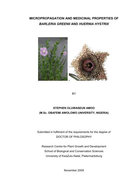
MICROPROPAGATION AND MEDICINAL PROPERTIES OF<br />
BARLERIA GREENII AND HUERNIA HYSTRIX<br />
BY<br />
STEPHEN OLUWASEUN AMOO<br />
(M.Sc. OBAFEMI AWOLOWO UNIVERSITY, NIGERIA)<br />
Submitted in fulfilment <strong>of</strong> the requirements for the degree <strong>of</strong><br />
DOCTOR OF PHILOSOPHY<br />
Research Centre for Plant Growth <strong>and</strong> Development<br />
School <strong>of</strong> Biological <strong>and</strong> Conservation Sciences<br />
University <strong>of</strong> KwaZulu-Natal, Pietermaritzburg<br />
November 2009
TABLE OF CONTENTS<br />
STUDENT DECLARATION ................................................................................... vii<br />
DECLARATION BY SUPERVISORS ................................................................... viii<br />
FACULTY OF SCIENCE & AGRICULTURE DECLARATION 1 - PLAGIARISM.... ix<br />
FACULTY OF SCIENCE & AGRICULTURE DECLARATION 2 - PUBLICATIONS x<br />
ACKNOWLEDGEMENTS ..................................................................................... xii<br />
LIST OF FIGURES ............................................................................................... xiii<br />
LIST OF TABLES .................................................................................................xvii<br />
LIST OF ABBREVIATIONS .................................................................................. xix<br />
ABSTRACT….. ....................................................................................................xxii<br />
Chapter 1 General introduction ........................................................................ 1<br />
1.1 Use <strong>of</strong> plants in horticulture <strong>and</strong> traditional medicine .......................... 1<br />
1.2 The need for conservation <strong>of</strong> plant species ........................................... 1<br />
1.3 Distribution, morphology, uses <strong>and</strong> conservation status <strong>of</strong> the<br />
studied plant species ............................................................................... 3<br />
1.3.1 <strong>Barleria</strong> <strong>greenii</strong> .................................................................................... 3<br />
1.3.2. Huernia hystrix..................................................................................... 5<br />
1.4 Value <strong>of</strong> tissue culture ............................................................................. 7<br />
1.5 Aims <strong>and</strong> objectives ................................................................................. 8<br />
1.6 General overview <strong>of</strong> the thesis ................................................................ 8<br />
Chapter 2 Literature review ............................................................................ 10<br />
2.1 <strong>Micropropagation</strong> ................................................................................... 10<br />
2.1.1 Introduction ........................................................................................ 10<br />
2.1.2 Stage 0: Selection <strong>and</strong> preparation <strong>of</strong> mother plants ......................... 10<br />
2.1.3 Stage I: Initiation <strong>and</strong> establishment <strong>of</strong> aseptic culture ...................... 11<br />
ii
2.1.4 Stage II: Proliferation or multiplication <strong>of</strong> propagules ......................... 15<br />
2.1.5 Stage III: Preparation <strong>of</strong> propagules for transfer to soil ..................... 17<br />
2.1.6 Stage IV: In vivo rooting <strong>and</strong> acclimatization for soil establishment... 19<br />
2.1.7 Effects <strong>of</strong> auxins <strong>and</strong> cytokinins on plant regeneration in<br />
micropropagation ............................................................................. 22<br />
2.1.8 Environmental factors affecting micropropagation ............................. 29<br />
2.1.8.1 Light ............................................................................................... 29<br />
2.1.8.2 Temperature ................................................................................... 31<br />
2.1.9 Tissue culture <strong>of</strong> the families: Acanthaceae <strong>and</strong> Asclepiadaceae ..... 32<br />
2.2 Pharmacological <strong>and</strong> phytochemical investigation <strong>of</strong> plant extracts 36<br />
2.2.1 Introduction ........................................................................................ 36<br />
2.2.2 Antimicrobial activity .......................................................................... 37<br />
2.2.3 Anti-inflammatory activity ................................................................... 39<br />
2.2.4 Acetylcholinesterase inhibition .......................................................... 42<br />
2.2.5 Antioxidant activity ............................................................................. 43<br />
2.2.6 Phytochemical property ..................................................................... 45<br />
Chapter 3 In vitro propagation <strong>of</strong> <strong>Barleria</strong> <strong>greenii</strong> ........................................ 48<br />
3.1 Introduction ............................................................................................ 48<br />
3.2 Materials <strong>and</strong> methods ........................................................................... 49<br />
3.2.1 Explant decontamination, selection <strong>and</strong> bulking ................................ 49<br />
3.2.2 Effects <strong>of</strong> BA <strong>and</strong> NAA on shoot multiplication .................................. 50<br />
3.2.3 Effects <strong>of</strong> types <strong>and</strong> concentrations <strong>of</strong> cytokinins on shoot<br />
multiplication .................................................................................... 50<br />
3.2.4 Effects <strong>of</strong> photoperiod on shoot multiplication ................................... 51<br />
3.2.5 In vitro rooting <strong>of</strong> regenerated shoots ................................................ 51<br />
3.2.6 Ex vitro rooting <strong>and</strong> acclimatization ................................................... 52<br />
3.2.7 Data analyses .................................................................................... 52<br />
iii
3.3 Results <strong>and</strong> discussion ......................................................................... 53<br />
3.3.1 Explant decontamination ................................................................... 54<br />
3.3.2 Effects <strong>of</strong> BA <strong>and</strong> NAA on shoot multiplication .................................. 55<br />
3.3.3 Effects <strong>of</strong> types <strong>and</strong> concentrations <strong>of</strong> cytokinins on shoot<br />
multiplication .................................................................................... 58<br />
3.3.4 Effects <strong>of</strong> photoperiod on shoot multiplication ................................... 62<br />
3.3.5 In vitro rooting <strong>of</strong> regenerated shoots ................................................ 63<br />
3.3.6 Ex vitro rooting <strong>and</strong> acclimatization ................................................... 65<br />
Chapter 4 In vitro propagation <strong>of</strong> Huernia hystrix ........................................ 67<br />
4.1 Introduction ............................................................................................ 67<br />
4.2 Materials <strong>and</strong> methods ........................................................................... 69<br />
4.2.1 Source material, decontamination <strong>and</strong> bulking <strong>of</strong> explants ................ 69<br />
4.2.2 Effects <strong>of</strong> BA <strong>and</strong> NAA on shoot multiplication .................................. 69<br />
4.2.3 Effects <strong>of</strong> temperature <strong>and</strong> photoperiod on shoot multiplication ........ 70<br />
4.2.4 Determination <strong>of</strong> titratable acidity ...................................................... 70<br />
4.2.5 Effects <strong>of</strong> culture vessel size on shoot multiplication ......................... 71<br />
4.2.6 Indirect organogenesis ...................................................................... 71<br />
4.2.7 Rooting <strong>and</strong> acclimatization ............................................................... 71<br />
4.2.8 Data analyses .................................................................................... 72<br />
4.3 Results <strong>and</strong> discussion ......................................................................... 72<br />
4.3.1 Explant decontamination ................................................................... 72<br />
4.3.2 Shoot <strong>and</strong> root organogenesis .......................................................... 74<br />
4.3.3 Effects <strong>of</strong> temperature <strong>and</strong> photoperiod on shoot multiplication ........ 77<br />
4.3.4 Titratable acidity ................................................................................ 82<br />
4.3.5 Effects <strong>of</strong> culture vessel size on shoot multiplication ......................... 84<br />
4.3.6 Indirect organogenesis ...................................................................... 86<br />
4.3.7 Rooting <strong>and</strong> acclimatization ............................................................... 87<br />
iv
Chapter 5 Pharmacological <strong>and</strong> phytochemical evaluation <strong>of</strong> <strong>Barleria</strong><br />
species <strong>and</strong> Huernia hystrix ......................................................... 90<br />
5.1 Introduction ............................................................................................ 90<br />
5.2 Materials <strong>and</strong> methods ........................................................................... 91<br />
5.2.1 Collection <strong>of</strong> plant materials .............................................................. 91<br />
5.2.2 Pharmacological evaluation ............................................................... 92<br />
5.2.2.1 Preparation <strong>of</strong> extracts ................................................................... 92<br />
5.2.2.2 Antibacterial activity ....................................................................... 92<br />
5.2.2.3 Antifungal activity ........................................................................... 93<br />
5.2.2.4 Anti-inflammatory activity ............................................................... 94<br />
5.2.2.5 Acetylcholinesterase (AChE) inhibition........................................... 95<br />
5.2.2.6 Antioxidant activity ......................................................................... 96<br />
5.2.2.6.1 DPPH radical-scavenging activity ............................................. 96<br />
5.2.2.6.2 Ferric reducing power activity .................................................... 97<br />
5.2.2.6.3 β-Carotene linoleic acid assay .................................................. 97<br />
5.2.3 Phytochemical evaluation .................................................................. 98<br />
5.2.3.1 Preparation <strong>of</strong> extracts ................................................................... 98<br />
5.2.3.2 Total phenolic content .................................................................... 98<br />
5.2.3.3 Total iridoid content ........................................................................ 99<br />
5.2.3.4 Flavonoid content ........................................................................... 99<br />
5.2.3.5 Gallotannin content ...................................................................... 100<br />
5.2.3.6 Condensed tannin (proanthocyanidin) content ............................. 100<br />
5.2.4 Data analyses .................................................................................. 101<br />
5.3 Results <strong>and</strong> discussion ....................................................................... 101<br />
5.3.1 Yield <strong>of</strong> plant extracts ...................................................................... 101<br />
5.3.2 Pharmacological evaluation ............................................................. 101<br />
5.3.2.1 Antibacterial activity ..................................................................... 101<br />
v
5.3.2.2 Antifungal activity ......................................................................... 107<br />
5.3.2.3 Anti-inflammatory activity ............................................................. 110<br />
5.3.2.4 Acetylcholinesterase inhibition ..................................................... 112<br />
5.3.2.5 Antioxidant activity ....................................................................... 115<br />
5.3.2.5.1 DPPH radical scavenging activity ............................................ 115<br />
5.3.2.5.2 Ferric ion reducing power activity ............................................ 118<br />
5.3.2.5.3 β-Carotene linoleic acid assay ................................................ 120<br />
5.3.3.1 Total phenolic content .................................................................. 124<br />
5.3.3.2 Total iridoid content ...................................................................... 126<br />
5.3.3.3 Flavonoid content ......................................................................... 128<br />
5.3.3.4 Gallotannin content ...................................................................... 130<br />
5.3.3.5 Condensed tannin (proanthocyanidin) content ............................. 132<br />
Chapter 6 General conclusions .................................................................... 134<br />
References ....................................................................................................... 137<br />
vi
STUDENT DECLARATION<br />
<strong>Micropropagation</strong> <strong>and</strong> <strong>medicinal</strong> <strong>properties</strong> <strong>of</strong> <strong>Barleria</strong> <strong>greenii</strong> <strong>and</strong><br />
Huernia hystrix<br />
I, Stephen Oluwaseun Amoo, Student Number 205527320<br />
declare that:<br />
(i) The research reported in this dissertation, except where otherwise<br />
indicated, is the result <strong>of</strong> my own endeavours in the Research Centre for<br />
Plant Growth <strong>and</strong> Development, School <strong>of</strong> Biological <strong>and</strong> Conservation<br />
Sciences, University <strong>of</strong> KwaZulu-Natal Pietermaritzburg;<br />
(ii) This dissertation has not been submitted for any degrees or examination<br />
at any other University;<br />
(iii) This thesis does not contain data, figures or writing, unless specifically<br />
acknowledged, copied from other researchers; <strong>and</strong><br />
(iv) Where I have reproduced a publication <strong>of</strong> which I am an author or co-<br />
author, I have indicated which part <strong>of</strong> the publication was contributed by<br />
me.<br />
Signed at Pietermaritzburg on the...…. day <strong>of</strong> March, 2010.<br />
__________________________<br />
SIGNATURE<br />
vii
DECLARATION BY SUPERVISORS<br />
We hereby declare that we acted as Supervisors for this PhD student:<br />
Student’s Full Name: Stephen Oluwaseun Amoo<br />
Student Number: 205527320<br />
Thesis Title: <strong>Micropropagation</strong> <strong>and</strong> <strong>medicinal</strong> <strong>properties</strong> <strong>of</strong> <strong>Barleria</strong> <strong>greenii</strong> <strong>and</strong><br />
Huernia hystrix<br />
Regular consultation took place between the student <strong>and</strong> ourselves throughout the<br />
investigation. We advised the student to the best <strong>of</strong> our ability <strong>and</strong> approved the<br />
final document for submission to the Faculty <strong>of</strong> Science <strong>and</strong> Agriculture Higher<br />
Degrees Office for examination by the University appointed Examiners.<br />
__________________________<br />
SUPERVISOR: PROFESSOR J VAN STADEN<br />
________________________<br />
CO-SUPERVISOR: DR JF FINNIE<br />
viii
FACULTY OF SCIENCE & AGRICULTURE DECLARATION 1 -<br />
I, Stephen Oluwaseun Amoo, declare that<br />
PLAGIARISM<br />
1. The research reported in this thesis, except where otherwise indicated,<br />
is my original research.<br />
2. This thesis has not been submitted for any degree or examination at any<br />
other university.<br />
3. This thesis does not contain other persons‟ data, pictures, graphs or<br />
other information, unless specifically acknowledged as being sourced<br />
from other persons.<br />
4. This thesis does not contain other persons' writing, unless specifically<br />
acknowledged as being sourced from other researchers. Where other<br />
written sources have been quoted, then:<br />
a. Their words have been re-written but the general information<br />
attributed to them has been referenced<br />
b. Where their exact words have been used, then their writing has been<br />
placed in italics <strong>and</strong> inside quotation marks, <strong>and</strong> referenced.<br />
5. This thesis does not contain text, graphics or tables copied <strong>and</strong> pasted<br />
Signed<br />
from the Internet, unless specifically acknowledged, <strong>and</strong> the source<br />
being detailed in the thesis <strong>and</strong> in the References sections.<br />
………………………………………………………………………………<br />
Declaration Plagiarism 22/05/08 FHDR Approved<br />
ix
FACULTY OF SCIENCE & AGRICULTURE DECLARATION 2 -<br />
PUBLICATIONS<br />
PUBLISHED ARTICLES FROM THIS THESIS<br />
AMOO, S.O., FINNIE, J.F. <strong>and</strong> VAN STADEN, J. 2009. Effects <strong>of</strong><br />
temperature, photoperiod <strong>and</strong> culture vessel size on adventitious shoot<br />
production <strong>of</strong> in vitro propagated Huernia hystrix. Plant Cell, Tissue <strong>and</strong><br />
Organ Culture 99: 233-238<br />
Contribution: Experimental work <strong>and</strong> writing <strong>of</strong> publication done by the first<br />
author under the supervision <strong>of</strong> the last two authors.<br />
AMOO, S.O., FINNIE, J.F. <strong>and</strong> VAN STADEN, J. 2009. In vitro<br />
pharmacological evaluation <strong>of</strong> three <strong>Barleria</strong> species. Journal <strong>of</strong><br />
Ethnopharmacology 121: 274-277<br />
Contribution: Experimental work <strong>and</strong> writing <strong>of</strong> publication done by the first<br />
author under the supervision <strong>of</strong> the last two authors.<br />
AMOO, S.O., FINNIE, J.F. <strong>and</strong> VAN STADEN, J. 2009. In vitro<br />
propagation <strong>of</strong> Huernia hystrix: an endangered <strong>medicinal</strong> <strong>and</strong> ornamental<br />
succulent. Plant Cell, Tissue <strong>and</strong> Organ Culture 96: 273-278<br />
Contribution: Experimental work <strong>and</strong> writing <strong>of</strong> publication done by the first<br />
author under the supervision <strong>of</strong> the last two authors.<br />
CONFERENCE CONTRIBUTIONS FROM THIS THESIS<br />
AMOO, S.O., FINNIE, J.F. <strong>and</strong> VAN STADEN, J. 2008. In vitro<br />
pharmacological evaluation <strong>of</strong> three <strong>Barleria</strong> species. Joint Conference <strong>of</strong><br />
the 34 th South African Association <strong>of</strong> Botanists (SAAB) <strong>and</strong> 7 th Southern<br />
African Society <strong>of</strong> Systematic Biology (SASSB). Drakensville Mountain<br />
Resort, South Africa. (Oral presentation by the first author)<br />
x
AMOO, S.O., FINNIE, J.F. <strong>and</strong> VAN STADEN, J. 2009. <strong>Micropropagation</strong><br />
Signed<br />
<strong>of</strong> an endangered valuable succulent: Huernia hystrix. 35 th Annual<br />
Conference <strong>of</strong> the South African Association <strong>of</strong> Botanists (SAAB) <strong>and</strong><br />
International workshop on “Phosphate as a limiting resource”. Stellenbosch<br />
University, South Africa. (Oral presentation by the first author)<br />
………………………………………………………………………………<br />
Declaration Publications FHDR 22/05/08 Approved<br />
xi
ACKNOWLEDGEMENTS<br />
I would like to express a special appreciation to my supervisor, Pr<strong>of</strong> J. van Staden<br />
for his invaluable support, guidance <strong>and</strong> encouragement throughout the duration<br />
<strong>of</strong> this study. I am very grateful for the financial support he provided in the form <strong>of</strong><br />
a postgraduate bursary.<br />
Many thanks to my co-supervisor, Dr J.F. Finnie, for his constructive advice, as<br />
well as constant support <strong>and</strong> encouragement.<br />
My sincere thanks to all members <strong>of</strong> the Research Centre for Plant Growth <strong>and</strong><br />
Development, for their extensive support. The administrative staff (Mrs Magnussen<br />
<strong>and</strong> Mrs Warren) <strong>and</strong> my Research Committee members (Dr Bairu <strong>and</strong> Dr<br />
Elgorashi) are especially thanked for their support <strong>and</strong> inputs. My sincere<br />
appreciation to Dr Gary Stafford <strong>and</strong> Mrs Alison Young for their help in the<br />
collection <strong>of</strong> plants used in this study. Dr Gary Stafford also kindly supplied the<br />
Huernia hystrix flower used on the cover page. Thanks to my colleagues in the lab,<br />
especially Ashwell Ndhlala <strong>and</strong> Mack Moyo, with whom I shared many<br />
brainstorming sessions related to some aspects <strong>of</strong> this research. The technical<br />
assistance provided by Mr Hampton <strong>and</strong> his team in the use <strong>of</strong> some equipment<br />
are much appreciated.<br />
My special thanks to my parents <strong>and</strong> brother as well as Grace, Afolad, Folake <strong>and</strong><br />
Bunmi for their underst<strong>and</strong>ing, love, support <strong>and</strong> encouragement. I miss you all.<br />
Above all, glory, praise <strong>and</strong> honour to Jehovah God, the omnipotent <strong>and</strong><br />
omniscient one, for his indescribable free gift.<br />
xii
LIST OF FIGURES<br />
Chapter 1<br />
Figure 1.1: <strong>Barleria</strong> <strong>greenii</strong>. (A) The plant during flowering (B) Calyx bearing the<br />
unopened, mature fruits (capsules). ................................................... 4<br />
Figure 1.2: Flowering Huernia hystrix potted in a small container. Bar = 10 mm. 6<br />
Chapter 2<br />
Figure 2.1: Molecular structures <strong>of</strong> some auxins commonly used in plant tissue<br />
culture. ............................................................................................. 23<br />
Figure 2.2: Molecular structures <strong>of</strong> some cytokinins commonly used in plant<br />
tissue culture. ................................................................................... 27<br />
Figure 2.3: The physiological <strong>and</strong> pathophysiological functions <strong>of</strong> COX-2 enzyme<br />
(STEINMEYER, 2000). .................................................................... 41<br />
Chapter 3<br />
Figure 3.1: In vitro propagation <strong>of</strong> <strong>Barleria</strong> <strong>greenii</strong>. (A) Stock plant. (B) Control<br />
(MS medium without PGR). (C) Shoot multiplication on MS medium<br />
supplemented with 3 µM BA. (D) Shoot multiplication on MS medium<br />
supplemented with 7 µM MemTR. (E) In vitro rooted regenerated<br />
shoot ready for acclimatization. (F) Three-month-old fully<br />
acclimatized plant. Bars = 10 mm. ................................................... 53<br />
Figure 3.2: Effects <strong>of</strong> sodium hypochlorite (NaOCl) solution treatments on<br />
explants decontamination. ............................................................... 54<br />
Figure 3.3: Effects <strong>of</strong> BA <strong>and</strong> NAA on adventitious shoot production <strong>of</strong> <strong>Barleria</strong><br />
<strong>greenii</strong> after four weeks <strong>of</strong> culture. Bars with different letters are<br />
significantly different (P = 0.05) according to DMRT. ....................... 56<br />
Figure 3.4: Effects <strong>of</strong> BA <strong>and</strong> NAA on adventitious shoot production <strong>of</strong> <strong>Barleria</strong><br />
<strong>greenii</strong> after six weeks <strong>of</strong> culture. Bars with different letters are<br />
significantly different (P = 0.05) according to DMRT. ....................... 57<br />
Figure 3.5: Effects <strong>of</strong> half strength MS medium with or without IBA treatment on<br />
in vitro rooting <strong>of</strong> regenerated shoots. Bars with different letters are<br />
significantly different (P = 0.05) according to DMRT. ....................... 64<br />
xiii
Figure 3.6: Effects <strong>of</strong> pulsing with different IBA concentrations on ex vitro<br />
acclimatization <strong>of</strong> regenerated shoots .............................................. 66<br />
Chapter 4<br />
Figure 4.1: In vitro propagation <strong>of</strong> Huernia hystrix. (A) Stock plant. (B) Control<br />
with root production (MS medium without plant growth regulators).<br />
(C) Multiple shoot production accompanied with root formation on MS<br />
medium supplemented with 5.37 µM NAA <strong>and</strong> 22.19 µM BA. (D)<br />
Two-month-old fully acclimatized plants (E) Green callus growth with<br />
root hairs on MS medium supplemented with 5.37 µM NAA. (F) Root<br />
regeneration from callus on MS medium supplemented with 2.69 µM<br />
NAA <strong>and</strong> 2.22 µM BA. Scale bar = 10 mm. ...................................... 73<br />
Figure 4.2: Frequencies <strong>of</strong> explants decontamination in different mercuric<br />
chloride solution treatments. ............................................................ 74<br />
Figure 4.3: Nocturnal titratable acidity in Huernia hystrix shoots cultured at<br />
different temperatures. 22:00, 02:00 <strong>and</strong> 06:00 h are the beginning,<br />
middle <strong>and</strong> end <strong>of</strong> 8 h dark period respectively. Bars with different<br />
letters are significantly different (Ρ = 0.05) according to DMRT. ...... 84<br />
Figure 4.4: Huernia hystrix adventitious shoot production from explants cultured<br />
in different culture vessels. (A) Screw cap jar [300 ml]. (B) Culture<br />
tube [40 ml]. Scale bar = 10 mm. ..................................................... 86<br />
Figure 4.5: Effects <strong>of</strong> combinations <strong>of</strong> NAA <strong>and</strong> BA concentrations on callus<br />
growth. Bars with different letters are significantly different (Ρ = 0.05)<br />
according to DMRT. ........................................................................... 87<br />
Chapter 5<br />
Figure 5.1: Anti-inflammatory activity <strong>of</strong> extracts from different parts <strong>of</strong> three<br />
<strong>Barleria</strong> species in COX-1 (on the left side) <strong>and</strong> COX-2 (on the right<br />
side) assays. B.p, B.a, <strong>and</strong> B.g are <strong>Barleria</strong> prionitis, B. albostellata<br />
<strong>and</strong> B. <strong>greenii</strong>, respectively. (A) <strong>and</strong> (B) are PE extracts. (C) <strong>and</strong> (D)<br />
are DCM extracts. (E) <strong>and</strong> (F) are EtOH extracts. Bars in the same<br />
graph bearing different letters are significantly different (P = 0.05)<br />
according to DMRT. Percentage inhibition by indomethacin in COX-1<br />
was 63.4 ± 1.98 <strong>and</strong> COX-2 was 73.6 ± 1.47. ................................ 111<br />
xiv
Figure 5.2: Anti-inflammatory activity <strong>of</strong> different extracts <strong>of</strong> Huernia hystrix. (A)<br />
COX-1 assay. (B) COX-2 assay. Bars with different letters in each<br />
graph are significantly different (P = 0.05) according to DMRT.<br />
Percentage inhibition by indomethacin in COX-1 was 54.73 ± 2.00<br />
<strong>and</strong> COX-2 was 63.44 ± 2.52. ........................................................ 112<br />
Figure 5.3: Dose-dependent acetylcholinesterase inhibition by different parts <strong>of</strong><br />
three <strong>Barleria</strong> species. B.p = <strong>Barleria</strong> prionitis, B.g = <strong>Barleria</strong> <strong>greenii</strong>,<br />
B.a = <strong>Barleria</strong> albostellata. Bars bearing different letters are<br />
significantly different (P = 0.05) according to DMRT. The AChE<br />
inhibition activities by galanthamine at 0.5, 1.0 <strong>and</strong> 2 µM were 49.24,<br />
59.81 <strong>and</strong> 77.03%, respectively. .................................................... 113<br />
Figure 5.4: Dose-dependent acetylcholinesterase inhibition by different parts <strong>of</strong><br />
Huernia hystrix. Bars bearing different letters are significantly<br />
different (P = 0.05) according to DMRT. The AChE inhibition<br />
activities by galanthamine at 0.5, 1.0 <strong>and</strong> 2 µM were 49.24, 59.81<br />
<strong>and</strong> 77.03%, respectively. .............................................................. 114<br />
Figure 5.5: Dose-dependent curve <strong>of</strong> DPPH radical scavenging activity <strong>of</strong><br />
different parts <strong>of</strong> three <strong>Barleria</strong> species. (A) Leaves (B) Stems (C)<br />
Roots. ............................................................................................. 116<br />
Figure 5.6: Dose-dependent curve <strong>of</strong> DPPH radical scavenging activity <strong>of</strong><br />
different parts <strong>of</strong> Huernia hystrix. ................................................... 117<br />
Figure 5.7: Ferric ion reducing power activity <strong>of</strong> different parts <strong>of</strong> <strong>Barleria</strong><br />
species. (A) Leaves (B) Stems (C) Roots. ..................................... 119<br />
Figure 5.8: Ferric ion reducing power activity <strong>of</strong> different parts <strong>of</strong> Huernia hystrix.<br />
....................................................................................................... 120<br />
Figure 5.9: Antioxidant activities <strong>of</strong> different parts <strong>of</strong> three <strong>Barleria</strong> species in β-<br />
carotene-linoleic acid model system. Bars bearing different letters in<br />
each graph are significantly different (P = 0.05) according to DMRT.<br />
(A) Antioxidant activity (ANT) based on the average β-carotene<br />
bleaching rate. (B) Oxidation rate ratio (ORR). (C) Antioxidant activity<br />
(AA) at t = 60 min. (D) Antioxidant activity (AA) at t = 120 min. ...... 122<br />
Figure 5.10: Antioxidant activities <strong>of</strong> different parts <strong>of</strong> Huernia hystrix in β-<br />
carotene-linoleic acid model system. Bars bearing different letters in<br />
each graph are significantly different (P = 0.05) according to DMRT.<br />
xv
(A) Antioxidant activity (ANT) based on the average β-carotene<br />
bleaching rate. (B) Oxidation rate ratio. (C) Antioxidant activity at 60<br />
min. (D) Antioxidant activity at 120 min. ......................................... 123<br />
Figure 5.11: Total phenolic content <strong>of</strong> different parts <strong>of</strong> three <strong>Barleria</strong> species.<br />
Bars bearing different letters are significantly different (P = 0.05)<br />
according to DMRT. ....................................................................... 125<br />
Figure 5.12: Total phenolic content <strong>of</strong> different parts <strong>of</strong> Huernia hystrix. Bars<br />
bearing different letters are significantly different (P = 0.05) according<br />
to DMRT. ........................................................................................ 125<br />
Figure 5.13: Total iridoid content <strong>of</strong> different parts <strong>of</strong> three <strong>Barleria</strong> species. Bars<br />
bearing different letters are significantly different (P = 0.05) according<br />
to DMRT. ........................................................................................ 127<br />
Figure 5.14: Total iridoid content <strong>of</strong> different parts <strong>of</strong> Huernia hystrix. Bars bearing<br />
different letters are significantly different (P = 0.05) according to<br />
DMRT. ............................................................................................ 127<br />
Figure 5.15: Flavonoid content <strong>of</strong> different parts <strong>of</strong> three <strong>Barleria</strong> species. Bars<br />
bearing different letters are significantly different (P = 0.05) according<br />
to DMRT. ........................................................................................ 129<br />
Figure 5.16: Flavonoid content <strong>of</strong> different parts <strong>of</strong> Huernia hystrix. Bars bearing<br />
different letters are significantly different (P = 0.05) according to<br />
DMRT. ............................................................................................ 129<br />
Figure 5.17: Gallotannin content <strong>of</strong> different parts <strong>of</strong> three <strong>Barleria</strong> species. Bars<br />
bearing different letters are significantly different (P = 0.05) according<br />
to DMRT. ........................................................................................ 131<br />
Figure 5.18: Gallotannin content <strong>of</strong> different parts <strong>of</strong> Huernia hystrix. Bars bearing<br />
different letters are significantly different (P = 0.05) according to<br />
DMRT. ............................................................................................ 131<br />
Figure 5.19: Condensed tannin content (as leucocyanidin equivalents) <strong>of</strong> different<br />
parts <strong>of</strong> three <strong>Barleria</strong> species. Bars bearing different letters are<br />
significantly different (P = 0.05) according to DMRT. ..................... 132<br />
xvi
LIST OF TABLES<br />
Chapter 2<br />
Table 2.1: List <strong>of</strong> some tissue cultured plant species in the Acanthaceae <strong>and</strong><br />
Asclepiadaceae families ..................................................................... 33<br />
Chapter 3<br />
Table 3.1: Effects <strong>of</strong> types <strong>and</strong> concentrations <strong>of</strong> cytokinins on adventitious shoot<br />
production <strong>of</strong> <strong>Barleria</strong> <strong>greenii</strong> ............................................................. 60<br />
Table 3.2: Effects <strong>of</strong> photoperiod on adventitious shoot production <strong>of</strong> <strong>Barleria</strong><br />
<strong>greenii</strong> after six weeks <strong>of</strong> culture ........................................................ 63<br />
Chapter 4<br />
Table 4.1: Effects <strong>of</strong> different combinations <strong>of</strong> NAA <strong>and</strong> BA on shoot <strong>and</strong> root<br />
regeneration <strong>of</strong> Huernia hystrix after nine weeks <strong>of</strong> culture ............... 75<br />
Table 4.2: Frequencies <strong>of</strong> shoot, root <strong>and</strong> basal callus production from treatments<br />
with different concentration combinations <strong>of</strong> NAA <strong>and</strong> BA ................. 76<br />
Table 4.3: Effects <strong>of</strong> different concentration combinations <strong>of</strong> NAA <strong>and</strong> BA on<br />
fresh weights <strong>of</strong> adventitious shoots <strong>and</strong> roots produced per explant 78<br />
Table 4.4: Effects <strong>of</strong> temperature <strong>and</strong> photoperiod on adventitious shoot<br />
production <strong>of</strong> Huernia hystrix after eight weeks <strong>of</strong> culture .................. 79<br />
Table 4.5: Effects <strong>of</strong> temperature <strong>and</strong> photoperiod on frequency <strong>and</strong> fresh weight<br />
<strong>of</strong> regenerated shoots per explant <strong>of</strong> Huernia hystrix after eight weeks<br />
<strong>of</strong> culture ............................................................................................ 80<br />
Table 4.6: Effects <strong>of</strong> culture vessel size on adventitious shoot production <strong>of</strong><br />
Huernia hystrix after eight weeks <strong>of</strong> culture........................................ 85<br />
Table 4.7: Effects <strong>of</strong> half-strength MS medium with or without IBA<br />
supplementation on rooting <strong>and</strong> acclimatization <strong>of</strong> regenerated plants<br />
........................................................................................................... 88<br />
Chapter 5<br />
Table 5.1: Yield (% w/w) <strong>of</strong> extracts prepared from different parts <strong>of</strong> three <strong>Barleria</strong><br />
species in terms <strong>of</strong> starting crude material ....................................... 103<br />
xvii
Table 5.2: Yield (% w/w) <strong>of</strong> extracts prepared from different parts <strong>of</strong> Huernia<br />
hystrix in terms <strong>of</strong> starting crude material ......................................... 104<br />
Table 5.3: Antibacterial activity (MIC <strong>and</strong> MID) <strong>of</strong> crude extracts from different<br />
parts <strong>of</strong> three <strong>Barleria</strong> species .......................................................... 105<br />
Table 5.4: Antibacterial activity (MIC <strong>and</strong> MBC) <strong>of</strong> crude extracts from different<br />
parts <strong>of</strong> Huernia hystrix .................................................................... 106<br />
Table 5.5: Antifungal activity <strong>of</strong> crude extracts from different parts <strong>of</strong> three<br />
<strong>Barleria</strong> species against C<strong>and</strong>ida albicans ....................................... 108<br />
Table 5.6: Antifungal activity <strong>of</strong> different parts <strong>of</strong> Huernia hystrix against C<strong>and</strong>ida<br />
albicans ............................................................................................ 109<br />
Table 5.7: DPPH radical scavenging activity <strong>of</strong> different parts <strong>of</strong> three <strong>Barleria</strong><br />
species ............................................................................................. 117<br />
Table 5.8: DPPH radical scavenging activity <strong>of</strong> different parts <strong>of</strong> Huernia hystrix<br />
......................................................................................................... 118<br />
xviii
LIST OF ABBREVIATIONS<br />
[3G]BA 6-Benzylamino-3-β-D-glucopyranosylpurine<br />
[7G]BA 6-Benzylamino-7-β-D-glucopyranosylpurine<br />
[9G]BA 6-Benzylamino-9-β-D-glucopyranosylpurine<br />
[9R]BA 6-Benzylamino-9-β-D-rib<strong>of</strong>uranosylpurine<br />
2,4-D 2,4-Dichlorophenoxyacetic acid<br />
AChE Acetylcholinesterase<br />
AD Alzheimer‟s disease<br />
AIDS Acquired immune deficiency syndrome<br />
ANOVA Analysis <strong>of</strong> variance<br />
ATCC American type culture collection<br />
ATCI Acetylthiocholine iodide<br />
ATP Adenosine triphosphate<br />
B5 B5 medium (GAMBORG et al., 1968)<br />
BA 6-Benzyladenine<br />
BHT Butylated hydroxytoluene (2,6-Di-tert.-butyl-p-cresol)<br />
CAM Crassulacean acid metabolism<br />
CNS Central nervous system<br />
COX Cyclooxygenase<br />
DCM Dichloromethane<br />
DHZ Dihydrozeatin<br />
DIF Difference in photoperiod <strong>and</strong> dark period temperatures<br />
DMRT Duncan‟s Multiple Range Test<br />
DPM Disintegrations per minute<br />
DPPH 1,1-Diphenyl-2-picrylhydrazyl<br />
DTNB 5,5-Dithiobis-2-nitrobenzoic acid<br />
DW Dry weight<br />
ET Electron transfer<br />
EtOH Ethanol<br />
Folin C Folin-Ciocalteu<br />
FRAP Ferric ion reducing power assay<br />
FSA Flora <strong>of</strong> southern Africa<br />
xix
GAE Gallic acid equivalents<br />
GST Glutathione S-transferase<br />
HAT Hydrogen atom transfer<br />
HE Harpagoside equivalents<br />
HPLC High-performance liquid chromatography<br />
IAA Indole-3-acetic acid<br />
IBA Indole-3-butyric acid<br />
iNOS Inducible nitric oxide synthase<br />
INT p-Iodonitrotetrazolium chloride<br />
iP N 6 -Isopentenyladenine<br />
IUCN International Union for the Conservation <strong>of</strong> Nature <strong>and</strong><br />
Natural Resources<br />
LOX 5-Lipoxygenase<br />
LTs Leukotrienes<br />
MBC Minimum bactericidal concentration<br />
MemTR meta-Methoxytopolin riboside<br />
MeOH Methanol<br />
MFC Minimum fungicidal concentration<br />
MFD Minimum fungicidal dilution<br />
MH Mueller-Hinton<br />
MIC Minimum inhibitory concentration<br />
MID Minimum inhibitory dilution<br />
MS MURASHIGE <strong>and</strong> SKOOG (1962)<br />
mT meta-Topolin<br />
mTR meta-Topolin riboside<br />
NAA α-Naphtalene acetic acid<br />
NADPH Reduced adenine dinucleotide phosphate<br />
ND Not determined<br />
NF-кB Nuclear factor-кB<br />
NSAIDs Nonsteroidal anti-inflammatory drugs<br />
OG O-glucosides<br />
OH Hydroxyl<br />
ORAC Oxygen radical absorbance capacity<br />
xx
PE Petroleum ether<br />
PEPC Phosphoenolpyruvate carboxylase<br />
PGR Plant growth regulators<br />
PPF Photosynthetic photon flux<br />
RNA Ribonucleic acid<br />
ROS Reactive oxygen species<br />
RSA Radical scavenging activity<br />
SH Schenk <strong>and</strong> Hilderbr<strong>and</strong>t (1972)<br />
TEAC Trolox equivalent antioxidant capacity<br />
TLC Thin-layer-chromatographic<br />
US United States<br />
WWF World Wildlife Fund<br />
YM Yeast Malt<br />
Z trans-Zeatin<br />
xxi
ABSTRACT<br />
The crisis <strong>of</strong> newly emerging diseases <strong>and</strong> the resistance <strong>of</strong> many pathogens to<br />
currently used drugs, coupled with the adverse side-effects <strong>of</strong> many <strong>of</strong> these drugs<br />
have necessitated the continuous search for new drugs that are potent <strong>and</strong><br />
efficacious with minimal or no adverse side-effects. The plant kingdom is known to<br />
contain many novel biologically active compounds, many <strong>of</strong> which could potentially<br />
have a higher <strong>medicinal</strong> value when compared to some <strong>of</strong> the current medications.<br />
Indeed, the use <strong>of</strong> plants in traditional medicine, especially in African communities,<br />
is gaining more importance due to their affordability <strong>and</strong> accessibility as well as<br />
their effectiveness. Exponential population growth rates in many developing<br />
countries has resulted in heavy exploitation <strong>of</strong> our plant resources for their<br />
<strong>medicinal</strong> values. In addition, plant habitat destruction arising from human<br />
developmental activities has contributed to the fragmentation or loss <strong>of</strong> many plant<br />
populations. Owing to these factors, many plant species with horticultural <strong>and</strong>/or<br />
<strong>medicinal</strong> potential have become either extinct or are threatened with extinction.<br />
These threatened species cut across different taxonomic categories including<br />
shrubs, trees <strong>and</strong> succulents. Without the application <strong>of</strong> effective conservation<br />
strategies, the <strong>medicinal</strong> <strong>and</strong>/or horticultural potential <strong>of</strong> such threatened species<br />
may be totally lost with time. The extinction <strong>of</strong> such species could lead to the loss<br />
<strong>of</strong> potential therapeutic compounds <strong>and</strong>/or genes capable <strong>of</strong> being exploited in the<br />
biosynthesis <strong>of</strong> new potent pharmaceutical compounds.<br />
The overall aims <strong>of</strong> this study were to establish efficient regeneration protocols<br />
<strong>and</strong> explore the <strong>medicinal</strong> <strong>properties</strong> <strong>of</strong> two threatened South African species<br />
belonging to different taxonomic categories: <strong>Barleria</strong> <strong>greenii</strong> (a shrub) <strong>and</strong> Huernia<br />
hystrix (a succulent). <strong>Barleria</strong> <strong>greenii</strong> is a perennial ornamental <strong>and</strong> a critically<br />
endangered shrub, endemic to a small area in KwaZulu-Natal province <strong>of</strong> South<br />
Africa. Its conventional propagation is hampered by high seed parasitism <strong>and</strong><br />
difficulty in rooting. Huernia hystrix is a dwarf perennial stem succulent heavily<br />
exploited for traditional medicine among the Zulu people in South Africa. It is<br />
considered vulnerable in KwaZulu-Natal province, an endangered endemic to the<br />
flora <strong>of</strong> the southern Africa region <strong>and</strong> is vulnerable in its global conservation<br />
xxii
status. Being a dwarf species, very limited cuttings can be taken from the mother<br />
plant for conventional propagation.<br />
In developing a micropropagation protocol for B. <strong>greenii</strong> using shoot-tip explants (5<br />
mm length), the effects <strong>of</strong> BA with or without NAA combinations, in MS medium<br />
were evaluated. Concentrations <strong>of</strong> 0.0, 0.5, <strong>and</strong> 1.0 µM <strong>of</strong> NAA were combined<br />
with 1.0, 2.0, 3.0, 4.0 <strong>and</strong> 5.0 µM BA in a 3 × 5 completely r<strong>and</strong>omised factorial<br />
design. The treatments with BA alone gave higher adventitious shoot production<br />
both after four <strong>and</strong> six weeks <strong>of</strong> culture when compared to the BA treatments<br />
supplemented with NAA concentrations. These results imply that the exogenous<br />
application <strong>of</strong> NAA is neither required nor beneficial for adventitious shoot<br />
induction or proliferation from the shoot-tip explants <strong>of</strong> this species. In evaluating<br />
the effects <strong>of</strong> different types <strong>and</strong> concentrations <strong>of</strong> cytokinins on shoot production,<br />
the treatments with kinetin generally gave a low shoot production whereas BA<br />
treatments gave increased adventitious shoot production with the optimum at a<br />
concentration <strong>of</strong> 3 µM. High abnormality indices were however, observed in all the<br />
treatments with BA. At higher concentrations (5 <strong>and</strong> 7 µM), the treatments with<br />
mTR <strong>and</strong> MemTR gave increased adventitious shoot production with abnormality<br />
indices less than that <strong>of</strong> the control. These results indicated that the topolins (mTR<br />
<strong>and</strong> MemTR) are less toxic <strong>and</strong> more effective in the micropropagation <strong>of</strong> this plant<br />
species. The abnormality indices recorded in the topolin treatments could possibly<br />
be carry-over effects <strong>of</strong> BA since the explants used were obtained from BA-treated<br />
cultures. Furthermore, cultures maintained under a 16 h photoperiod gave a<br />
significantly higher production <strong>of</strong> adventitious shoots with lengths greater than 10<br />
mm than those placed under continuous light. Regenerated shoots were ex vitro<br />
rooted after an IBA pulse-treatment for five hours <strong>and</strong> acclimatized successfully<br />
with 65% survival. This developed protocol could potentially produce over 60,000<br />
transplantable shoots per year from a single shoot-tip explant.<br />
An efficient <strong>and</strong> rapid micropropagation protocol was successfully developed for H.<br />
hystrix. Adventitious shoots were regenerated from stem explants (10 mm length)<br />
cultured on MS medium supplemented with a range <strong>of</strong> NAA (0.00, 2.69, 5.37 <strong>and</strong><br />
8.06 µM) <strong>and</strong> BA (4.44, 13.32 <strong>and</strong> 22.19 µM BA) concentrations. The treatments<br />
with a combination <strong>of</strong> BA <strong>and</strong> NAA demonstrated a synergistic effect on<br />
xxiii
adventitious shoot production. A 100% shoot regeneration frequency with a<br />
production <strong>of</strong> four adventitious shoots per explant was obtained on MS medium<br />
containing 5.37 µM NAA <strong>and</strong> 22.19 µM BA. Callus produced at the base <strong>of</strong> the<br />
explant on the same medium showed root organogenic potential. The effects <strong>of</strong><br />
photoperiod <strong>and</strong> temperature were further evaluated in optimizing this<br />
micropropagation protocol. Significant increases in shoot proliferation were<br />
observed with increased temperature in cultures maintained under a 16 h<br />
photoperiod. Slow growth observed at low temperatures (15 <strong>and</strong> 20°C) <strong>of</strong>fers a<br />
potential strategy for cost-effective in vitro storage <strong>of</strong> H. hystrix germplasm. The<br />
maximum number <strong>of</strong> adventitious shoots produced per explant <strong>and</strong> shoot<br />
regeneration frequency were observed in cultures maintained at 35ºC, the<br />
optimum temperature for photosynthesis in plants possessing CAM. The nocturnal<br />
accumulation <strong>of</strong> organic acids in cultures incubated under a 16 h photoperiod<br />
further suggest the presence <strong>of</strong> CAM in this species. On the other h<strong>and</strong>, cultures<br />
kept under continuous light appear to shift to a C-3 photosynthetic pathway. With<br />
an increase in temperature under continuous light, there was a significant<br />
decrease in the fresh weight <strong>of</strong> adventitious shoots regenerated per explant. The<br />
use <strong>of</strong> larger culture vessels further increased the shoot proliferation to 5.6 shoots<br />
per explant with a potential production <strong>of</strong> 3429 shoots per m 2 in the growth room<br />
compared to 2750 shoots per m 2 using culture tubes. Regenerated shoots<br />
produced roots when transferred to half strength MS medium with or without auxin.<br />
The micropropagated plants were easily acclimatized within two months under<br />
greenhouse conditions when potted in a soil <strong>and</strong> s<strong>and</strong> mixture (1:1; v/v) treated<br />
with a fungicide (Benlate, 0.01%). More than 95% survival with no observable<br />
morphological variations was obtained. The developed protocol provides a simple,<br />
cost-effective means for the conservation <strong>of</strong> endangered H. hystrix by clonal<br />
propagation within a short time.<br />
The dual biological <strong>and</strong> chemical screening approach was used to evaluate the<br />
<strong>medicinal</strong> <strong>properties</strong> <strong>of</strong> three <strong>Barleria</strong> species (including B. <strong>greenii</strong>) <strong>and</strong> H. hystrix.<br />
Different extracts <strong>of</strong> these species demonstrated antibacterial, antifungal, anti-<br />
inflammatory, antioxidant <strong>and</strong> AChE inhibition activities. The observed<br />
pharmacological activities might be largely due to their relatively high flavonoid<br />
content, with a contributing effect from their iridoid <strong>and</strong> tannin compounds. The<br />
xxiv
activities shown by H. hystrix extracts might possibly explain its heavy exploitation<br />
in traditional medicine. Extracts from <strong>Barleria</strong> species generally had comparatively<br />
higher pharmacological activities <strong>and</strong> phytochemical content. The concept <strong>of</strong><br />
substituting plant parts (such as leaves <strong>and</strong> stems for roots) for sustainable<br />
exploitation was found to be dependent on the species <strong>and</strong>/or biological activity<br />
evaluated. The substantial high activities observed with some B. <strong>greenii</strong> extracts in<br />
the pharmacological assays used further highlight the need to conserve our plant<br />
resources before they become extinct, since some <strong>of</strong> them could be<br />
pharmacologically active <strong>and</strong> perhaps contain novel compounds that are<br />
biologically active against some treatment-resistant infections.<br />
xxv
Chapter 1 General introduction<br />
1.1 Use <strong>of</strong> plants in horticulture <strong>and</strong> traditional medicine<br />
Worldwide, there is an ever-growing dem<strong>and</strong> for valuable plants many <strong>of</strong> which<br />
are used for horticultural <strong>and</strong> <strong>medicinal</strong> purposes. Flowering plant species that can<br />
be easily potted, are frost-hardy, cold or drought tolerant <strong>and</strong> requiring little<br />
maintenance are sought after by collectors due to their high ornamental value.<br />
Some <strong>of</strong> these ornamental species are also known to be <strong>medicinal</strong>, <strong>and</strong> <strong>medicinal</strong><br />
plants are important in meeting the human need for good healthcare. A report by<br />
the World Health Organization, for example, shows that about 80% <strong>of</strong> the world‟s<br />
population depends on traditional medicines to meet at least some <strong>of</strong> their primary<br />
healthcare needs (WHO, 2004). The use <strong>of</strong> plants in indigenous or traditional<br />
medicine is more affordable <strong>and</strong> accessible to most <strong>of</strong> the population especially in<br />
African communities, <strong>and</strong> it is generally believed to be effective. FENNELL (2002)<br />
observed that about two-thirds <strong>of</strong> the South African population still use plants for<br />
traditional medicines. The majority <strong>of</strong> plants traditionally used for <strong>medicinal</strong><br />
purposes in South Africa are harvested from the wild <strong>and</strong> are yet to be fully<br />
analyzed for their bioactive compounds (TAYLOR <strong>and</strong> VAN STADEN, 2001). In<br />
addition, the exponential population growth rates in developing countries since the<br />
latter half <strong>of</strong> the twentieth century has resulted in increased dem<strong>and</strong> for plant<br />
resources (JÄGER <strong>and</strong> VAN STADEN, 2000). This increasing growth rate has<br />
also resulted in plant habitat destruction to allow for agricultural <strong>and</strong> settlement<br />
l<strong>and</strong> among other developmental activities (JÄGER <strong>and</strong> VAN STADEN, 2000).<br />
Owing to their over-exploitation, coupled with destructive harvesting methods,<br />
habitat loss, habitat change <strong>and</strong> other human activities, many plant species with<br />
horticultural <strong>and</strong>/or <strong>medicinal</strong> potentials have become either extinct or are<br />
threatened with extinction.<br />
1.2 The need for conservation <strong>of</strong> plant species<br />
According to SARASAN et al. (2006), more than eight thous<strong>and</strong> plant species<br />
were added to the International Union for the Conservation <strong>of</strong> Nature <strong>and</strong> Natural<br />
Resources (IUCN) Red List <strong>of</strong> Threatened Species during the period 1996 -2004.<br />
1
During this same period, these authors noted that the number <strong>of</strong> plants recorded<br />
as „critically endangered‟ increased by over 60%. The International Union for<br />
Conservation <strong>of</strong> Nature (IUCN) <strong>and</strong> the World Wildlife Fund (WWF) estimated that<br />
up to 60,000 higher plant species could become extinct or nearly extinct by the<br />
year 2050 if the current trends <strong>of</strong> utilization continue (ETKIN, 1998). Despite<br />
increased governmental regulation, destructive <strong>and</strong> indiscriminate harvesting <strong>of</strong><br />
<strong>medicinal</strong> plants continues unabated especially in Africa, where the collection <strong>of</strong><br />
<strong>medicinal</strong> plants mainly from the wild has become a form <strong>of</strong> rural self-employment<br />
(AFOLAYAN <strong>and</strong> ADEBOLA, 2004). The rising rate <strong>of</strong> unemployment coupled<br />
with the recent global economic recession might even worsen the current situation.<br />
Although South Africa is very rich in floral biodiversity, many <strong>of</strong> the species are<br />
highly endemic, heavily exploited <strong>and</strong> are thus facing the risk <strong>of</strong> becoming extinct.<br />
Added to this problem is the fact that some <strong>of</strong> the species involved are slow-<br />
growing, not readily cultivated <strong>and</strong> with a high habitat specificity. These threatened<br />
species cut across different taxonomic categories including shrubs, trees <strong>and</strong><br />
succulents.<br />
In the light <strong>of</strong> the current increasing dem<strong>and</strong>s (which far exceeds supply), the<br />
<strong>medicinal</strong> <strong>and</strong>/or horticultural potentials <strong>of</strong> such species may be totally lost if<br />
efforts are not geared towards their conservation. Their extinction would mean,<br />
amongst other things, the loss <strong>of</strong> genes which could be used for plant<br />
improvement or in the biosynthesis <strong>of</strong> new compounds (RATES, 2001). It would<br />
also mean the loss <strong>of</strong> interesting chemical compounds (RATES, 2001) with<br />
pharmaceutical or nutraceutical potential. CUNNINGHAM (1993) observed that<br />
the majority <strong>of</strong> plants used in traditional medicines have not been adequately<br />
screened for active ingredients. He therefore suggested that conservation efforts<br />
be directed at all species vulnerable to being over-exploited. Moreover, the<br />
conservation <strong>of</strong> plant species is crucial to the survival <strong>of</strong> other life-forms since<br />
plants contribute to the integrity <strong>of</strong> our environment (ABOEL-NIL, 1997). For<br />
instance, it has been estimated that a disappearing plant (due to extinction) can<br />
take with it ten to thirty other species such as insects, higher animals, <strong>and</strong> even<br />
plants that depend directly or indirectly on it (WOCHOK, 1981).<br />
2
1.3 Distribution, morphology, uses <strong>and</strong> conservation status <strong>of</strong> the studied<br />
plant species<br />
1.3.1 <strong>Barleria</strong> <strong>greenii</strong><br />
The genus <strong>Barleria</strong>, belonging to the Acanthaceae family, is a large genus <strong>of</strong><br />
herbs <strong>and</strong> shrubs comprising about 300 species worldwide (MAKHOLELA et al.,<br />
2003). The richest representation is in Africa where there are two centers <strong>of</strong><br />
diversity, one in tropical east Africa (about 80 species) <strong>and</strong> the other in southern<br />
Africa (about 70 species) (BALKWILL <strong>and</strong> BALKWILL, 1998). Most species in<br />
this genus show a high degree <strong>of</strong> regional endemism. For example, the Indian<br />
subcontinent, West Africa, southern Africa <strong>and</strong> East Africa are reported to have<br />
75, 72, 65 <strong>and</strong> 56% endemism, respectively (BALKWILL <strong>and</strong> BALKWILL, 1998).<br />
<strong>Barleria</strong> <strong>greenii</strong> M.-J. Balkwill & K. Balkwill is one such species endemic to South<br />
Africa. The first population <strong>of</strong> B. <strong>greenii</strong> was discovered in 1984 by Dave Green, a<br />
farmer <strong>and</strong> amateur botanist from the Estcourt district <strong>of</strong> Natal (BALKWILL et al.,<br />
1990). It is extremely localized <strong>and</strong> extremely restricted in distribution, occurring in<br />
eight localities on three farms near Estcourt, South Africa (MAKHOLELA et al.,<br />
2003). Plants belonging to this species are found in open, rocky areas on<br />
moderately sloping north-facing aspects, mostly between the 1200 m <strong>and</strong> 1260 m<br />
contours (BALKWILL et al., 1990). The ballistic seed dispersal occurring over<br />
short distances further affects its distribution such that long-range dispersal to new<br />
suitable habitats occurs rather rarely (BALKWILL et al., 1990).<br />
<strong>Barleria</strong> <strong>greenii</strong> is a perennial, pr<strong>of</strong>usely branched woody shrub (Figure 1.1) up to<br />
1.8 m high (BALKWILL et al., 1990). Its growth form is affected by light intensity<br />
<strong>and</strong> the frequency with which its habitat area is burnt (BALKWILL et al., 1990).<br />
BALKWILL et al. (1990), for example, observed that plants growing in the shade<br />
are much less robust <strong>and</strong> have broader leaves than those growing in full sun. They<br />
noted that plants burnt less <strong>of</strong>ten are extremely robust, woody, attaining heights <strong>of</strong><br />
almost 2 m, whereas those burnt <strong>of</strong>ten are less robust, attaining a greater<br />
diameter with more vigorous branching but a height <strong>of</strong> only 0.8 m. <strong>Barleria</strong> <strong>greenii</strong><br />
flowers from mid-to late summer, usually over a period <strong>of</strong> a few weeks<br />
(MAKHOLELA et al., 2003). The attractive flowers, ranging from pure white to<br />
3
dark pink with magenta streaks on the corolla lobes, emit a strong, sweet<br />
fragrance at night <strong>and</strong> produce large quantities <strong>of</strong> nectar (MAKHOLELA et al.,<br />
2003). The fruits, which are produced from early to late autumn, are a 4-seeded<br />
capsule, green when young, black when mature (Figure 1.1) <strong>and</strong> dehiscing<br />
explosively (BALKWILL et al., 1990). The seeds are discoid, greyish-black, <strong>and</strong><br />
covered in hygroscopic hairs (BALKWILL et al., 1990).<br />
<strong>Barleria</strong> <strong>greenii</strong> is a beautiful garden plant, flowering prolifically. It grows under a<br />
wide range <strong>of</strong> conditions <strong>and</strong> is frost hardy (TURNER, 2001). SCOTT-SHAW<br />
(1999) described it as a successful <strong>and</strong> popular garden plant with attractive<br />
flowers. Other <strong>Barleria</strong> species grown as ornamentals include B. cristata, B.<br />
repens <strong>and</strong> B. prionitis. Although B. <strong>greenii</strong> has no recorded usage in traditional<br />
medicine, many <strong>Barleria</strong> species have been reportedly used in folk medicine <strong>and</strong><br />
validated to contain compounds possessing biological effects such as anti-<br />
inflammatory, analgesic, antileukemic, antitumor, antihyperglycemic, anti-amoebic,<br />
virucidal <strong>and</strong> antibiotic activities.<br />
Figure 1.1: <strong>Barleria</strong> <strong>greenii</strong>. (A) The plant during flowering (B) Calyx bearing<br />
the unopened, mature fruits (capsules).<br />
4
Out <strong>of</strong> eighteen <strong>Barleria</strong> species listed by HILTON-TAYLOR (1996) in the Red<br />
Data List <strong>of</strong> Southern Africa plants, thirteen are endemic to the flora <strong>of</strong> southern<br />
Africa (FSA) region. These include <strong>Barleria</strong> natalensis (extinct in its global<br />
conservation status), B. argillicola <strong>and</strong> B. <strong>greenii</strong> (global conservation status:<br />
vulnerable), as well as B. dolomiticola <strong>and</strong> B. solitaria (global conservation status:<br />
rare). SCOTT-SHAW (1999) described B. <strong>greenii</strong> as a very rare species with<br />
narrow distribution, low abundance <strong>and</strong> high habitat specificity, <strong>and</strong> endangered in<br />
its conservation status. Currently, B. <strong>greenii</strong> is listed as „critically endangered‟ in<br />
the National Red List <strong>of</strong> South African plants (SANBI, 2009).<br />
1.3.2. Huernia hystrix<br />
Huernia (Family: Asclepiadaceae) is a genus <strong>of</strong> about sixty-four species found in<br />
eastern <strong>and</strong> southern Africa, Ethiopia <strong>and</strong> Arabia (HODGKISS, 2004). HILTON-<br />
TAYLOR (1996) listed sixteen Huernia species including H. hystrix as endemic to<br />
the FSA region. Huernia hystrix has three varieties, which are hystrix, nova, <strong>and</strong><br />
parvula. This succulent species is reported to be very rare, <strong>of</strong> narrow distribution<br />
<strong>and</strong> low abundance, especially the parvula variety (SCOTT-SHAW, 1999).<br />
The plants are dwarf, perennial stem succulents which are normally mat forming or<br />
creeping, rarely pendulous (LIEDE-SCHUMANN <strong>and</strong> MEVE, 2006). The grey-<br />
green, five-ridged, prickled stems <strong>of</strong>ten branch from the base (Figure 1.2) forming<br />
large clusters. Their attractive flowers with short stalks have a five-angled margin<br />
or are five-lobed with a characteristic small lobe in the angle between the main<br />
lobes (HODGKISS, 2004). The plants are free-flowering in late summer; the<br />
flowers being quite frilly <strong>and</strong> particularly attractive (HODGKISS, 2004).<br />
Huernia hystrix is in the category <strong>of</strong> Cactus <strong>and</strong> succulent plants. Being drought-<br />
tolerant, it is suitable for xeriscaping <strong>and</strong> can easily be grown in small containers.<br />
AL-TURKI (2002) described it as an excellent ornamental for rock gardens.<br />
Huernia species (likely H. hystrix) are reportedly consumed as typical famine-food<br />
plants in southern Ethiopia (GUINAND <strong>and</strong> LEMESSA, 2000). According to<br />
SCOTT-SHAW (1999), the whole plant <strong>of</strong> H. hystrix is heavily exploited for<br />
5
traditional medicine (muthi) among the Zulu people in South Africa, though an<br />
assessment <strong>of</strong> its <strong>medicinal</strong> usage has to be done. It is traded in traditional<br />
medicine system <strong>and</strong> commonly known as toad plant or ililo elinsundu (Zulu). The<br />
infusions <strong>of</strong> the plant are reportedly used as protective charms (HUTCHINGS et<br />
al., 1996). In Swazil<strong>and</strong>, the stem <strong>of</strong> H. hystrix is said to be used for sexual<br />
stimulation (LONG, 2005). Other Huernia species reported to be <strong>medicinal</strong> include<br />
H. stapelioides <strong>and</strong> H. zebrina, though their <strong>medicinal</strong> usages were not specified<br />
(LONG, 2005).<br />
Of the sixteen endemic Huernia species in the Red Data List <strong>of</strong> southern African<br />
plants, two are extinct, one endangered, two vulnerable, <strong>and</strong> five rare (HILTON-<br />
TAYLOR, 1996). Huernia hystrix is considered vulnerable in KwaZulu-Natal,<br />
endangered endemic to FSA region <strong>and</strong> vulnerable in its global conservation<br />
status (HILTON-TAYLOR, 1996; SCOTT-SHAW, 1999). It is threatened by its<br />
heavy exploitation for traditional medicine. Its collection or harvesting is destructive<br />
since the whole plant is <strong>of</strong>ten used. Furthermore, the plants are susceptible to<br />
stem <strong>and</strong> root mealy bugs, <strong>and</strong> damage from these may well facilitate fungal<br />
attack (HODGKISS, 2004).<br />
Figure 1.2: Flowering Huernia hystrix potted in a small container. Bar = 10 mm<br />
6
1.4 Value <strong>of</strong> tissue culture<br />
The application <strong>of</strong> tissue culture as a biotechnological tool in the conservation <strong>of</strong><br />
threatened economic plants has gained tremendous impetus in the last two<br />
decades. The tissue culture technique is a powerful tool for plant germplasm<br />
conservation <strong>and</strong> can be a viable alternative to conventional propagation <strong>of</strong> slow<br />
growing species or species that produce recalcitrant or few viable seeds (ABOEL-<br />
NIL, 1997). Rapid <strong>and</strong> mass propagation <strong>of</strong> plant species <strong>and</strong> their long-term<br />
germplasm storage are achievable in a small space <strong>and</strong> short time, with no<br />
damage to the existing population. Plant material can be produced throughout the<br />
year without any seasonal limitation. Due to the aseptic nature <strong>of</strong> tissue culture<br />
technique, large numbers <strong>of</strong> uniform <strong>and</strong> disease-free plants can be produced<br />
from very small portions <strong>of</strong> the mother plant. The sterile nature <strong>of</strong> in vitro cultures<br />
facilitates the exchange <strong>of</strong> germplasm or plant materials even at international level<br />
(SALIH et al., 2001).<br />
Furthermore, plant tissue culture systems <strong>of</strong>ten serve as model systems in the<br />
study <strong>of</strong> physiological, biochemical, genetic <strong>and</strong> structural problems related to<br />
plants (TORRES, 1989). The use <strong>of</strong> in vitro culture technique has found<br />
applications in genetic breeding <strong>and</strong> improvement, production <strong>of</strong> new hybrids <strong>and</strong><br />
overcoming incompatibility during crosses. Somaclonal variation, <strong>of</strong>ten referred to<br />
as „<strong>of</strong>f-types‟ is sometimes considered to be an unwanted side-effect <strong>of</strong> in vitro<br />
culture technique. However, depending on the research objective, this could be<br />
advantageous in increasing the pool <strong>of</strong> variability. The variability can result in<br />
superior plants or plants with adaptive advantage that can be selected <strong>and</strong> further<br />
exploited in genetic improvement programs. Moreover, the control <strong>of</strong> chemical <strong>and</strong><br />
physical conditions <strong>of</strong> in vitro cultures makes it possible to optimize conditions<br />
needed for enhanced production <strong>of</strong> secondary metabolites in target cells or tissues<br />
(ABOEL-NIL, 1997). In vitro production <strong>of</strong> secondary metabolites, in turn, <strong>of</strong>fers a<br />
steady <strong>and</strong> reliable source <strong>of</strong> supply for pharmaceutical or nutraceutical industries<br />
(GIULIETTI <strong>and</strong> ERTOLA, 1999).<br />
7
1.5 Aims <strong>and</strong> objectives<br />
Endemic <strong>and</strong> threatened South African species fall in different taxonomic<br />
categories including succulents, shrubs <strong>and</strong> trees. The overall aims <strong>of</strong> this study<br />
were to establish efficient regeneration protocols <strong>and</strong> explore the <strong>medicinal</strong><br />
<strong>properties</strong> <strong>of</strong> two threatened South African species belonging to different<br />
taxonomic categories: <strong>Barleria</strong> <strong>greenii</strong> (a shrub) <strong>and</strong> Huernia hystrix (a succulent).<br />
The specific objectives were to:<br />
Determine the appropriate chemical (nutritional) <strong>and</strong> environmental<br />
conditions for in vitro propagation <strong>of</strong> each <strong>of</strong> the two species;<br />
Investigate the acetylcholinesterase inhibition, antimicrobial, anti-<br />
inflammatory <strong>and</strong> anti-oxidant activities <strong>of</strong> different extracts <strong>of</strong> these<br />
species;<br />
Investigate the possibility <strong>of</strong> plant-part substitution as a conservation<br />
strategy against destructive harvesting <strong>of</strong> these species for <strong>medicinal</strong><br />
purpose;<br />
Explore the phytochemical <strong>properties</strong> <strong>of</strong> different parts <strong>of</strong> these species.<br />
1.6 General overview <strong>of</strong> the thesis<br />
Chapter 2 provides background information from literature, relating to<br />
micropropagation techniques <strong>and</strong> major factors affecting the success <strong>of</strong> the<br />
technique. It also provides an insight into the fundamental principles underlying the<br />
pharmacological activities evaluated in this study.<br />
Chapter 3 gives an insight into the development <strong>of</strong> the micropropagation protocol<br />
established for B. <strong>greenii</strong>. The results suggest that exogenous application <strong>of</strong> NAA<br />
is neither required nor beneficial for in vitro shoot induction or multiplication from<br />
shoot-tip explants <strong>of</strong> this species. The effects <strong>of</strong> types <strong>and</strong> concentrations <strong>of</strong><br />
aromatic cytokinins as well as the effects <strong>of</strong> different light periods were<br />
investigated <strong>and</strong> will be discussed.<br />
8
Chapter 4 describes the simple, rapid <strong>and</strong> cost-effective clonal propagation<br />
protocol developed for H. hystrix. The findings indicate that both BA (cytokinin)<br />
<strong>and</strong> NAA (auxin) have a synergistic effect on shoot multiplication from stem<br />
explants <strong>of</strong> this species. The chapter further highlights the need to investigate the<br />
effects <strong>of</strong> environmental conditions when developing efficient micropropagation<br />
protocols, especially for commercial purposes. Optimizing environmental<br />
conditions could increase growth rate, reduce labour costs <strong>and</strong> thus subsequent<br />
production costs.<br />
Chapter 5 presents the pharmacological <strong>and</strong> phytochemical <strong>properties</strong> <strong>of</strong> extracts<br />
from different parts <strong>of</strong> B. <strong>greenii</strong> <strong>and</strong> H. hystrix. For comparison purpose, extracts<br />
from two other <strong>Barleria</strong> species (B. albostellata <strong>and</strong> B. prionitis) were included.<br />
The results indicate the <strong>medicinal</strong> potential <strong>of</strong> the studied plants. Different extracts<br />
<strong>of</strong> different plant parts show different potencies in the activities evaluated. The<br />
possibility <strong>of</strong> plant part substitution as a conservation measure is discussed.<br />
Chapter 6 summarizes the major conclusions drawn from this study.<br />
The section „References‟ provides an alphabetical list <strong>of</strong> publications or materials<br />
cited in this thesis.<br />
9
2.1 <strong>Micropropagation</strong><br />
2.1.1 Introduction<br />
Chapter 2 Literature review<br />
The term „micropropagation‟ also known as „in vitro propagation‟ generally involves<br />
aseptic culturing <strong>of</strong> excised small plant parts (explants) in an artificial medium<br />
under appropriate physical conditions for the purpose <strong>of</strong> producing clonal plants<br />
capable <strong>of</strong> surviving in a natural environment. Although it is not different in<br />
principle from the conventional propagation <strong>of</strong> some species by cuttings, the use<br />
<strong>of</strong> smaller propagules, provision <strong>of</strong> aseptic <strong>and</strong> controlled environmental<br />
conditions, heterotrophic development, <strong>and</strong> faster plant multiplication makes the<br />
micropropagation technique more effective (MURASHIGE, 1978). The process <strong>of</strong><br />
micropropagation is <strong>of</strong>ten accomplished in different but sequential stages.<br />
MURASHIGE (1978) proposed four widely accepted stages (I – IV) while<br />
DEBERGH <strong>and</strong> MAENE (1981) suggested an additional stage 0. These stages<br />
are <strong>of</strong>ten used worldwide in research <strong>and</strong> commercial laboratories for plant<br />
propagation through tissue culture. The objectives <strong>of</strong> each <strong>of</strong> the stages <strong>and</strong> what<br />
they involve are discussed in the following sections.<br />
2.1.2 Stage 0: Selection <strong>and</strong> preparation <strong>of</strong> mother plants<br />
The objective <strong>of</strong> this first stage in micropropagation is to select mother (stock)<br />
plants that could provide healthy explants capable <strong>of</strong> being uniformly initiated into<br />
culture (DEBERGH <strong>and</strong> MAENE, 1981). Plants that are healthy, apparently free<br />
from any disease symptom <strong>and</strong> vigorously growing are <strong>of</strong>ten selected as stock<br />
plants (CONSTABEL <strong>and</strong> SHYLUK, 1994). In some cases, however, selected<br />
stock plants are preconditioned by growing them in the greenhouse at a relatively<br />
low humidity <strong>and</strong> avoiding overhead watering (DEBERGH <strong>and</strong> MAENE, 1981). In<br />
addition, subjecting the stock plants sometimes to antibiotic, fungicidal <strong>and</strong><br />
antiviral treatments could help reduce the contamination level <strong>of</strong> explants taken<br />
subsequently from them (MURASHIGE, 1978). In any case, the careful selection<br />
10
<strong>and</strong> preparation <strong>of</strong> stock plants would not only allow for the availability <strong>of</strong><br />
st<strong>and</strong>ardized <strong>and</strong> healthy explants, but also increase the rate <strong>of</strong> explant survival<br />
during culture initiation (DEBERGH <strong>and</strong> MAENE, 1981).<br />
2.1.3 Stage I: Initiation <strong>and</strong> establishment <strong>of</strong> aseptic culture<br />
Owing to the inherent totipotentiality <strong>of</strong> plant cells (MURASHIGE, 1978), explants<br />
obtained from different plant tissues have a potential to regenerate plantlets when<br />
given appropriate environmental <strong>and</strong> nutrient conditions. It is important that the<br />
explants be able to grow well in an aseptic in vitro environment free from any<br />
obvious infection (MURASHIGE, 1974). In order to achieve this objective, the<br />
choice <strong>of</strong> explants <strong>and</strong> their decontamination are given serious attention during<br />
this stage.<br />
MURASHIGE (1974) listed five important factors to be considered when choosing<br />
suitable explants. These include (i) the organ serving as explant source; (ii) the<br />
physiological or ontogenic age <strong>of</strong> the organ; (iii) the season in which the explant is<br />
obtained; (iv) the size <strong>of</strong> the explant; <strong>and</strong> (v) the overall quality <strong>of</strong> the stock plant.<br />
Nearly all plant organs or tissues can serve as a source <strong>of</strong> explants in<br />
micropropagation. However, explants taken from different plant parts <strong>of</strong>ten<br />
manifest different regeneration <strong>and</strong> morphogenic responses. KOMALAVALLI <strong>and</strong><br />
RAO (2000), for example, reported different propagation rates <strong>and</strong> morphogenic<br />
responses in axillary node, shoot-tip, cotyledonary node, leaf, petiole, root <strong>and</strong><br />
internode explants <strong>of</strong> Gymnema sylvestre. They observed that only axillary node,<br />
cotyledonary node <strong>and</strong> shoot-tip explants readily regenerated multiple shoots<br />
while other explants produced only callus. These observations were attributed to<br />
the variation in endogenous levels <strong>of</strong> plant growth regulators in the explants at the<br />
time <strong>of</strong> excision. Similar results have been reported by other researchers in<br />
different plant species (NHUT et al., 2004; ISLAM et al., 2005; CONDE et al.,<br />
2008).<br />
The regenerative <strong>and</strong> morphogenic capacities <strong>of</strong> explants are sometimes affected<br />
by the physiological age <strong>of</strong> the explant <strong>and</strong> the degree <strong>of</strong> differentiation among<br />
11
their constituent cells (MURASHIGE, 1974). SUDHA et al. (1998), while<br />
investigating the regeneration potential <strong>of</strong> shoot-tip, terminal <strong>and</strong> basal node<br />
explants in Holostemma annulare, observed more rapid multiple shoot formation<br />
from basal node explants. They concluded that this variation in regenerative<br />
response might be due to the differences in endogenous growth regulator level,<br />
nutrient availability <strong>and</strong> physiological status <strong>of</strong> the explants. In general, explants<br />
obtained from juvenile plants <strong>of</strong>ten have a better regenerative capacity compared<br />
to those from older plants, especially in trees <strong>and</strong> shrubs (NHUT et al., 2007),<br />
perhaps due to their meristematic cell types <strong>and</strong> other endogenous factors<br />
(FENNELL, 2002). BECERRA et al. (2004) reported an inverse linear<br />
regeneration capacity in relation to the age <strong>of</strong> donor plants in Passiflora edulis.<br />
They noted that leaf explants taken from juvenile plants (1- to 6-month-old)<br />
showed a statistically significant higher shoot regeneration compared to explants<br />
taken from reinvigorated adult plants (shoots emerging next to the base after<br />
severe pruning <strong>of</strong> 1-year-old adult plants). Even among the juvenile explant<br />
sources, the same inverse linear regeneration capacity with age was observed. In<br />
Gerbera jamesonii, flower bud age was reported to significantly affect the survival<br />
rate <strong>and</strong> shoot regeneration capacity <strong>of</strong> the receptacle transverse thin cell layers,<br />
with the optimum observed in 10-day-old flower buds (NHUT et al., 2007). The<br />
authors further observed that the position <strong>of</strong> receptacle transverse thin cell layers<br />
significantly affected the receptacle morphogenic ability. They noted that shoot<br />
production <strong>and</strong> regeneration frequency decreased from the middle to exterior<br />
receptacle layers, possibly due to more nutrient reserves in the middle layers.<br />
The regenerative capacity <strong>of</strong> explants in tissue culture could also be affected by<br />
the season in which the explant is obtained due to seasonal influences on the<br />
plant developmental stages. PRAKASH <strong>and</strong> VAN STADEN (2008) observed that<br />
<strong>of</strong>fshoots <strong>of</strong> Searsia dentata collected during the vegetative stage (April - May)<br />
exhibited a better morphogenic response compared to those collected during the<br />
flowering stage (October – November). Similarly, young <strong>of</strong>fshoots <strong>of</strong> Hoslundia<br />
opposita collected during the months <strong>of</strong> March <strong>and</strong> April were reported to elicit a<br />
better morphogenic response compared to those collected during other months <strong>of</strong><br />
the year (PRAKASH <strong>and</strong> VAN STADEN, 2007). Other researchers have reported<br />
12
similar differential seasonal responses in explants <strong>of</strong> different plant species (LITZ<br />
<strong>and</strong> CANOVER, 1981; RAZDAN, 2003).<br />
The size <strong>of</strong> explants has been reported to influence their survival <strong>and</strong> growth rates<br />
in vitro. Very small shoot-tip explants such as meristem tips are said to have a low<br />
survival <strong>and</strong> initial growth rates (RAZDAN, 2003) <strong>and</strong> are therefore not practical<br />
for achieving rapid clonal multiplication (MURASHIGE, 1974). Nevertheless, these<br />
authors noted that very small explants (such as submillimetre shoot-tips) are very<br />
useful in obtaining virus-free plants from an infected individual. STROSSE et al.<br />
(2008) observed that 5 mm sized explants <strong>of</strong> banana produced two to three times<br />
more shoots in 3-month-old cultures than 1 mm sized shoot-tips. On the other<br />
h<strong>and</strong>, OTHMANI et al. (2009) reported a decrease in embryogenic callus<br />
frequency in date palm with an increase in explant size. They observed that leaf<br />
explants (5 – 10 mm) had the highest embryogenic callus frequency while in larger<br />
explants (15 – 20 mm), the peripheral parts either turned brown or produced<br />
hyperhydric non-embryogenic callus. A similar observation was reported in<br />
Triticum turgidum with small explants showing a higher morphogenic capacity<br />
(BENKIRANE et al., 2000). According to the authors, larger explants have more<br />
normal tissue interactions than the smaller ones <strong>and</strong> such interactions have a<br />
tendency to inhibit cell division.<br />
As regards the overall quality <strong>of</strong> the donor plants, explants obtained from healthy<br />
plants are generally known to respond better than those from diseased plants<br />
(MURASHIGE, 1974). LIU <strong>and</strong> PIJUT (2008) observed that only selected leaf<br />
explants from Prunus serotina shoots in an active state <strong>of</strong> growth <strong>and</strong> with no sign<br />
<strong>of</strong> chlorosis regenerated adventitious shoots with a high frequency. MURASHIGE<br />
(1974) noted that an explant taken soon after heavy fertilization <strong>of</strong> its source plant<br />
may produce a different response in the same culture medium compared with<br />
explants taken later from the same plant or from an unfertilized plant. Such<br />
differential responses could be largely due to the differences in the physiological<br />
status <strong>of</strong> the parent plants.<br />
In addition to making a good choice <strong>of</strong> explants, the decontamination treatments<br />
given to the explants are crucial for their successful initiation into culture. Since the<br />
13
media are sterile, the explants mainly need to be free <strong>of</strong> contaminants in order to<br />
establish an aseptic culture. Culture contamination <strong>of</strong>ten results in explant death<br />
as contaminants rapidly outgrow the cultured explants, exploit the nutrient-rich<br />
medium, starve the explants, or even produce phytotoxic substances. Surface-<br />
decontamination <strong>of</strong> explants is <strong>of</strong>ten done by soaking the explants in „sterilants‟<br />
such as sodium hypochlorite, calcium hypochlorite, hydrogen peroxide, silver<br />
nitrate <strong>and</strong> mercuric chloride solutions for some seconds or minutes, followed by<br />
thorough rinsing with sterile distilled water under aseptic conditions (TORRES,<br />
1989). MURASHIGE (1978) highlighted other measures that could enhance the<br />
effectiveness <strong>of</strong> the disinfectant. These include the addition <strong>of</strong> small quantities <strong>of</strong><br />
detergent (such as Tween 20 which acts as a surfactant), performing the<br />
disinfestations under gentle vacuum or with constant <strong>and</strong> relatively vigorous<br />
agitation <strong>and</strong> prewashing the plant material with alcohol or detergent. In a situation<br />
where the explant is internally infested by bacteria, fungi or viruses, the inclusion<br />
<strong>of</strong> antibiotics, fungicides or anti-viral agents respectively in the preliminary culture<br />
media has been suggested (MURASHIGE, 1978; PEIXOTO et al., 2006).<br />
Another problem usually experienced during culture initiation <strong>of</strong> some explants is<br />
explant browning, which <strong>of</strong>ten results in necrosis or tissue death. Browning <strong>of</strong><br />
explants is most severe in high-tannin or other hydroxyphenol containing species<br />
(TORRES, 1989). The browning is attributed to the action <strong>of</strong> copper-containing<br />
oxidase enzymes (such as polyphenol oxidase <strong>and</strong> tyrosinase), which are<br />
produced <strong>and</strong>/or released due to wounding during the excision <strong>of</strong> the tissue<br />
(TORRES, 1989). The incorporation <strong>of</strong> antioxidants (such as ascorbic or citric<br />
acid), <strong>and</strong>/or absorbents (such as activated charcoal or polyvinylpyrrolidone) into<br />
the culture medium as well as regular subculturing to a fresh medium, culturing in<br />
the dark, or excising the explant tissues under sterile distilled water are various<br />
suggested measures to minimize or prevent this problem (RAZDAN, 2003).<br />
Although the use <strong>of</strong> activated charcoal is said to be inhibitory in some cultures due<br />
to its absorption <strong>of</strong> plant growth regulators, its stimulatory effect has nevertheless<br />
been reported in some other cultures (TORRES, 1989; RAZDAN, 2003).<br />
14
2.1.4 Stage II: Proliferation or multiplication <strong>of</strong> propagules<br />
This stage aims at optimizing the production <strong>of</strong> propagules which can potentially<br />
give rise to whole plants. Multiplication can be brought about by axillary shoot<br />
production, adventitious shoot proliferation as well as asexual or somatic<br />
embryogenesis. The latter two regeneration methods can occur either directly from<br />
the cultured explants or indirectly via callus production. Although a particular<br />
predetermined regeneration pathway may be inherent within a specific tissue, the<br />
type <strong>and</strong> concentration <strong>of</strong> exogenous plant growth regulators as well as the culture<br />
environmental conditions <strong>of</strong>ten affect <strong>and</strong> can modify the expression <strong>of</strong> a<br />
regeneration method. In deciding the most appropriate multiplication method,<br />
however, factors such as the rate <strong>and</strong> frequency <strong>of</strong> multiplication as well as the<br />
probability <strong>of</strong> producing aberrant plants should be seriously considered<br />
(MURASHIGE, 1978).<br />
During axillary shoot proliferation, axillary <strong>and</strong> terminal buds (quiescent or active)<br />
from the nodal or shoot-tip segments cultured on appropriate media grow into<br />
axillary shoots. The newly produced shoots with buds along their axis can, in turn<br />
be used to regenerate more shoots until a satisfactory number is achieved.<br />
Although the axillary shoot proliferation method is considerably slower than other<br />
multiplication methods, it allows a yearly multiplication rate that is much faster than<br />
conventional propagation by cuttings (MURASHIGE, 1978). In addition, it has<br />
great potential for shoot multiplication in woody plant species that could not be<br />
successfully regenerated through adventitious shoot proliferation <strong>and</strong> somatic<br />
embryogenesis (TORRES, 1989). Regeneration through axillary shoot production<br />
has been reported in many plant species such as Opuntia spp. (GARCÍA-<br />
SAUCEDO et al., 2005), Cedrela fissilis (NUNES et al., 2002) <strong>and</strong> Chimonanthus<br />
praecox (KOZOMARA et al., 2008), which are succulents, a tree <strong>and</strong> a shrub,<br />
respectively.<br />
Adventitious shoots can be regenerated from nearly all plant tissues or organs<br />
either directly or indirectly (via callus production). They <strong>of</strong>ten develop from sites<br />
where meristems do not exist (RAZDAN, 2003). Whereas this regeneration<br />
method is more rapid, the rate <strong>of</strong> producing genetically or epigenetically altered<br />
15
plants is higher (MURASHIGE, 1978) especially when the regeneration is indirect<br />
through callus production. Minimizing the number <strong>of</strong> subcultures, especially on<br />
media containing plant growth regulators, is therefore encouraged since both the<br />
loss <strong>of</strong> morphogenic capacity <strong>and</strong> abnormality or variation frequency increase<br />
progressively with each subculture (MURASHIGE, 1978; TORRES, 1989; BAIRU<br />
et al., 2008). Nevertheless, the maintenance <strong>of</strong> regenerative ability, for example,<br />
<strong>of</strong> a 5-year-old callus subcultured at 6-weekly intervals has been reported (SATO<br />
et al., 1995). Some researchers have recorded simultaneous shoot multiplication<br />
through axillary <strong>and</strong> adventitious pathways in some cultures (PIERIK, 1987; DE<br />
FÁTIMA et al., 1996; NUNES et al., 2002; DÁVILA-FIGUEROA et al., 2005;<br />
PRAKASH <strong>and</strong> VAN STADEN, 2008).<br />
Somatic embryogenesis involves the production <strong>of</strong> embryos from vegetative or<br />
somatic cells. These embryos could, in turn develop into whole plants. Direct<br />
somatic embryogenesis occurs when an embryo develops directly from a cell or<br />
tissue without the callus phase. The cells giving rise to the embryos, in this case,<br />
are said to be pre-embryonic determined (PIERIK, 1987). On the other h<strong>and</strong>, an<br />
unorganized mass <strong>of</strong> relatively undifferentiated cells (callus) with proembryogenic<br />
masses may be formed from an explant (SRIVASTAVA, 2002). When subcultured<br />
on media without auxin, these proembryogenic masses could develop through<br />
different embryo stages into whole plants. Somatic embryogenesis is said to have<br />
the greatest potential for achieving rapid clonal micropropagation (TORRES,<br />
1989). It has major application in genetic transformation <strong>and</strong> biolistic gene transfer<br />
(OTHMANI, et al., 2009). In addition, the formation <strong>of</strong> synthetic seeds through<br />
somatic embryogenesis could facilitate the development <strong>of</strong> compact storage,<br />
packaging <strong>and</strong> distribution methods for superior varieties (PAREEK <strong>and</strong><br />
KOTHARI, 2003). However, somatic embryogenesis is not <strong>of</strong>ten used in practice<br />
as a means <strong>of</strong> propagation due to some problems as listed by PIERIK (1987) <strong>and</strong><br />
OTHMANI et al. (2009). These problems include the recalcitrance <strong>of</strong> some<br />
species to this method, the high probability <strong>of</strong> developing mutations, abnormal<br />
development, low maturation <strong>and</strong> germination frequencies <strong>of</strong> somatic embryos,<br />
<strong>and</strong> the increasing probability <strong>of</strong> losing regenerative capacity with repeated<br />
subculture. Nevertheless, many researchers have reported successful<br />
regeneration through direct or indirect somatic embryogenesis from explants <strong>of</strong><br />
16
different plant species (GILL et al., 1995; GOGATE <strong>and</strong> NADGAUDA, 2003;<br />
PAREEK <strong>and</strong> KOTHARI, 2003; GOMES et al., 2006; RAI et al., 2007; AZAD et<br />
al., 2009; OTHMANI et al., 2009).<br />
2.1.5 Stage III: Preparation <strong>of</strong> propagules for transfer to soil<br />
Regenerated shoots or plantlets produced during the immediate previous stage<br />
(stage II) are <strong>of</strong>ten too small or <strong>of</strong> low vigour to be able to survive in soil. The<br />
objective <strong>of</strong> this third micropropagation stage is therefore to prepare regenerated<br />
shoots or plantlets for successful transfer to soil (MURASHIGE, 1978). This stage<br />
involves the in vitro rooting <strong>of</strong> individual regenerated shoots, hardening <strong>of</strong> plants to<br />
impart some disease resistance <strong>and</strong> tolerance to moisture stress, as well as<br />
rendering the plants capable <strong>of</strong> autotrophic development (MURASHIGE, 1978).<br />
Although in vitro rooting is generally an expensive, labour-consuming process,<br />
especially from a commercial perspective, it may be the only practical method <strong>of</strong><br />
rooting plantlets in some species (TORRES, 1989). DE KLERK (2002) reported<br />
that in vitro rooting <strong>of</strong> apple microcuttings strongly increased plantlet size during<br />
the rooting treatment, enhanced acclimatization by improved water uptake during<br />
early acclimatization <strong>and</strong> facilitated the addition <strong>of</strong> extra nutrients <strong>and</strong> protective<br />
compounds that may strongly improve performance. In vitro rooting is <strong>of</strong>ten<br />
achieved by culturing shoot cuttings on medium with half-strength concentrations<br />
<strong>of</strong> macro- <strong>and</strong> micronutrients, with or without auxins (TORRES, 1989;<br />
MONCOUSIN, 1991). This however, is <strong>of</strong>ten dependent on the plant species. SUN<br />
et al., (2008), for example, observed a significantly higher rooting frequency in<br />
Zygophyllum xanthoxylon shoots cultured on full-strength MS medium compared<br />
to those cultured on half-strength medium. On the other h<strong>and</strong>, PATIL (1998)<br />
reported an inverse relationship between media strength <strong>and</strong> rooting frequency in<br />
three different species <strong>of</strong> Ceropegia. Indeed, many other researchers have<br />
reported better in vitro rooting on low-strength media compared to full-strength<br />
(SAXENA <strong>and</strong> BHOJWANI, 1993; PUROHIT et al., 1994; ANDRADE et al.,<br />
1999; BEENA et al., 2003). The beneficial effect <strong>of</strong> low medium strength on<br />
rooting has been reported to be possibly due to a reduced requirement <strong>of</strong> nitrogen<br />
by the plantlets for root formation (HYNDMAN et al., 1982). In some plant species,<br />
17
adventitious roots are produced without the application <strong>of</strong> exogenous auxin. This<br />
response is reportedly triggered by the endogenous auxin produced in the shoot<br />
apex <strong>and</strong> transported basipetally to the cut surface (DE KLERK et al., 1999).<br />
NORDSTRÖM <strong>and</strong> ELIASSON (1991) observed that the removal <strong>of</strong> the apex in<br />
pea cuttings resulted in a reduction <strong>of</strong> both the endogenous auxin level in the<br />
basal part <strong>of</strong> the cutting as well as the number <strong>of</strong> roots produced.<br />
In situations where auxins are exogenously applied for rooting purposes, the<br />
auxins frequently added to the media include IAA (0.1 – 10.0 mg l -1 ), IBA (0.5 – 3.0<br />
mg l -1 ) <strong>and</strong> NAA (0.05 – 1.0 mg l -1 ) (TORRES, 1989). BEENA et al. (2003)<br />
observed that IBA was the best for inducing roots in Ceropegia c<strong>and</strong>elabrum,<br />
followed by IAA whereas treatments with NAA showed callus formation <strong>and</strong> poor<br />
root induction. Similarly, CHOFFE et al. (2000) reported that IAA was less<br />
effective than IBA for induction <strong>of</strong> root organogenesis from Echinacea purpurea<br />
hypocotyl explants, while treatments with NAA were ineffective. The superiority <strong>of</strong><br />
IAA <strong>and</strong> IBA over NAA was also reported in Metrosideros excelsa (IAPICHINO<br />
<strong>and</strong> AIRÒ, 2008). According to FOGAÇA <strong>and</strong> FETT-NETO (2005), the efficacy <strong>of</strong><br />
IBA in inducing adventitious rooting in Eucalyptus globulus <strong>and</strong> E. saligna might be<br />
due to its capacity to be converted to IAA (EPSTEIN <strong>and</strong> LAVEE, 1984), thus<br />
serving as a slow release reservoir <strong>of</strong> a more easily metabolized auxin.<br />
Conversely, these authors attributed the negative response <strong>of</strong> NAA to its longer<br />
persistence or stability in plant tissues, resulting in inhibition <strong>of</strong> root growth. DE<br />
KLERK et al. (1997) suggested that NAA might be the preferable auxin<br />
(particularly when applied as a short term initial treatment) for rooting plants with a<br />
high activity <strong>of</strong> auxin-oxidase, due to the non-destruction <strong>of</strong> NAA by auxin-oxidase.<br />
High auxin concentrations are generally known to stimulate root induction but<br />
inhibit root initiation or emergence (DE KLERK et al., 1997, 1999). Ultimately, the<br />
effectiveness <strong>of</strong> auxins in rooting processes depends on the affinity <strong>of</strong> the auxin<br />
receptor involved in rooting <strong>and</strong> the concentration <strong>of</strong> free auxin that is reached in<br />
the target cells (DE KLERK et al., 1999). The authors stated further that the<br />
concentration reached in the target cells depends on the amount <strong>of</strong> endogenous<br />
auxin as well as the uptake, transport <strong>and</strong> metabolism <strong>of</strong> the exogenously applied<br />
auxin.<br />
18
Other medium additives such as phloroglucinol <strong>and</strong> activated charcoal could be<br />
beneficial in the rooting <strong>of</strong> some species. BOPANA <strong>and</strong> SAXENA (2008), for<br />
instance, observed that the inclusion <strong>of</strong> phloroglucinol was critical in the rooting <strong>of</strong><br />
Asparagus racemosus, as it enhanced the rooting frequency considerably.<br />
According to these authors, phloroglucinol acts as an auxin synergist during the<br />
auxin sensitive phase <strong>of</strong> root induction. The root-promoting effects <strong>of</strong> activated<br />
charcoal have been extensively reviewed by PAN <strong>and</strong> VAN STADEN (1998).<br />
In addition to reducing nutrient level, hardening <strong>and</strong> preparation <strong>of</strong> plants for<br />
autotrophic development can be accomplished by the use <strong>of</strong> growth retardants, by<br />
reducing relative humidity as well as increasing the photosynthetic photon flux <strong>and</strong><br />
the carbon dioxide concentration in cultures (MURASHIGE 1978; DEBERGH,<br />
1991; HAZARIKA, 2003). Measures for reducing relative humidity as listed by<br />
HAZARIKA (2003) in his review include the use <strong>of</strong> desiccants, opening the<br />
culture containers, adjusting culture closures or using special closures that<br />
facilitate water loss.<br />
2.1.6 Stage IV: In vivo rooting <strong>and</strong> acclimatization for soil establishment<br />
DEBERGH <strong>and</strong> MAENE (1981) listed four problems associated with in vitro<br />
rooting <strong>of</strong> regenerated shoots:<br />
(i) The process <strong>of</strong> rooting regenerated shoots in vitro is the most labour-<br />
intensive stage in micropropagation, <strong>and</strong> has been estimated to be<br />
responsible for approximately 35 to 75% <strong>of</strong> the total cost <strong>of</strong><br />
micropropagation in different species;<br />
(ii) The root system produced in vitro is usually non-functional in a normal<br />
substrate <strong>and</strong> thus require formation <strong>of</strong> new roots after in vivo planting. The<br />
production <strong>of</strong> such new roots is accompanied by a cessation <strong>of</strong> growth;<br />
(iii) Optimal root formation requires an auxin concentration for the induction<br />
phase. This auxin concentration could, when present beyond the induction<br />
phase, inhibit subsequent root elongation; <strong>and</strong><br />
(iv) Damage to the root system during the transplanting process to the soil<br />
creates openings for the entrance <strong>of</strong> micro-organisms, resulting in root<br />
<strong>and</strong>/or stem diseases.<br />
19
DEBERGH <strong>and</strong> MAENE (1981) therefore proposed in vivo rooting <strong>of</strong> tissue<br />
cultured shoots as a viable alternative. In vivo rooting is <strong>of</strong>ten accomplished either<br />
by pulse-treating the shoots for some hours, days or weeks with an auxin<br />
concentration followed by direct planting on normal substrate, dipping the shoots<br />
in an auxin powder or planting the shoots directly in a rooting mixture previously<br />
saturated with an auxin solution (DEBERGH <strong>and</strong> MAENE, 1981; DEBERGH,<br />
1991). Rooting mixtures include such materials as peat, vermiculite, perlite, bark,<br />
pumice, rockwool, s<strong>and</strong>, soil, or their mixtures, with or without supplementation<br />
with small amount <strong>of</strong> lime or fertilizer (DEBERGH <strong>and</strong> MAENE, 1981; TORRES,<br />
1989). MAENE <strong>and</strong> DEBERGH (1983) observed that the choice <strong>of</strong> a substrate,<br />
the concentration <strong>and</strong> duration <strong>of</strong> IBA treatment, <strong>and</strong> the size <strong>of</strong> the shoots could<br />
affect in vivo rooting <strong>of</strong> regenerated shoots as demonstrated in Cordyline<br />
terminalis <strong>and</strong> Ficus benjamina. SHARMA et al. (1999) reported that the<br />
optimization <strong>of</strong> harvesting time <strong>of</strong> microshoots, shoot size, soil pH, plant growth<br />
regulator treatment, CO2 enrichment <strong>and</strong> light conditions in specially designed<br />
hardening chambers significantly affected the success rate <strong>of</strong> hardening Camellia<br />
sinensis microshoots.<br />
The success <strong>of</strong> micropropagation ultimately lies not only in the production <strong>of</strong> large<br />
numbers <strong>of</strong> plantlets but also on their survival rate in field conditions. As stated by<br />
RAZDAN (2003), tissue cultured plants generally manifest some structural <strong>and</strong><br />
physiological abnormalities which make them vulnerable to transplantation shocks.<br />
The author listed such abnormalities to include noticeable decrease in epicuticular<br />
wax, abnormal leaf morphology <strong>and</strong> anatomy, poor photosynthetic efficiency,<br />
malfunctioning <strong>of</strong> stomata, <strong>and</strong> hypolignified stems. These abnormalities <strong>of</strong>ten<br />
lead to high water loss, high susceptibility to leaf scorch by sun <strong>and</strong> poor survival<br />
<strong>of</strong> micropropagated plants. SANTAMARIA et al. (1993) however, reported that<br />
high rates <strong>of</strong> water loss from micropropagated Delphinium leaves were not due to<br />
high rates <strong>of</strong> cuticular transpiration but linked to a lack <strong>of</strong> stomatal control. In a<br />
similar vein, SANTAMARIA <strong>and</strong> KERSTIENS (1994) strongly argued <strong>and</strong><br />
concluded that the lack <strong>of</strong> control <strong>of</strong> water loss shown by various micropropagated<br />
plants is not associated with poor cuticular development, but rather related to the<br />
failure <strong>of</strong> stomata to close in response to leaf dehydration. Nevertheless,<br />
micropropagated plants are <strong>of</strong>ten placed in conditions <strong>of</strong> high relative humidity,<br />
20
partial shade <strong>and</strong> good even temperatures for acclimatization before their transfer<br />
to field conditions (TORRES, 1989). A condition <strong>of</strong> high relative humidity could be<br />
created by placing the plants under a transparent plastic tunnel or an intermittent<br />
mist system (DEBERGH <strong>and</strong> MAENE, 1981). The relative humidity could then be<br />
reduced gradually by gradually slitting the plastic bag or reducing the amount <strong>of</strong><br />
mist received by the plants (TORRES, 1989). MURASHIGE (1978) suggested the<br />
use <strong>of</strong> foliar feeding until an effective rooting system is developed. According to<br />
RAZDAN (2003), partial defoliation <strong>and</strong> the application <strong>of</strong> transpirants (1%<br />
Acropol, v/v) in the initial stages <strong>of</strong> transpiration have been reported to improve the<br />
survival frequency by reducing water loss. On the other h<strong>and</strong>, the use <strong>of</strong><br />
antitranspirants to reduce water loss during acclimatization, though suggested as<br />
a positive measure by some researchers, is said to have mixed results (see the<br />
review by HAZARIKA, 2003).<br />
From the above review <strong>of</strong> the stages in micropropagation, it is clear that success<br />
in micropropagation depends on a host <strong>of</strong> factors. The factors could possibly be<br />
classified into three aspects, which are: (i) the chemical composition <strong>and</strong> physical<br />
qualities <strong>of</strong> the medium (MURASHIGE, 1974), (ii) the culture environment qualities<br />
(MURASHIGE, 1974), <strong>and</strong> (iii) the physiology <strong>of</strong> the plant species. A suitable in<br />
vitro medium contains all essential nutrients (macro <strong>and</strong> micro) in appropriate<br />
proportions, a carbohydrate source (such as sucrose <strong>and</strong> glucose), vitamins (like<br />
nicotinic acid <strong>and</strong> pyridoxine) <strong>and</strong> plant growth regulators (mainly auxins <strong>and</strong><br />
cytokinins). Some st<strong>and</strong>ard basal media formulated <strong>and</strong> commonly used include<br />
MS medium (MURASHIGE <strong>and</strong> SKOOG, 1962), B5 medium (GAMBORG et al.,<br />
1968), White‟s medium (WHITE, 1963), as well as SH medium (SCHENK <strong>and</strong><br />
HILDERBRANDT, 1972). Of these, MS medium remains the most widely used.<br />
MURASHIGE (1974) observed that the types <strong>and</strong> concentrations <strong>of</strong> auxin <strong>and</strong><br />
cytokinin are the most critical organic components <strong>of</strong> plant propagation media. The<br />
active but hidden role <strong>of</strong> tissue endogenous growth regulators in plant growth <strong>and</strong><br />
development must however, not be ignored since the exogenously applied growth<br />
regulators <strong>of</strong>ten interact with the endogenous ones, which may even be <strong>of</strong> a<br />
different group (such as abscisic acid, ethylene <strong>and</strong> gibberellins) (GASPAR et al.,<br />
1996). MURASHIGE (1974) further observed that light <strong>and</strong> temperature are the<br />
major environmental factors affecting tissue culture. The effects <strong>of</strong> auxins <strong>and</strong><br />
21
cytokinins as well as these major environmental factors (light <strong>and</strong> temperature) on<br />
in vitro plant regeneration are reviewed in the next two sections <strong>of</strong> this Chapter.<br />
2.1.7 Effects <strong>of</strong> auxins <strong>and</strong> cytokinins on plant regeneration in micropropagation<br />
Auxins <strong>and</strong> cytokinins are usually required for growth <strong>and</strong> morphogenesis <strong>and</strong> are<br />
therefore frequently added to the culture media. According to DAVIES (2004), the<br />
regulatory role <strong>of</strong> plant growth regulators in growth <strong>and</strong> development depends on<br />
three main factors. These are: (i) the amount present – a factor that is regulated by<br />
biosynthesis, degradation <strong>and</strong> conjugation (ii) the location <strong>of</strong> the growth regulator –<br />
a factor affected by movement or transport (iii) the sensitivity (or responsiveness)<br />
<strong>of</strong> the tissue – a factor which involves the presence <strong>of</strong> receptors <strong>and</strong> signal-<br />
transduction chain components. The exogenous application <strong>of</strong> many growth<br />
regulators may however modify the synthesis, degradation, activation,<br />
sequestration or translocation <strong>of</strong>, as well as sensitivity to endogenous growth<br />
regulators <strong>of</strong> the same or different type (GASPAR et al., 1996; KAMĺNEK et al.,<br />
1997).<br />
Figure 2.1 presents the structure <strong>of</strong> some frequently used auxins in plant tissue<br />
culture. IAA is regarded as the most common naturally occurring auxin in plants<br />
(MUDAY <strong>and</strong> DELONG, 2001). In general, auxins exist in plant cells either as free<br />
acids or in conjugated forms. They can be conjugated to sugars (such as<br />
monosaccharides <strong>and</strong> high molecular weight polysaccharides) <strong>and</strong> sugar alcohols<br />
(such as myo-inositol) through ester linkages or to single amino acids, peptides<br />
<strong>and</strong> proteins through amide linkages (NORMANLY et al., 2004; BAJGUZ <strong>and</strong><br />
PIOTROWSKA, 2009). The formation <strong>of</strong> auxin conjugates is a mechanism<br />
involved in the homeostatic control <strong>of</strong> auxin levels in plant cells (BAJGUZ <strong>and</strong><br />
PIOTROWSKA, 2009). Free auxins, when in excess, can be stored <strong>and</strong><br />
transported in conjugate forms. Such conjugated molecules are protected from<br />
oxidative breakdown <strong>and</strong> may be later hydrolysed enzymatically to release free<br />
auxin when required (GASPAR et al., 1996). The formation <strong>and</strong> hydrolysis <strong>of</strong><br />
auxin conjugates as well as the conjugate pr<strong>of</strong>ile <strong>of</strong> endogenous auxin differ<br />
among plant tissues <strong>and</strong> are controlled by the developmental stage <strong>of</strong> the plant<br />
(RAMPEY et al., 2004; BAJGUZ <strong>and</strong> PIOTROWSKA, 2009). The exogenous<br />
22
application <strong>of</strong> different auxins may lead to a distinct physiological <strong>and</strong>/or<br />
morphogenic response in different explants due to variation in their endogenous<br />
growth regulator pr<strong>of</strong>ile at the time <strong>of</strong> excision, determined by their growth <strong>and</strong><br />
developmental stage.<br />
Indole-3-acetic acid (IAA)<br />
Indole-3-butyric acid (IBA)<br />
α-Naphtalene acetic acid (NAA)<br />
2,4-Dichlorophenoxyacetic acid (2,4-D)<br />
Figure 2.1: Molecular structures <strong>of</strong> some auxins commonly used in plant tissue<br />
culture.<br />
23
Auxins are known to play a crucial role in regulating or influencing growth <strong>and</strong><br />
developmental processes such as cell expansion, cell wall acidification, initiation <strong>of</strong><br />
cell division, differentiation <strong>of</strong> vascular tissue <strong>and</strong> organization <strong>of</strong> meristems giving<br />
rise to either unorganized tissue (callus) or defined organs (GASPAR et al., 1996;<br />
MUDAY <strong>and</strong> DELONG, 2001). Responses such as callus production, induction <strong>of</strong><br />
somatic embryogenesis, shoot production, rooting <strong>and</strong> secondary metabolite<br />
production in growth media supplemented with different auxin concentrations have<br />
been reported by many researchers (NIGRA et al., 1987; DE KLERK et al., 1997;<br />
SUN et al., 2008; AZAD et al., 2009; BASKARAN et al., 2009). Depending on<br />
the objective, PIERIK (1987) encouraged limiting the use <strong>of</strong> 2,4–D due to its ability<br />
to induce mutations <strong>and</strong>/or inhibit photosynthesis. GASPAR et al. (1996) noted<br />
that, in addition to the modifying effect <strong>of</strong> other growth regulators (ALONI, 1995;<br />
LIU <strong>and</strong> REID, 1992), the activity <strong>of</strong> auxins can be affected by the free availability<br />
<strong>of</strong> boron. They noted that both the translocation <strong>of</strong> IAA <strong>and</strong> nuclear RNA synthesis<br />
in response to auxin treatment can be inhibited in boron-deficient plants.<br />
Furthermore, the endogenous level <strong>of</strong> IAA can be regulated by its photo-oxidation<br />
(RAY, 1958) when cultures are over-exposed to light. The rapid metabolism <strong>of</strong><br />
auxins such as IAA <strong>and</strong> IBA within plant tissues may be useful in automatically<br />
changing the auxin:cytokinin ratio in culture (for example, during callus production<br />
followed by organogenesis) or when less auxin is required for a subsequent<br />
developmental phase in the same culture (for example, during root elongation<br />
following root induction) (GASPAR et al., 1996).<br />
The structures <strong>of</strong> some commonly used cytokinins in tissue culture are shown in<br />
Figure 2.2. Cytokinins such as zeatin <strong>and</strong> isopentenyladenine with isoprenoid<br />
derived chains attached at their N 6 terminal are <strong>of</strong>ten described as isoprenoid<br />
cytokinins. The aromatic cytokinins (such as benzyladenine <strong>and</strong> topolins) have an<br />
aromatic derivative side chain at their N 6 terminal. Cytokinins can be found in<br />
different forms such as free bases, ribosides, nucleotides, N-glucosides <strong>and</strong> O-<br />
glucosides (OG) in plant cells (LETHAM <strong>and</strong> PALNI, 1983) <strong>and</strong> their endogenous<br />
levels <strong>of</strong>ten regulate their physiological activities (KAMĺNEK et al., 1997). The<br />
exogenous application <strong>of</strong> cytokinins, their uptake <strong>and</strong> metabolism in turn influence<br />
the endogenous cytokinin levels in plant cells (KAMĺNEK et al., 1997). STRNAD<br />
(1997) classified the metabolism <strong>of</strong> cytokinins from a physiological perspective into<br />
24
four main categories. These include: (i) their interconversion, (ii) hydroxylation, (iii)<br />
conjugation, <strong>and</strong> (iv) degradation.<br />
According to LETHAM <strong>and</strong> PALNI (1983), exogenous cytokinin bases are<br />
converted by plant tissues into various types <strong>of</strong> metabolites. Such metabolites<br />
include products <strong>of</strong> ring substitution (ribosides, nucleotides, N-glucosides), <strong>and</strong><br />
products <strong>of</strong> isoprenoid side chain cleavage (adenine, adenosine, adenosine-5'-<br />
monophosphate) <strong>and</strong> substitution (O-glucosides) (LETHAM <strong>and</strong> PALNI, 1983).<br />
The authors suggested that these metabolites could be active forms <strong>of</strong> cytokinin,<br />
translocation forms, storage forms (releasing free cytokinin when needed), or<br />
detoxification products (formed when exogenous levels become too high <strong>and</strong><br />
toxic), amongst others. According to STRNAD (1997), there is indirect evidence<br />
suggesting that the free cytokinin bases are most likely the biologically active<br />
forms <strong>of</strong> cytokinins. That being the case, the interconversion <strong>of</strong> cytokinin bases,<br />
nucleosides <strong>and</strong> nucleotides is an important process in the regulation <strong>of</strong> cytokinin<br />
activity (CHEN, 1997; BAJGUZ <strong>and</strong> PIOTROWSKA, 2009). The stereo- <strong>and</strong>/or<br />
regiospecific hydroxylation <strong>of</strong> isoprenoid <strong>and</strong> aromatic cytokinins, respectively, is<br />
another factor that could regulate cytokinin activity (KAMĺNEK et al., 1979;<br />
KAMĺNEK et al., 1987a; STRNAD, 1997). KAMĺNEK et al. (1987a) observed that<br />
the hydroxylation <strong>of</strong> the phenyl ring <strong>of</strong> N 6 -benzyladenosine in meta-position<br />
increased cytokinin activity in tobacco callus <strong>and</strong> wheat leaf chlorophyll retention<br />
bioassays. On the other h<strong>and</strong>, they noted that the hydroxylation <strong>of</strong> the phenyl ring<br />
in ortho- <strong>and</strong> para-positions significantly decreased cytokinin activity, suggesting a<br />
possible regulation <strong>of</strong> cytokinin biological activity by a position specific<br />
hydroxylation <strong>of</strong> the side chain.<br />
25
6-Benzyladenine (BA)<br />
6-Furfurylaminopurine (Kinetin)<br />
trans-Zeatin (Z)<br />
N 6 -Isopentenyladenine (iP)<br />
Dihydrozeatin (DHZ)<br />
meta-Topolin (mT)<br />
meta-Topolin riboside (mTR)<br />
26
meta-Methoxytopolin riboside (MemTR)<br />
Figure 2.2: Molecular structures <strong>of</strong> some cytokinins commonly used in plant<br />
tissue culture.<br />
STRNAD (1997) noted that the glucosides, unlike the ribosyl <strong>and</strong> alanyl<br />
conjugates, are not limited to the N 9 position <strong>of</strong> the purine ring, hence they appear<br />
to be the most prominent among the cytokinin conjugates. Owing to this, according<br />
to the author, cytokinin glycosides are widespread in the plant kingdom <strong>and</strong> exist<br />
in different metabolic forms. They play indispensable roles in plant growth <strong>and</strong><br />
developmental processes. In many experiments involving the exogenous<br />
application <strong>of</strong> BA, for example, N-glucoside metabolites such as [3G]BA, [7G]BA<br />
<strong>and</strong> [9G]BA have been identified in plant cells <strong>and</strong> tissues, the latter two usually<br />
being the major ones (LETHAM <strong>and</strong> PALNI, 1983; WERBROUCK et al., 1995;<br />
VAN STADEN <strong>and</strong> CROUCH, 1996). BA is an aromatic cytokinin most widely<br />
used in the micropropagation industry due to its effectiveness <strong>and</strong><br />
inexpensiveness (BAIRU et al., 2007). The [3G]BA metabolite reportedly exhibited<br />
a markedly greater cytokinin-like activity in different bioassays compared to<br />
[7G]BA <strong>and</strong> [9G]BA, but a weak activity when compared to its corresponding base<br />
(see the review by VAN STADEN <strong>and</strong> CROUCH, 1996). As demonstrated in<br />
radish cotyledons (LETHAM <strong>and</strong> GOLLNOW, 1985) <strong>and</strong> soybean callus (VAN<br />
27
STADEN <strong>and</strong> DREWES, 1992), the relatively high activity <strong>of</strong> [3G]BA is likely due<br />
to cleavage <strong>of</strong> the 3-glucoside moiety to release free BA (VAN STADEN <strong>and</strong><br />
CROUCH, 1996). On the other h<strong>and</strong>, the metabolites [7G]BA <strong>and</strong> [9G]BA are said<br />
to be products <strong>of</strong> irreversible inactivation <strong>of</strong> cytokinins which regulate levels <strong>of</strong> the<br />
active free bases (STRNAD, 1997). They are extremely stable in plant tissues <strong>and</strong><br />
biologically relatively inactive, especially [9G]BA (LALOUE, 1977; AUER et al.,<br />
1992). Indeed, the accumulation <strong>of</strong> [9G]BA in the basal part <strong>of</strong> Spathiphyllum<br />
floribundum was reported to be responsible for heterogeneity in growth <strong>and</strong><br />
inhibition <strong>of</strong> rooting during the acclimatization <strong>of</strong> this species (WERBROUCK et<br />
al., 1995). BAIRU et al (2007) were <strong>of</strong> a similar opinion that the negative effect <strong>of</strong><br />
BA on ex vitro growth <strong>of</strong> Aloe polyphylla could be due to the accumulation <strong>of</strong> the<br />
stable derivative, [9G]BA.<br />
WERBROUCK et al. (1996) suggested two alternatives to the use <strong>of</strong> BA in<br />
micropropagation systems due to the problems associated with its exogenous<br />
application. One is the use <strong>of</strong> BA derivatives which are already conjugated at the<br />
N 9 position (such as [9R]BA) as they are better protected against N 9 -glucosylation.<br />
The second alternative is the use <strong>of</strong> hydroxylated BA analogues, mainly the<br />
topolins. Many researchers have indeed demonstrated the superiority <strong>of</strong> certain<br />
topolins over some commonly used cytokinins in micropropagation <strong>of</strong> different<br />
species. KUBALÁKOVÁ <strong>and</strong> STRNAD (1992), for example, observed a high<br />
shoot formation frequency in sugar beet on medium containing meta-topolin (mT)<br />
compared to BA <strong>and</strong> zeatin. WERBROUCK et al. (1996) reported improved<br />
rooting in Spathiphyllum floribundum with the use <strong>of</strong> mT compared to BA. The use<br />
<strong>of</strong> mT improved shoot multiplication <strong>of</strong> Curcuma longa compared to BA, kinetin<br />
<strong>and</strong> zeatin (SALVI et al., 2002). Other reported effects <strong>of</strong> topolins include<br />
improved survival rate <strong>of</strong> in vitro grown potato (BAROJA-FERNÁNDEZ et al.,<br />
2002), higher multiplication rates <strong>of</strong> plantain by mT (ESCALONA et al., 2003),<br />
histogenic stability <strong>of</strong> Petunia meristem <strong>and</strong> anti-senescing activity <strong>of</strong> rose cultures<br />
by MemTR (BOGAERT et al., 2006), improved multiplication <strong>and</strong> control <strong>of</strong><br />
hyperhydricity in Aloe polyphylla by mT (BAIRU et al., 2007), superior<br />
multiplication rates by mT <strong>and</strong> mTR in two banana cultivars (BAIRU et al., 2008)<br />
<strong>and</strong> lower percentages <strong>of</strong> necrotic shoot-tips in Harpagophytum procumbens in<br />
treatments containing mTR (BAIRU et al., 2009).<br />
28
In general, the effectiveness <strong>of</strong> topolins has been attributed to its distinct<br />
metabolism from that <strong>of</strong> BA (STRNAD, 1997). Their metabolites in plant tissues<br />
are mainly the O-glucosides, a glucosylation made possible by the hydroxy group<br />
on the benzyl ring (WERBROUCK et al., 1996; MALÁ et al., 2009). STRNAD<br />
(1997) stated that these metabolites appear to be biologically active intrinsically or<br />
as products <strong>of</strong> β-glucosidase activity (LETHAM <strong>and</strong> PALNI, 1983) <strong>and</strong> are not<br />
clear substrates for cytokinin oxidase. Furthermore, studies have revealed that the<br />
O-glucosides may be storage forms <strong>of</strong> cytokinin, stable under certain conditions<br />
<strong>and</strong> rapidly converted to biologically active forms when required (PARKER et al.,<br />
1978; WERBROUCK et al., 1996; STRNAD, 1997). STRNAD (1997) therefore<br />
concluded that the reversible sequestration <strong>of</strong> aromatic cytokinin O-glucosides<br />
may confer stability <strong>and</strong> continuous availability over an extended period <strong>of</strong> time at<br />
a physiologically active level, resulting in improved shoot multiplication in plants<br />
cultured in vitro.<br />
Many developmental processes in plants such as cell growth, cell division <strong>and</strong><br />
differentiation as well as organogenesis in tissue <strong>and</strong> organ cultures are controlled<br />
by an interaction between cytokinins <strong>and</strong> auxins (GASPAR et al., 1996). For<br />
instance, it is well known that a high cytokinin to auxin ratio generally favours<br />
shoot production whereas a high auxin to cytokinin ratio favours root formation. It<br />
should be noted however, that the cytokinin levels could be regulated by auxin<br />
levels (KAMĺNEK et al., 1997). These authors noted that auxin may down regulate<br />
cytokinin levels on one h<strong>and</strong>, by inhibiting cytokinin biosynthesis. On the other<br />
h<strong>and</strong>, auxin may promote cytokinin metabolic inactivation by N-glucosylation or<br />
degradation by cytokinin oxidase. Either way, auxin can strikingly decrease<br />
cytokinin to auxin ratio <strong>and</strong> induce certain physiological events (KAMĺNEK et al.,<br />
1997).<br />
2.1.8 Environmental factors affecting micropropagation<br />
2.1.8.1 Light<br />
Light duration (photoperiod), intensity (photosynthetic photon flux, PPF) <strong>and</strong><br />
spectral quality are among the important environmental factors affecting plant<br />
29
growth <strong>and</strong> development throughout the culture period (MURASHIGE, 1974).<br />
They regulate photomorphogenetic processes in tissue cultures <strong>and</strong> can lead to<br />
improved plant quality <strong>and</strong> growth rate (ECONOMOU <strong>and</strong> READ, 1987). Their<br />
influence on the levels <strong>of</strong> endogenous plant growth regulators may elicit different<br />
morphological responses in different plant species (MACHÁČKOVA et al., 1992;<br />
TAPINGKAE <strong>and</strong> TAJI, 2000). KAMADA et al. (1995) reported adventitious bud<br />
formation in root explants <strong>of</strong> horseradish treated with auxin alone <strong>and</strong> cultured<br />
under a 16 h photoperiod. No adventitious buds were produced when the cultures<br />
were incubated under continuous darkness. They further observed that treatment<br />
with cytokinin alone induced bud formation under both continuous dark <strong>and</strong> 16 h<br />
light conditions. They attributed their results to light inducing either the activation <strong>of</strong><br />
cytokinin biosynthesis or reduction <strong>of</strong> cytokinin degradation, resulting in an<br />
increased endogenous cytokinin level relative to that <strong>of</strong> auxin.<br />
Shoot multiplication reportedly improved in shoot-tip cultures <strong>of</strong> Pelargonium<br />
species (DEBERGH <strong>and</strong> MAENE, 1977) <strong>and</strong> leaf explants <strong>of</strong> Peperomia<br />
sc<strong>and</strong>ens (KUKLCZANKA et al., 1977) maintained under continuous light. On the<br />
other h<strong>and</strong>, ECONOMOU <strong>and</strong> READ (1986) observed a decrease in the shoot<br />
length <strong>and</strong> quality during the first reculture (transfer without subdivision), <strong>and</strong> in<br />
number <strong>of</strong> shoots during the second reculture <strong>of</strong> azalea (Rhododendron species)<br />
placed under continuous light. JO et al. (2008) noted that the shoot length, root<br />
length, number <strong>of</strong> roots, leaf area, plant fresh weight, dry weight, chlorophyll <strong>and</strong><br />
carotenoid content in Alocasia amazonica increased under shorter day (8/16 h)<br />
<strong>and</strong> equinoctial (12/12 h) (light/dark cycle) photoperiods. MORINI et al. (1991)<br />
however, observed that although shoot production in plum (Prunus cerasifera) was<br />
not statistically different between 12 <strong>and</strong> 16 h photoperiods, the 8 h photoperiod<br />
gave a much lower shoot production rate. In addition, PATERSON <strong>and</strong> ROSE<br />
(1979) reported better shoot production in leaf explants <strong>of</strong> Crassula argenta<br />
cultured under constant darkness. It is therefore evident that different photoperiods<br />
may elicit different morphogenic response in different species or explants.<br />
ECONOMOU <strong>and</strong> READ (1987), in their review, showed that root formation may<br />
be inhibited by light duration in some species. They listed some factors likely to be<br />
responsible for such responses which include insufficient IAA synthesis or IAA<br />
30
eakdown, photo-inactivation <strong>of</strong> factors promoting root formation, inhibition <strong>of</strong><br />
rooting c<strong>of</strong>actor synthesis (phenolic compounds) or stimulation <strong>of</strong> c<strong>of</strong>actor<br />
breakdown as well as production <strong>of</strong> anatomical or histological barriers.<br />
JO et al. (2008) observed that the shoot length, bulb size, leaf area, as well as<br />
fresh <strong>and</strong> dry matter yields in plants regenerated from Alocasia amazonica leaf<br />
explants, cultured under very low PPF (15 <strong>and</strong> 30 µmol m -2 s -1 ) were more or less<br />
the same. These values however decreased in plants cultured at higher PPF (60<br />
<strong>and</strong> 90 µmol m -2 s -1 ). Conversely, ECONOMOU <strong>and</strong> READ (1986) reported<br />
increased number, length <strong>and</strong> quality <strong>of</strong> shoots obtained from in vitro-derived<br />
shoot explants <strong>of</strong> Rhododendron species cultured under PPF <strong>of</strong> 30 <strong>and</strong> 75 µmol<br />
m -2 s -1 compared to those cultured under a 10 µmol m -2 s -1 PPF. In a similar vein,<br />
TAPINGKAE <strong>and</strong> TAJI (2000) recorded a significant increase in the dry weight <strong>of</strong><br />
Anigozanthos bicolor roots with an increase in PPF (up to 200 µmol m -2 s -1 ). They<br />
further observed the highest number <strong>of</strong> roots under a white light while yellow <strong>and</strong><br />
red lights significantly increased root length, suggesting the participation <strong>of</strong> the<br />
phytochrome system in the rhizogenesis <strong>of</strong> this species. The authors were <strong>of</strong> the<br />
opinion that the rate <strong>of</strong> auxin breakdown in the media over time may be faster<br />
under red <strong>and</strong> yellow light conditions, resulting in increased root elongation.<br />
2.1.8.2 Temperature<br />
In order to derive the most benefits from the tissue culture propagation <strong>of</strong> a<br />
particular plant species, its precise temperature needs must be met<br />
(MURASHIGE, 1974). This is particularly important as the activities <strong>of</strong> the<br />
enzymes involved in many biochemical reactions increase at optimum<br />
temperatures. On the other h<strong>and</strong>, low temperature could modify the levels <strong>of</strong> plant<br />
growth regulators by stimulating the activity <strong>of</strong> cytokinin oxidase (VESELOVA et<br />
al., 2005), prolong cell cycle process <strong>and</strong> activate cold acclimation signalling<br />
pathways (XIA et al., 2009). In general, in vitro cultures are <strong>of</strong>ten maintained at a<br />
constant temperature <strong>of</strong> approximately 25°C in many micropropagation protocols.<br />
In an experiment carried out on four accessions <strong>of</strong> mint plants (Mentha spp.),<br />
ISLAM et al. (2005) observed that both the apical <strong>and</strong> nodal explants cultured at<br />
25°C exhibited better in vitro growth compared to those cultured at 20°C.<br />
31
HASEGAWA et al (1973) reported a constant temperature <strong>of</strong> 27°C as the<br />
optimum temperature for in vitro plant formation <strong>of</strong> Asparagus <strong>of</strong>ficinalis through<br />
shoot apex culture. Although many annual <strong>and</strong> tropical species whose life cycles<br />
are completed during a period <strong>of</strong> relatively uniform temperature conditions may<br />
respond well under constant temperatures, some other species such as those<br />
adapted to temperate <strong>and</strong> desert climates may respond better under periodically<br />
varied temperature conditions (MURASHIGE, 1974). KOZAI et al (1995) reported<br />
increases in stem length <strong>and</strong> internode length <strong>of</strong> Solanum tuberosum cultures as<br />
the difference in photoperiod <strong>and</strong> dark period temperatures (DIF) increased. An<br />
increase in plantlet growth with increased DIF was similarly reported in Rehmannia<br />
glutinosa (CUI et al., 2001).<br />
2.1.9 Tissue culture <strong>of</strong> the families: Acanthaceae <strong>and</strong> Asclepiadaceae<br />
With the exception <strong>of</strong> published articles from this research, the available literature<br />
reveals no published reports on the in vitro propagation <strong>of</strong> <strong>Barleria</strong> species <strong>and</strong><br />
Huernia species. However, successful micropropagation has been reported in the<br />
families Acanthaceae <strong>and</strong> Asclepiadaceae (Table 2.1), to which the genera<br />
<strong>Barleria</strong> <strong>and</strong> Huernia belong. As Table 2.1 shows, different parts <strong>of</strong> a plant can<br />
serve as explant sources <strong>and</strong> regeneration can be accomplished through axillary<br />
shoot proliferation, adventitious shoot production <strong>and</strong>/or somatic embryogenesis.<br />
In addition, the production <strong>of</strong> valuable secondary metabolites was reported in<br />
induced callus in some cases.<br />
32
Table 2.1: List <strong>of</strong> some tissue cultured plant species in the Acanthaceae <strong>and</strong> Asclepiadaceae families<br />
Plant species Explant Response Reference<br />
Acanthaceae<br />
Adhatoda beddomei Node Axillary shoots, callus SUDHA <strong>and</strong> SEENI (1994)<br />
Adhatoda vasica Shoot-tip Axillary shoots ANAND <strong>and</strong> BANSAL (1998)<br />
Adhatoda zeylanica Leaf, petiole Callus, somatic embryos, roots JAYAPAUL et al. (2005)<br />
Andrographis paniculata Leaf, internode Callus, somatic embryos MARTIN (2004a)<br />
Aphel<strong>and</strong>ra sinclairiana Anther, root, leaf Callus NEZBEDOVÁ et al. (1999)<br />
Aphel<strong>and</strong>ra tetragona Petiole, leaf Callus NEZBEDOVÁ et al. (1999)<br />
Cross<strong>and</strong>ra infundibuliformis Bud Axillary shoots GIRIJA et al. (1999)<br />
Justicia gendarussa Leaf, node Callus, adventitious shoots RAGHUVANSHI et al. (1994), AGASTIAN et<br />
Nilgirianthus ciliatus Leaf, node,<br />
Asclepiadaceae<br />
internode<br />
al. (2006)<br />
Callus, adventitious shoots DEVI <strong>and</strong> KAMALAM (2007)<br />
Asclepias curassavica Node Shoots, roots, callus PRAMANIK <strong>and</strong> DATTA (1986)<br />
Calotropis gigantean Hypocotyl,<br />
immature embryo,<br />
stem, leaf<br />
Callus, adventitious shoots ROY et al. (2000), ROY <strong>and</strong> DE (1990)<br />
Calotropis procera Cotyledon Callus, laticifer differentiation SURI <strong>and</strong> RAMAWAT (1996)<br />
33
Table 2.1 continued<br />
Plant species Explant Response Reference<br />
Ceropegia bulbosa<br />
var. bulbosa<br />
Ceropegia bulbosa<br />
var. lushii<br />
Node, stem, leaf,<br />
bud<br />
Ceropegia c<strong>and</strong>elabrum Node, leaf,<br />
Axillary shoots, adventitious shoots,<br />
callus, flowering, microtubers,<br />
somatic embryos<br />
Node, stem, leaf Axillary shoots, callus, microtubers,<br />
internode<br />
somatic embryos<br />
Axillary shoots, callus, somatic<br />
embryos<br />
Ceropegia hirsuta Bud Axillary shoots, adventitious shoots,<br />
flowering<br />
Ceropegia jainii Node, stem, leaf Axillary shoots, callus, microtubers,<br />
somatic embryos, flowering<br />
Ceropegia juncea Node, internode Axillary shoots, callus, adventitious<br />
shots, roots<br />
Ceropegia lawii Bud Axillary shoots, adventitious shoots,<br />
flowering<br />
Ceropegia maccannii Bud Axillary shoots, adventitious shoots,<br />
flowering<br />
Ceropegia oculata Bud Axillary shoots, adventitious shoots,<br />
flowering<br />
Ceropegia sahyadrica Bud Axillary shoots, adventitious shoots,<br />
flowering<br />
PATIL (1998), NAIR et al. (2007), BRITTO et<br />
al. (2003)<br />
PATIL (1998)<br />
BEENA et al. (2003), BEENA <strong>and</strong> MARTIN<br />
(2003)<br />
NAIR et al. (2007)<br />
PATIL (1998)<br />
NIKAM <strong>and</strong> SAVANT (2009)<br />
NAIR et al. (2007)<br />
NAIR et al. (2007)<br />
NAIR et al. (2007)<br />
NAIR et al. (2007)<br />
34
Table 2.1 continued<br />
Plant species Explant Response Reference<br />
Cryptolepis buchanani Leaf, internode Callus VENKATESWARA et al. (1987)<br />
Decalepis hamiltonii Leaf, shoot-tip Callus, somatic embryos,<br />
Gymnema sylvestre Node, hypocotyl,<br />
cotyledon, leaf<br />
Hemidesmus indicus Leaf, root, node,<br />
shoot-tip, root<br />
Holostemma ada-kodien Node, internode,<br />
leaf<br />
adventitious shoots<br />
Axillary shoots, callus,somatic<br />
embryos<br />
Axillary shoots, callus, somatic<br />
embryogenesis, adventitious shoots<br />
Axillary shoots, callus, adventitious<br />
shoots, somatic embryos<br />
GIRIDHAR et al. (2004, 2005)<br />
KOMALAVALLI <strong>and</strong> RAO (2000), ASHOK<br />
KUMAR et al. (2002)<br />
PATNAIK <strong>and</strong> DEBATA (1996), SARASAN<br />
et al. (1994), MISRA et al. (2005), RAMULU<br />
et al. (2003)<br />
MARTIN (2002), MARTIN (2003)<br />
Holostemma annulare Shoot-tip, node Axillary shoots, callus SUDHA et al. (1998)<br />
Leptadenia reticulata Node, leaf,<br />
internode, shoot tip<br />
Axillary shoots, callus, somatic<br />
embryos<br />
ARYA et al. (2003), MARTIN (2004b)<br />
Pergularia daemia Shoot-tip, node Shoots KIRANMAI et al. (2007)<br />
Sarcostemma brevistigma Node Axillary shoots, callus, adventitious<br />
Tylophora indica Stem, leaf, node,<br />
petiole<br />
shoots<br />
Callus, somatic embryos, axillary<br />
shoots, adventitious shoots<br />
THOMAS <strong>and</strong> SHANKAR (2009)<br />
RAO <strong>and</strong> NARAYANASWAMI (1972),<br />
MANJULA et al. (2000), SHARMA <strong>and</strong><br />
CHANDEL (1992), THOMAS <strong>and</strong> PHILIP<br />
(2005), FAISAL et al. (2005)<br />
35
2.2 Pharmacological <strong>and</strong> phytochemical investigation <strong>of</strong> plant extracts<br />
2.2.1 Introduction<br />
The use <strong>of</strong> natural products in disease prevention <strong>and</strong> control as well as in drug<br />
development has received increased attention in recent times. According to<br />
RATES (2001), about 25% <strong>of</strong> the globally prescribed drugs are obtained from<br />
plants. The author observed that 11% <strong>of</strong> the 252 drugs considered as basic <strong>and</strong><br />
essential by the World Health Organisation are solely <strong>of</strong> plant origin. This growing<br />
interest in the therapeutic use <strong>of</strong> drugs <strong>of</strong> plant origin has been attributed to some<br />
factors such as (i) the side effects <strong>and</strong> ineffective therapy <strong>of</strong> conventional<br />
medicine, (ii) the incorrect <strong>and</strong>/or abusive use <strong>of</strong> synthetic drugs resulting in<br />
undesired side effects <strong>and</strong> other problems, (iii) the inaccessibility <strong>of</strong> a large part <strong>of</strong><br />
the world‟s population to conventional pharmacological treatment, <strong>and</strong> (iv) the<br />
suggestion created by folk medicine <strong>and</strong> ecological awareness that natural<br />
products are <strong>of</strong>ten harmless (RATES, 2001). In many parts <strong>of</strong> the world, especially<br />
in Africa, the increasing use <strong>of</strong> plants in traditional medicines by a growing<br />
population, among other reasons, has resulted in the over-exploitation <strong>of</strong> many <strong>of</strong><br />
our plant resources.<br />
Plants, in addition to their therapeutic use in herbal preparations, can serve as<br />
important sources <strong>of</strong> new drugs, new drug leads <strong>and</strong> new chemical entities<br />
(BALUNAS <strong>and</strong> KINGHORN, 2005). However, a large percentage <strong>of</strong> the<br />
estimated 350, 000 plant species on earth is yet to be investigated for their<br />
pharmacological <strong>and</strong> phytochemical potential (HOSTETTMAN <strong>and</strong> MARSTON,<br />
2002). SHAI et al. (2008) for example, noted that some South African indigenous<br />
plant species are at risk <strong>of</strong> becoming extinct before the investigation <strong>and</strong><br />
application <strong>of</strong> their potential as sources <strong>of</strong> therapeutic drugs can be carried out.<br />
HOSTETTMANN <strong>and</strong> MARSTON (2002) recommended that urgent attention be<br />
given to the exploration <strong>of</strong> this „green inheritance‟ for their pharmacological <strong>and</strong><br />
phytochemical potential, especially due to the rapid disappearance <strong>of</strong> our tropical<br />
forests <strong>and</strong> other important vegetation areas. Some <strong>of</strong> these plant resources<br />
(including those rapidly disappearing) could contain novel, active chemotypes that<br />
can serve as leads for effective drug development (CRAGG et al., 1997).<br />
36
The dual chemical-biological screening approach has been recommended as the<br />
fastest method <strong>of</strong> discovering important plant-derived bioactive compounds<br />
(HOSTETTMAN <strong>and</strong> MARSTON, 2002). The localisation <strong>of</strong> such active<br />
compounds however, requires the use <strong>of</strong> simple but sensitive target-specific<br />
bioassays, since plant extract <strong>of</strong>ten contains low concentrations <strong>of</strong> active<br />
compounds (RATES, 2001; HOSTETTMAN <strong>and</strong> MARSTON, 2002). The main<br />
targets for biological tests can be lower organisms (bacteria, fungi <strong>and</strong> viruses),<br />
invertebrates (insects, crustaceans <strong>and</strong> molluscs), isolated subcellular systems<br />
(enzymes <strong>and</strong> receptors), animal or human cell cultures, isolated organs <strong>of</strong><br />
vertebrates, or whole animals (HOSTETTMAN <strong>and</strong> MARSTON, 2002).<br />
Furthermore, since plants <strong>of</strong>ten contain a large number <strong>of</strong> promising compounds,<br />
there is a need to use a variety <strong>of</strong> test systems in order to detect other potential<br />
bioactivities (RATES, 2001; HOUGHTON et al., 2007). The use <strong>of</strong> solvents <strong>of</strong><br />
increasing polarity during extraction processes has also been recommended,<br />
especially when the chemical composition is unknown (WILLIAMSON et al., 1996;<br />
cited by RATES, 2001).<br />
2.2.2 Antimicrobial activity<br />
Some bacteria <strong>and</strong> fungi have been implicated in the pathology <strong>of</strong> many diseases.<br />
For example, the fungus C<strong>and</strong>ida albicans is known to be a pathogenic organism<br />
causing c<strong>and</strong>idosis (SHAI et al., 2008), while bacteria such as Eschericha coli,<br />
Staphylococcus aureus <strong>and</strong> Klebsiella pneumoniae are known to be involved in<br />
gastroenteritis or respiratory infections (MOISE <strong>and</strong> SCHENTAG, 2000; SUN et<br />
al., 2006). The occurrence <strong>of</strong> new <strong>and</strong> re-emerging infectious diseases with no<br />
effective therapies as well as the development <strong>of</strong> resistant pathogen strains to<br />
some currently used drugs necessitate the urgent <strong>and</strong> continuous search for<br />
potent <strong>and</strong> efficacious antimicrobial compounds (CRAGG et al., 1997).<br />
In the biological screening <strong>of</strong> plants for antibacterial activity, COS et al. (2006)<br />
recommended the use <strong>of</strong> a panel <strong>of</strong> test organisms consisting <strong>of</strong>, at least, a Gram-<br />
positive (such as S. aureus <strong>and</strong> Bacillus subtilis) <strong>and</strong> a Gram-negative (such as E.<br />
coli <strong>and</strong> K. pneumonia) bacterium. The use <strong>of</strong> yeasts (such as C. albicans),<br />
dermatophytes (such as Trichophyton mentagrophytes) <strong>and</strong> opportunistic<br />
37
filamentous fungi (such as Fusarium solani), are among the recommended fungi in<br />
the screening for antifungal activity (COS et al., 2006). These authors further<br />
suggested the use <strong>of</strong> American type culture collection (ATCC) strains as they are<br />
well-characterized.<br />
Besides the choice <strong>of</strong> test organisms, <strong>of</strong> vital importance is the choice <strong>of</strong> an<br />
appropriate screening method. The three commonly used antimicrobial screening<br />
methods are (i) agar-diffusion, (ii) bio-autographic, <strong>and</strong> (iii) dilution assays (COS et<br />
al., 2006; VAN VUUREN, 2008). The agar-diffusion method is said to be simple<br />
<strong>and</strong> can be used to screen a large number <strong>of</strong> test samples against one micro-<br />
organism (RĺOS <strong>and</strong> RECIO, 2005; VAN VUUREN, 2008). However, the<br />
differences in physical <strong>properties</strong> <strong>of</strong> test samples, such as the solubility, volatility<br />
<strong>and</strong> diffusion characteristics in agar, make it difficult to effectively compare the<br />
antimicrobial activity <strong>of</strong> different samples using the agar-diffusion method (COS et<br />
al., 2006). The bio-autographic assay, although highly sensitive, requires the<br />
complete removal <strong>of</strong> residual low volatile solvents <strong>and</strong> is applicable mainly to<br />
micro-organisms that easily grow on thin-layer-chromatographic (TLC) plates<br />
(COS et al., 2006). It is therefore mainly used in the isolation <strong>of</strong> bioactive<br />
antimicrobial compounds (VAN VUUREN, 2008). The dilution assay allows for the<br />
quantitative determination <strong>of</strong> the minimum inhibitory concentrations (MIC) for<br />
comparing the antimicrobial potency <strong>of</strong> test samples (VAN VUUREN, 2008). In<br />
addition to its usefulness in assaying polar <strong>and</strong> non-polar extracts as well as<br />
isolated compounds, the dilution assay method can be used to determine whether<br />
an extract or a compound has a lethal (-cidal) or static action at a particular<br />
concentration (COS et al., 2006). In general, as benchmarks for determining the<br />
potency <strong>of</strong> extracts, extracts with MIC values below 8 g/ml are considered as<br />
having some antimicrobial activity (FABRY et al., 1998) while those with MIC<br />
values less than 1 mg/ml are regarded as exhibiting interesting activity (RĺOS <strong>and</strong><br />
RECIO, 2005).<br />
Antimicrobial activity has been reported in some <strong>Barleria</strong> species. For example,<br />
MAREGESI et al. (2008) reported a MIC value <strong>of</strong> 1 mg/ml in B. eranthemoides<br />
root methanolic extract tested against Bacillus cereus. CHOMNAWANG et al.<br />
(2009) reported MIC values <strong>of</strong> 5.0, 2.5 <strong>and</strong> 5 mg/ml in B. lupulina ethanolic extract<br />
38
tested against S. aureus, S. epidermidis <strong>and</strong> methicillin-resistant S. aureus,<br />
respectively. Unspecified extracts <strong>of</strong> B. lupulina were reported to have minimum<br />
bactericidal concentration (MBC) values <strong>of</strong> 1.25 <strong>and</strong> 5.0 mg/ml against<br />
Propionibacterium acnes <strong>and</strong> S. epidermidis, respectively (CHOMNAWANG et al.,<br />
2005). KOSMULALAGE et al. (2007) reported antibacterial activity <strong>of</strong> crude<br />
ethanolic extract <strong>of</strong> the aerial part <strong>of</strong> B. prionitis against S. aureus <strong>and</strong><br />
Pseudomonas aeruginosa using the paper disk diffusion method. The authors<br />
further reported the antibacterial activity <strong>of</strong> three compounds isolated from this<br />
ethanolic extract against Bacillus cereus <strong>and</strong> P. aeruginosa (25 μg/disk) using the<br />
same method. As far as can be ascertained from the available literature however,<br />
no report has been made (with the exception <strong>of</strong> published materials from this<br />
thesis) on the antibacterial <strong>and</strong> antifungal activities <strong>of</strong> the different parts <strong>of</strong> the<br />
three <strong>Barleria</strong> species in the present study as well as <strong>of</strong> any Huernia species.<br />
2.2.3 Anti-inflammatory activity<br />
Inflammation is a process implicated in the pathogenesis <strong>of</strong> many diseases like<br />
Alzheimer‟s disease, asthma <strong>and</strong> auto-immune diseases such as rheumatoid<br />
arthritis, multiple sclerosis <strong>and</strong> ulcerative colitis (HOWES <strong>and</strong> HOUGHTON, 2003;<br />
POLYA, 2003). It is an immunological response <strong>of</strong>ten elicited by tissue injury from<br />
bacterial <strong>and</strong> viral infection, wounding <strong>and</strong> other sources <strong>of</strong> damage (POLYA,<br />
2003; BYEON et al., 2008). Currently, nonsteroidal anti-inflammatory drugs<br />
(NSAIDs) are commonly used in the symptomatic treatment <strong>of</strong> inflammation (VIJI<br />
<strong>and</strong> HELEN, 2008). In fact, NSAIDs are reported to be among the most widely<br />
used drugs worldwide (FIORUCCI et al., 2001; STEINMEYER <strong>and</strong> KONTTINEN,<br />
2006). Nevertheless, the unwanted side effects such as gastric intolerance, water<br />
<strong>and</strong> electrolyte retention, bone marrow depression <strong>and</strong> cardiovascular risks<br />
associated with the use <strong>of</strong> these drugs (XIAO et al., 2005; STEINMEYER <strong>and</strong><br />
KONTTINEN, 2006) have necessitated further search for „classical‟ anti-<br />
inflammatory drugs with reduced or no side effects.<br />
Inflammation is a complex process involving the release <strong>of</strong> highly active pro-<br />
inflammatory mediators such as prostagl<strong>and</strong>ins, leukotrienes, histamine, cytokines<br />
<strong>and</strong> free radicals (ZSCHOCKE et al., 2000a). Prostagl<strong>and</strong>ins, such as PGE2,<br />
39
PGI2, PGF2α, are known to sensitize primary afferent nociceptive nerve fibers,<br />
which contributes to inflammatory pain (KONTTINEN et al., 1994). The release <strong>of</strong><br />
prostagl<strong>and</strong>ins from arachidonic acid is catalysed by the action <strong>of</strong> the<br />
cyclooxygenase (COX) enzyme (also known as prostagl<strong>and</strong>in-H2-synthase, PGH2<br />
synthase) (STEINMEYER, 2000). The bi-functional COX enzyme catalyses the<br />
oxygenation <strong>of</strong> arachidonic acid (hydrolytically released from membrane<br />
phospholipids) to form the cyclic prostagl<strong>and</strong>in endoperoxide PGG2, followed by a<br />
peroxidase reaction which results in the formation <strong>of</strong> PGH2 (FRÖLICH, 1997;<br />
STEINMEYER, 2000). PGH2 is the precursor <strong>of</strong> other bioactive prostagl<strong>and</strong>ins <strong>and</strong><br />
thromboxanes (STEINMEYER, 2000). NSAIDs are known to inhibit the production<br />
<strong>of</strong> prostagl<strong>and</strong>ins mainly by inhibiting COX activity (VANE, 1994).<br />
The COX enzyme is known to exist mainly in two is<strong>of</strong>orms (COX-1 <strong>and</strong> COX-2),<br />
although the possible existence <strong>of</strong> a third is<strong>of</strong>orm (COX-3) has been hypothesized<br />
(BERENBAUM, 2004). The COX-1 <strong>and</strong> COX-2 isoenzymes are said to have<br />
approximately 60% homology in their nucleic acid <strong>and</strong> amino acid structures<br />
(VANE, 1994). COX-1 is identified to be constitutively expressed in most tissues<br />
with prostagl<strong>and</strong>in synthesis for maintaining some physiological functions such as<br />
gastric mucosa protection <strong>and</strong> renal perfusion maintenance (LI et al., 2006). On<br />
the other h<strong>and</strong>, COX-2 was thought to be induced only under pathological<br />
conditions <strong>and</strong> thus is solely responsible for the production <strong>of</strong> inflammatory<br />
prostanoid mediators (STEINMEYER <strong>and</strong> KONTTINEN, 2006). This concept led<br />
many researchers to start focussing on selective COX-2 inhibitors. However,<br />
recent published studies have shown that COX-2 is also constitutively expressed<br />
in some body parts such as the brain, spinal cord, kidney <strong>and</strong> uterus<br />
(STEINMEYER <strong>and</strong> KONTTINEN, 2006). As illustrated in Figure 2.3, COX-2<br />
is<strong>of</strong>orm has both physiological <strong>and</strong> pathophysiological roles. Furthermore, the use<br />
<strong>of</strong> some selective COX-2 inhibitors have been reported to carry a risk <strong>of</strong> gastro-<br />
intestinal toxicity in some patients (MACAULAY <strong>and</strong> BLACKBURN, 2002;<br />
BERTIN, 2004; WARNER <strong>and</strong> MITCHELL, 2008). STEINMEYER <strong>and</strong><br />
KONTTINEN (2006) further stated some undesirable side effects such as water<br />
<strong>and</strong> electrolyte retention, delayed wound healing (in animal experiments) <strong>and</strong><br />
cardiovascular risks reported with the use <strong>of</strong> selective COX-2 inhibitors. According<br />
to these authors, several selective COX-2 inhibitors have already been withdrawn<br />
40
from the market due to their cardiovascular risk <strong>and</strong> at present, all COX-2 selective<br />
NSAIDs are surmised to be likely causing heart attacks, strokes <strong>and</strong> thromboses.<br />
They therefore recommended the classical combination <strong>of</strong> a non-selective NSAID<br />
with a proton pump inhibitor (such as omeprazol) or misoprostol as a viable, low-<br />
cost alternative to COX-2 selective NSAIDs in the prophylaxis <strong>of</strong> the NSAID-<br />
gastropathy.<br />
On the other h<strong>and</strong>, the inhibition <strong>of</strong> COX enzyme is known to up-regulate the<br />
production <strong>of</strong> leukotrienes (LTs) from arachidonic acid through the 5-lipoxygenase<br />
(LOX) enzyme pathway (FIORUCCI et al., 2001). Leukotrienes are involved in the<br />
pathophysiology <strong>of</strong> chronic inflammation <strong>and</strong> allergic diseases such as rheumatoid<br />
arthritis, asthma as well as skin diseases like psoriasis (ZSCHOCKE et al.,<br />
2000a). The leukotriene LTB4, in particular, has been reported to have a<br />
pathophysiological function in the development <strong>of</strong> gastrointestinal ulcers (ASAKO<br />
et al., 1992; cited by FIORUCCI et al., 2001). Many authors have therefore<br />
suggested that the use <strong>of</strong> compounds that are dual inhibitors <strong>of</strong> COX <strong>and</strong> LOX<br />
enzymes could enhance anti-inflammatory effects with reduced undesirable side<br />
effects (ZSCHOCKE et al., 2000a; FIORUCCI et al., 2001; LI et al., 2006; VIJI<br />
<strong>and</strong> HELEN, 2008). These authors were <strong>of</strong> the opinion that dual inhibitors <strong>of</strong> COX<br />
<strong>and</strong> LOX enzymes could be a potential viable alternative to st<strong>and</strong>ard NSAIDs <strong>and</strong><br />
selective COX-2 inhibitors.<br />
Figure 2.3: The physiological <strong>and</strong> pathophysiological functions <strong>of</strong> COX-2<br />
enzyme (STEINMEYER, 2000).<br />
41
Many researchers have reported anti-inflammatory activity in plant extracts using<br />
different in vivo animal models in their evaluation. SINGH et al. (2003)<br />
demonstrated the anti-inflammatory activity <strong>of</strong> the n-butanol <strong>and</strong> aqueous fractions<br />
<strong>of</strong> the whole B. prionitis plant against carrageenan-induced oedema in rats.<br />
WANIKIAT et al. (2008) reported the anti-inflammatory effect <strong>of</strong> B. lupulina<br />
methanolic extract in carrageenan-induced paw oedema <strong>and</strong> ethyl<br />
phenylpropiolate-induced ear oedema in rats. However, according to TALHOUK<br />
et al. (2007), studies reporting anti-inflammatory activities <strong>of</strong> crude plant extracts<br />
in whole animal models demonstrate gross anti-inflammatory activities but typically<br />
fall short <strong>of</strong> identifying possible intracellular targets. Some <strong>of</strong> the common key<br />
molecules at the cellular level in the inflammatory cascade that are targeted by<br />
plant extracts include nuclear factor-κB, cytokines <strong>and</strong> COX (TALHOUK et al.,<br />
2007). Assays involving enzymes are generally known to be highly specific, very<br />
sensitive <strong>and</strong> mechanism-based, <strong>and</strong> they are valuable in screening large<br />
numbers <strong>of</strong> samples as they are relatively easy <strong>and</strong> small amounts <strong>of</strong> material are<br />
required (HOSTETTMANN <strong>and</strong> MARSTON, 2002).<br />
2.2.4 Acetylcholinesterase inhibition<br />
Acetylcholinesterase (AChE) is an enzyme involved in the hydrolysis <strong>of</strong><br />
acetylcholine into a choline <strong>and</strong> acetyl group (DOHI et al., 2009). Acetylcholine is<br />
a neurotransmitter at all preganglionic autonomic, parasympathetic <strong>and</strong> some<br />
sympathetic postganglionic nerve endings, neuromuscular junctions <strong>and</strong> at some<br />
central nervous system (CNS) synapses (KOSMULALAGE et al., 2007).<br />
Neurotransmitter disturbances <strong>and</strong> low cholinergic function are among the<br />
pathological features involved in CNS disorders (HOWES <strong>and</strong> HOUGHTON,<br />
2003). The inhibition <strong>of</strong> the AChE enzyme, leading to the maintenance <strong>of</strong><br />
acetylcholine level <strong>and</strong> enhanced cholinergic function has become the st<strong>and</strong>ard<br />
approach in the symptomatic treatment <strong>of</strong> Alzheimer‟s disease (AD) (HOWES <strong>and</strong><br />
HOUGHTON, 2003; VINUTHA et al., 2007). AD is the most common type <strong>of</strong><br />
neurodegenerative disease, characterized primarily by impaired memory <strong>and</strong><br />
cognitive dysfunction, <strong>and</strong> at advanced stages, language deficit, depression,<br />
agitation, mood disturbances <strong>and</strong> psychosis (HOUGHTON et al., 2007). The<br />
undesirable side effects such as hepatotoxicity in the therapeutic use <strong>of</strong> some<br />
42
AChE inhibitors (like tacrine) coupled with their limitation to symptom alleviation<br />
has necessitated further search for more potent drugs (LÓPEZ et al., 2002;<br />
HOWES <strong>and</strong> HOUGHTON, 2003; FERREIRA et al., 2006).<br />
KOSMULALAGE et al. (2007) recently reported moderate AChE inhibitory<br />
activities <strong>of</strong> four compounds isolated from ethanolic extract <strong>of</strong> B. prionitis. These<br />
compounds were identified to be balarenone, pipataline, lupeol <strong>and</strong> 13,14-seco-<br />
stigmasta-5,14-diene-3-α-ol. They also reported a significant AChE inhibitory<br />
activity with an IC50 value <strong>of</strong> 36.8 µM in the compound 8-amino-7-<br />
hydroxypipataline, a synthetic derivative <strong>of</strong> pipataline. ATA et al. (2009) reported<br />
the isolation <strong>of</strong> a new phenylethanoid glycoside (barlerinoside) along with six<br />
known iridoid glycosides (shanzhiside methyl ester, 6-O-trans-p-coumaroyl-8-O-<br />
acetylshanzhiside methyl ester, barlerin, acetylbarlerin, 7-methoxydiderroside <strong>and</strong><br />
lupulinoside) from the ethanolic extract <strong>of</strong> B. prionitis aerial parts. These<br />
compounds were reported to exhibit weak AChE inhibitory activities with IC50<br />
values <strong>of</strong> 175.9, 133.5, 150.0, 100.0, 145.8, 190.0 <strong>and</strong> 178.9 µM, respectively. The<br />
possibility <strong>of</strong> a synergistic AChE inhibition <strong>of</strong> two or more <strong>of</strong> these compounds<br />
cannot be completely ruled out. Synergistic interaction was reported to be<br />
responsible for stronger AChE inhibition in Salvia lav<strong>and</strong>ulaefolia essential oil<br />
compared with that <strong>of</strong> its constituent terpenes (SAVELEV et al., 2003). The<br />
authors also reported both synergistic <strong>and</strong> antagonistic responses in some<br />
combinations <strong>of</strong> this plant terpene constituent. Taken together however, the<br />
reported results on B. prionitis perhaps suggest the possibility <strong>of</strong> discovering<br />
potential effective AChE inhibitors or leads from <strong>Barleria</strong> species. At present, there<br />
is no report available on the AChE inhibitory activities <strong>of</strong> the individual plant parts<br />
<strong>of</strong> B. prionitis <strong>and</strong> other <strong>Barleria</strong> species.<br />
2.2.5 Antioxidant activity<br />
Free radical reactions have been implicated in the pathology <strong>of</strong> a large number <strong>of</strong><br />
disease states such as cancer, AD, diabetes, inflammation <strong>and</strong> several<br />
cardiovascular diseases (HOUGHTON et al., 2007). According to HOUGHTON<br />
<strong>and</strong> HOWES (2003), the use <strong>of</strong> antioxidants has been shown to slow AD<br />
progression <strong>and</strong> neuronal degeneration. HOUGHTON et al. (2007) suggested the<br />
43
inclusion <strong>of</strong> antioxidant <strong>and</strong> free radical scavenging tests, among others, in the<br />
biological screening <strong>of</strong> plants used for wound healing. This is due to the fact that<br />
the wound healing process is impaired by microbial infection <strong>and</strong> destruction <strong>of</strong><br />
cells <strong>and</strong> tissues by reactive oxygen species (ROS) (HOUGHTON et al., 2007;<br />
PATTANAYAK <strong>and</strong> SUNITA, 2008). ROS can damage cellular components <strong>and</strong><br />
act as secondary messengers in the inflammation process (FERREIRA et al.,<br />
2006). Antioxidants however, have the ability to scavenge ROS, attenuate<br />
inflammation pathways, reduce cancer, heart disease <strong>and</strong> other degenerative<br />
problems associated with aging (HENRY et al., 2002; FERREIRA et al., 2006).<br />
The realization <strong>of</strong> antioxidant roles in the management <strong>of</strong> some disease states has<br />
led to the inclusion <strong>of</strong> antioxidant tests in many pharmacological screenings <strong>of</strong><br />
plant extracts <strong>and</strong> isolated compounds. According to HUANG et al. (2005), the<br />
major antioxidant assays can be divided into two categories, based on the<br />
chemical reactions involved. These two categories are (i) hydrogen atom transfer<br />
(HAT) reaction based assays <strong>and</strong> (ii) single electron transfer (ET) reaction based<br />
assays. In the HAT based assays, a synthetic free radical generator, an oxidizable<br />
molecular probe <strong>and</strong> an antioxidant are generally involved, <strong>and</strong> the assays<br />
measure the competitive kinetics <strong>of</strong> the reaction (HUANG et al., 2005). The ET<br />
based assays measure the antioxidant capacity <strong>of</strong> an extract or a compound in the<br />
reduction <strong>of</strong> an oxidant, a probe indicating the reaction endpoint (HUANG et al.,<br />
2005). Some examples <strong>of</strong> HAT based assays include inhibition <strong>of</strong> linoleic acid<br />
oxidation, ORAC (oxygen radical absorbance capacity) <strong>and</strong> Crocin bleaching<br />
assays while the ET based assays include FRAP (ferric ion reducing power<br />
assay), DPPH (1,1-diphenyl-2-picrylhydrazyl) <strong>and</strong> TEAC (Trolox equivalent<br />
antioxidant capacity) assays (HUANG et al., 2005). The use <strong>of</strong> the DPPH assay in<br />
antioxidant screening is very common due to its simplicity <strong>and</strong> reproducibility<br />
(KATSUBE et al., 2004). Nevertheless, due to the fact that antioxidant processes<br />
are complex, the use <strong>of</strong> at least two different types <strong>of</strong> assays in antioxidant activity<br />
study has been recommended (MOON <strong>and</strong> SHIBAMOTO, 2009). These authors<br />
recommended the combination <strong>of</strong> assays for scavenging electrons or radicals with<br />
assays associated with lipid peroxidations.<br />
44
2.2.6 Phytochemical property<br />
Plants <strong>of</strong>ten contain secondary metabolites, many <strong>of</strong> which are known to<br />
demonstrate good biological activity in pharmacological screening. The common<br />
groups <strong>of</strong> secondary metabolites in plants include the phenolics, terpenes <strong>and</strong><br />
alkaloids. Phenolic compounds are a class <strong>of</strong> secondary metabolites generally<br />
having an aromatic ring with one or more hydroxyl substituents (ROBARDS et al.,<br />
1999). ROBARDS et al. (1999) defined phenolics based on their metabolic origin<br />
as substances derived from the shikimate pathway <strong>and</strong> phenylpropanoid<br />
metabolism. They are extremely diverse both in their structures <strong>and</strong> functions<br />
(POLYA et al., 2003). The presence <strong>of</strong> -OH groups in the phenolic ring system<br />
allows for the polarity <strong>and</strong> water-solubility <strong>of</strong> phenolic compounds as well as their<br />
capacity for hydrogen bonding (POLYA, 2003). The capacity <strong>of</strong> the phenolic group<br />
to be deprotonated <strong>and</strong> oxidised explains their biologically important antioxidant<br />
<strong>properties</strong> (POLYA, 2003). Through covalent reaction with free radicals, especially<br />
the ROS, many phenolics can serve as good anti-inflammatory <strong>and</strong> antioxidant<br />
agents (POLYA, 2003). Some common phenolics in plants include tannins<br />
(hydrolysable tannins <strong>and</strong> condensed tannins) <strong>and</strong> flavonoids (the most<br />
widespread <strong>and</strong> diverse <strong>of</strong> the phenolics) (ROBARDS et al., 1999; POLYA,<br />
2003).<br />
Tannins are widely distributed defensive compounds in plants (POLYA, 2003).<br />
According to NIEMETZ <strong>and</strong> GROSS (2005), they are commonly classified into two<br />
categories: (i) condensed tannins (also known as proanthocyanidins), <strong>and</strong> (ii)<br />
hydrolysable tannins. Condensed tannins are essentially <strong>of</strong> flavonoid origin while<br />
hydrolysable tannins are further divided into two subclasses: (i) gallotannins, which<br />
involve a glucose esterified to gallic acid, <strong>and</strong> (ii) ellagitannins, which are ellagic<br />
acid-derived hexahydroxydiphenic acid (POLYA, 2003). The distribution <strong>of</strong><br />
gallotannins is rather limited in nature while ellagitannins are commonly found in<br />
many plant families (NIEMETZ <strong>and</strong> GROSS, 2005). OKUDA (2005) stated that<br />
the pr<strong>of</strong>ile <strong>of</strong> hydrolysable tannin in a plant species is generally stable throughout<br />
the year. AKIYAMA et al. (2001) reported the antibacterial activity <strong>of</strong> several<br />
tannins against 18 strains <strong>of</strong> S. aureus, including 11 strains that are methicillin-<br />
resistant. ENGELS et al. (2009) <strong>and</strong> TIAN et al. (2009a, b) observed antimicrobial<br />
45
activity <strong>of</strong> gallotannins from Mangifera indica <strong>and</strong> Galla chinensis, respectively.<br />
The antimicrobial activity <strong>of</strong> hydrolysable tannins have been linked to (i) their<br />
ability to interact with proteins <strong>and</strong> inhibit enzyme activities, (ii) the damage <strong>of</strong> lipid<br />
bilayer membranes as demonstrated in Helicobacter pylori, <strong>and</strong> (iii) their ability to<br />
complex metal ions (such as iron) (ENGELS et al., 2009). Furthermore, OKUDA<br />
(2005) stated that tannins are generally not mutagenic (based on Ames‟ test), but<br />
instead, they showed antimutagenic activity against certain mutagens. Their<br />
radical scavenging ability as well as the inhibition <strong>of</strong> lipid peroxidation <strong>and</strong> enzyme<br />
activities (such as LOX) are said to be the activities underlying the antioxidant <strong>and</strong><br />
anti-inflammatory effects <strong>of</strong> tannins (OKUDA, 2005).<br />
Flavonoids are well characterized in plant extracts as the main compounds<br />
possessing anti-inflammatory activity (TALHOUK et al., 2007). They inhibit a<br />
variety <strong>of</strong> molecules such as nuclear factor-кB (NF-кB), inducible nitric oxide<br />
synthase (iNOS), cytokines, COX, LOX <strong>and</strong> matrix metalloproteinases (TALHOUK<br />
et al., 2007). CHI et al. (2001) reported various degrees <strong>of</strong> inhibition <strong>of</strong> COX-1,<br />
COX-2, 5-LOX <strong>and</strong>/or 12-LOX by 19 naturally occurring flavonoids isolated from<br />
<strong>medicinal</strong> plants. TUNALIER et al. (2007) reported both antioxidant <strong>and</strong> anti-<br />
inflammatory activities in flavonoid rich polar extracts <strong>of</strong> Lythrum salicaria.<br />
PATTANAYAK <strong>and</strong> SUNITA (2008) observed antibacterial <strong>and</strong> antifungal<br />
activities <strong>of</strong> Dendrophthoe falcata flavonoid-containing extracts, using 12 <strong>and</strong> 5<br />
different bacterial <strong>and</strong> fungal strains, respectively. They concluded that the marked<br />
activity recorded was due to the presence <strong>of</strong> flavonoids <strong>and</strong> terpenes in the<br />
extracts.<br />
Many authors have reported the isolation <strong>of</strong> iridoids in some <strong>Barleria</strong> species. ATA<br />
et al. (2009) reported the isolation <strong>of</strong> seven iridoids from the aerial parts <strong>of</strong> B.<br />
prionitis, with different levels <strong>of</strong> biological activity in glutathione S-transferase<br />
(GST) inhibition, AChE inhibition <strong>and</strong> free radical scavenging assays. CHEN et al.<br />
(1998) reported the isolation <strong>of</strong> some iridoids from B. prionitis with antiviral potency<br />
against the respiratory syncytial virus. SINGH et al. (2004) observed a significant<br />
<strong>and</strong> concentration-dependent hepatoprotective potential <strong>of</strong> the iridoid enriched<br />
fraction from B. prionitis extract. The isolation <strong>and</strong> characterization <strong>of</strong> iridoids have<br />
also been reported in other <strong>Barleria</strong> species such as B. strigosa<br />
46
(KANCHANAPOOM et al., 2004) <strong>and</strong> B. lupulina (KANCHANAPOOM et al.,<br />
2001). The anti-inflammatory activity <strong>of</strong> some plant extracts has been attributed to<br />
the presence <strong>of</strong> iridoids (LEVIEILLE <strong>and</strong> WILSON, 2002). BOLZANI et al. (1997)<br />
have reported the antifungal property <strong>of</strong> certain iridoids such as gardiols. Iridoids<br />
generally are monoterpenes deriving biosynthetically from geranylpyrophosphate<br />
<strong>and</strong> can be found in free or glycoside form in plant extracts (POLYA, 2003).<br />
47
3.1 Introduction<br />
Chapter 3 In vitro propagation <strong>of</strong> <strong>Barleria</strong> <strong>greenii</strong><br />
The application <strong>of</strong> tissue culture techniques in the propagation <strong>of</strong> ornamental<br />
plants has recorded a big stride in recent years <strong>and</strong> has become widely accepted<br />
as a st<strong>and</strong>ard practice in the horticultural industry (READ <strong>and</strong> PREECE, 2009). At<br />
present, more than one billion traded ornamental plants are reportedly produced<br />
through tissue culture (PRAKASH, 2009). A large percentage <strong>of</strong> this figure<br />
focuses on cut flowers <strong>and</strong> pot plants whereas ornamental perennials <strong>and</strong> garden<br />
plants account for only 2% <strong>of</strong> the total ornamental plants produced through tissue<br />
culture (PRAKASH, 2009). It has been estimated that the global trade <strong>of</strong><br />
ornamental perennials alone is about 8 billion US dollars per year compared to 90<br />
<strong>and</strong> 60 billion US dollars for cut flower <strong>and</strong> pot plants, respectively (PRAKASH,<br />
2009). There is indeed a great need as well as a great potential for the application<br />
<strong>of</strong> tissue culture techniques in propagating ornamental perennials <strong>and</strong> garden<br />
plants.<br />
The major challenges or setback in the application <strong>of</strong> tissue culture techniques to<br />
ornamental perennials <strong>and</strong> garden plants include the recalcitrant nature <strong>of</strong> plant<br />
tissues, <strong>of</strong>ten related to seasonal dormancy, establishment <strong>of</strong> clean explants, slow<br />
multiplication rate, poor rooting frequency, morphological aberrations <strong>and</strong> high<br />
cost <strong>of</strong> production (PRAKASH, 2009). The choice <strong>of</strong> plant growth regulators has<br />
however been reported to play a crucial role in solving or alleviating most <strong>of</strong> these<br />
problems (WERBROUCK et al., 1995, 1996; BAIRU et al., 2007, 2008, 2009).<br />
The use <strong>of</strong> topolins, for example, has been reported to increase shoot<br />
multiplication, maintain histogenic stability, improve rooting efficiency <strong>and</strong><br />
subsequently reduce the production cost (KUBALÁKOVÁ <strong>and</strong> STRNAD, 1992;<br />
WERBROUCK et al., 1996; BOGAERT et al., 2006). It is therefore important that<br />
the choice <strong>of</strong> appropriate plant growth regulators, amongst others, be given close<br />
attention when developing micropropagation protocols especially suitable for<br />
commercial application.<br />
48
<strong>Barleria</strong> <strong>greenii</strong> (Family: Acanthaceae) is a perennial ornamental shrub, endemic<br />
to a specific area in the KwaZulu-Natal province, South Africa. SCOTT-SHAW<br />
(1999) described it as a successful garden plant with horticultural potential. This<br />
plant species was discovered in 1984 (BALKWILL et al., 1990) <strong>and</strong> is known to<br />
be found only in eight localities on three farms near Estcourt, South Africa<br />
(MAKHOLELA et al., 2003). It is currently listed in the National Red List <strong>of</strong> South<br />
African plants as critically endangered (SANBI, 2009). <strong>Barleria</strong> <strong>greenii</strong> can be<br />
grown from seed as well as from cuttings. The propagation from seed is <strong>of</strong>ten<br />
affected by low seed viability as a result <strong>of</strong> high seed parasitism. MAKHOLELA et<br />
al. (2003) observed that in instances where there might have been successful<br />
pollination <strong>and</strong> seed production, survival <strong>of</strong> the seed into the next generation is<br />
<strong>of</strong>ten prohibited by seed parasites, resulting in a highly reduced number <strong>of</strong> viable<br />
seeds. SCOTT-SHAW (1999) therefore suggested that where viable seeds are not<br />
available, propagation by cuttings, although difficult, is the best option.<br />
This study was aimed at developing an effective micropropagation protocol, which<br />
can potentially provide a conservation measure for this endangered plant species.<br />
The effects <strong>of</strong> different cytokinins <strong>and</strong> photoperiods were also investigated in this<br />
study. As far as can be ascertained from the available literatures, there is no report<br />
to date on micropropagation <strong>of</strong> any <strong>Barleria</strong> species.<br />
3.2 Materials <strong>and</strong> methods<br />
3.2.1 Explant decontamination, selection <strong>and</strong> bulking<br />
Shoot-tip <strong>and</strong> nodal explants were excised from stock plants (Figure 3.1A)<br />
maintained in the shade house in the University <strong>of</strong> KwaZulu-Natal Botanical<br />
Garden. The explants were thoroughly rinsed under running tap water before they<br />
were subjected to surface decontamination treatments. The treatments involved<br />
soaking the plant materials in 70% ethanol for 60 sec followed by 3.0% sodium<br />
hypochlorite solution for 20 or 30 min, or 3.5% for 20 min. In each solution, a few<br />
drops <strong>of</strong> Tween 20 were added as a surfactant. The surface decontaminated<br />
explants were cut into 10 mm lengths <strong>and</strong> cultured individually on 10 ml (in culture<br />
tubes, 100 mm × 25 mm, 40 ml volume) <strong>of</strong> full strength MS medium supplemented<br />
49
with 30 g l -1 sucrose, 0.1 g l -1 myo-inositol, 3.0 µM BA <strong>and</strong> solidified with 8 g l -1<br />
agar (Bacteriological agar–Oxoid Ltd., Basingstoke, Hampshire, Engl<strong>and</strong>). The pH<br />
<strong>of</strong> the medium was adjusted to 5.7 with KOH or HCl before autoclaving at 121 o C<br />
<strong>and</strong> 103 kPa for 20 min. Cultures were incubated in a growth room with 16 h<br />
photoperiod <strong>and</strong> PPF <strong>of</strong> 60 µmol m -2 s -1 at 25 ± 2 o C. The frequency <strong>of</strong><br />
decontaminated surviving explants, expressed in percentage, in each<br />
decontamination treatment was recorded. Shoot-tip explants exhibited a high<br />
decontamination frequency <strong>and</strong> were therefore selected for all the subsequent<br />
experiments. Due to the low availability <strong>of</strong> explants as a result <strong>of</strong> dormancy<br />
imposed during the winter season, plant materials were bulked up by subculturing<br />
on the same medium using screw cap jars (110 mm × 60 mm, approximately 300<br />
ml volume). Shoot-tip explants obtained from these cultures were used in the next<br />
two experiments (sections 3.2.2 <strong>and</strong> 3.2.3)<br />
3.2.2 Effects <strong>of</strong> BA <strong>and</strong> NAA on shoot multiplication<br />
After bulking up sufficient plant material, an experiment investigating the effects <strong>of</strong><br />
BA with or without NAA supplementation on shoot multiplication was conducted<br />
using shoot-tip explants (5 mm length). Concentrations <strong>of</strong> 0.0, 0.5, <strong>and</strong> 1.0 µM <strong>of</strong><br />
NAA were combined with 1.0, 2.0, 3.0, 4.0 <strong>and</strong> 5.0 µM BA in a 3 × 5 completely<br />
r<strong>and</strong>omised factorial design. MS medium without any plant growth regulator was<br />
included as a control. Three explants were cultured on 30 ml <strong>of</strong> medium contained<br />
in each screw cap jar, with a total replicate (observation unit) <strong>of</strong> twenty-seven<br />
explants per treatment. Cultures were incubated in a growth room with 16 h light/8<br />
h dark conditions <strong>and</strong> 60 µmol m -2 s -1 PPF at 25 ± 2 o C. The total number <strong>of</strong><br />
adventitious shoots produced per cultured explant, number <strong>of</strong> adventitious shoots<br />
greater than 10 mm in length, number <strong>of</strong> adventitious shoots 5 – 10 mm long, <strong>and</strong><br />
percentage <strong>of</strong> cultured explants producing shoots were recorded after four <strong>and</strong> six<br />
weeks <strong>of</strong> culture.<br />
3.2.3 Effects <strong>of</strong> types <strong>and</strong> concentrations <strong>of</strong> cytokinins on shoot multiplication<br />
Concentrations <strong>of</strong> 1.0, 3.0, 5.0 <strong>and</strong> 7.0 µM <strong>of</strong> different aromatic cytokinins (BA,<br />
Kinetin, mT, mTR, <strong>and</strong> MemTR) were used in a completely r<strong>and</strong>omised design.<br />
50
BA <strong>and</strong> Kinetin were purchased from SIGMA (USA) while mT, mTR, <strong>and</strong> MemTR<br />
were obtained from the Laboratory <strong>of</strong> Growth Regulators, Palacky University <strong>and</strong><br />
Institute <strong>of</strong> Experimental Botany AS CR, Czech Republic. Three explants were<br />
cultured on 30 ml medium contained in each screw cap jar, with a total replicate<br />
(observation unit) <strong>of</strong> twenty-four explants per treatment. Cultures were incubated<br />
in a growth room with 16 h photoperiod <strong>and</strong> 60 µmol m -2 s -1 PPF at 25 ± 2 o C. The<br />
total number <strong>of</strong> adventitious shoots produced per cultured explant, number <strong>of</strong><br />
adventitious shoots greater than 10 mm in length, number <strong>of</strong> adventitious shoots 5<br />
– 10 mm long, <strong>and</strong> percentage <strong>of</strong> cultured explants producing shoots were<br />
recorded after four <strong>and</strong> six weeks <strong>of</strong> culture. In addition, abnormality index,<br />
calculated as the ratio <strong>of</strong> abnormal (hyperhydric shoots <strong>and</strong> shoots with shoot-tip<br />
necrosis) to normal shoots (BAIRU et al., 2008), was recorded after six weeks <strong>of</strong><br />
culture.<br />
3.2.4 Effects <strong>of</strong> photoperiod on shoot multiplication<br />
Shoot-tip explants used for this experiment were excised from regenerated shoots<br />
in cultures containing MS medium supplemented with 7 µM MemTR. Based on the<br />
results obtained from the preceding experiment (section 3.2.3), three shoot-tip<br />
explants were cultured on 30 ml <strong>of</strong> MS medium supplemented with 7 µM MemTR<br />
in screw cap jar. The cultures were placed at 25°C under two different<br />
photoperiods: continuous light <strong>and</strong> 16 h light (60 µmol m -2 s -1 PPF in each case).<br />
Each treatment had a total <strong>of</strong> twenty-four replicates. The total number <strong>of</strong><br />
adventitious shoots produced per cultured explant, number <strong>of</strong> adventitious shoots<br />
greater than 10 mm in length, number <strong>of</strong> adventitious shoots 5 – 10 mm long, <strong>and</strong><br />
percentage <strong>of</strong> cultured explants producing shoots were recorded after six weeks <strong>of</strong><br />
culture.<br />
3.2.5 In vitro rooting <strong>of</strong> regenerated shoots<br />
For in vitro rooting <strong>of</strong> individual regenerated shoots, shoot clusters produced in the<br />
shoot multiplication stage from each optimal cytokinin concentration were carefully<br />
separated <strong>and</strong> cultured on half-strength MS medium with or without 2.5 µM IBA<br />
supplementation, contained in screw cap jars. The cultures were completely<br />
51
<strong>and</strong>omised <strong>and</strong> maintained in a growth room at 25°C under a 16 h photoperiod<br />
(60 µmol m -2 s -1 PPF). After four weeks <strong>of</strong> culture, the number <strong>of</strong> roots produced<br />
per shoot, root length <strong>and</strong> percentage <strong>of</strong> cultured shoots producing roots were<br />
recorded.<br />
3.2.6 Ex vitro rooting <strong>and</strong> acclimatization<br />
Regenerated shoots obtained from MS medium supplemented with 7 µM MemTR<br />
were used in this experiment. Ex vitro rooting <strong>of</strong> regenerated shoots was<br />
investigated by pulsing regenerated shoots in different IBA concentrations for five<br />
hours in the light at 25°C. The IBA concentrations used were 10 -3 , 10 -4 , 10 -5 <strong>and</strong><br />
10 -6 M while distilled water served as the control. After pulsing, the shoots were<br />
potted in vermiculite <strong>and</strong> placed in a mist house with about 90% relative humidity<br />
for three weeks. The acclimatized plants were subsequently transferred to a<br />
greenhouse <strong>and</strong> the percentage <strong>of</strong> surviving plants was recorded in each <strong>of</strong> the<br />
treatments after another two weeks.<br />
For the purpose <strong>of</strong> acclimatization, agar was carefully washed <strong>of</strong>f the in vitro<br />
rooted shoots (regenerated on medium with 7 µM MemTR) <strong>and</strong> the plants then<br />
potted in a mixture <strong>of</strong> s<strong>and</strong>:soil:vermiculite (1:1:1, v/v). The potted plants were<br />
maintained in the mist house for two weeks before being transferred to a<br />
greenhouse. The percentage <strong>of</strong> surviving plants was recorded after an additional<br />
two weeks.<br />
3.2.7 Data analyses<br />
Mean values <strong>of</strong> the various treatments were subjected, as appropriate, to either<br />
one way analysis <strong>of</strong> variance (ANOVA) or student‟s t-test using SPSS version 15.0<br />
or SigmaPlot 8.0, respectively. The significance level was determined at P = 0.05.<br />
Where there were significant differences, the means were separated using<br />
Duncan‟s Multiple Range Test (DMRT).<br />
52
3.3 Results <strong>and</strong> discussion<br />
Figure 3.1 shows the different stages involved in the successful micropropagation<br />
<strong>of</strong> <strong>Barleria</strong> <strong>greenii</strong>. These include explant selection from the stock plant, culture<br />
initiation, adventitious shoot regeneration, rooting <strong>of</strong> regenerated shoots <strong>and</strong> their<br />
acclimatization.<br />
Figure 3.1: In vitro propagation <strong>of</strong> <strong>Barleria</strong> <strong>greenii</strong>. (A) Stock plant. (B) Control<br />
(MS medium without PGR). (C) Shoot multiplication on MS medium<br />
supplemented with 3 µM BA. (D) Shoot multiplication on MS<br />
medium supplemented with 7 µM MemTR. (E) In vitro rooted<br />
regenerated shoot ready for acclimatization. (F) Three-month-old<br />
fully acclimatized plant. Bars = 10 mm.<br />
53
3.3.1 Explant decontamination<br />
Figure 3.2 shows the effects <strong>of</strong> different sodium hypochlorite solution treatments<br />
on decontamination <strong>of</strong> nodal <strong>and</strong> shoot-tip explants. The decontamination<br />
frequency recorded in the nodal explants was generally low, the highest being<br />
47% decontamination. On the other h<strong>and</strong>, shoot-tip explants treated with 3.0%<br />
NaOCl for 30 min <strong>and</strong> those treated with 3.5% NaOCl for 20 min showed high<br />
decontamination frequencies <strong>of</strong> 72 <strong>and</strong> 74%, respectively. The soaking <strong>of</strong> the<br />
shoot-tips in 70% ethanol for 60 sec followed by 3.5% for 20 min proved to be the<br />
best surface decontamination treatment.<br />
Frequency <strong>of</strong> decontamination (%)<br />
80<br />
60<br />
40<br />
20<br />
0<br />
3% NaOCl for 20 min<br />
3% NaOCl for 30 min<br />
3.5% NaOCl for 20 min<br />
Figure 3.2: Effects <strong>of</strong> sodium hypochlorite (NaOCl) solution treatments on<br />
explants decontamination.<br />
Nodal Shoot-tip<br />
Explant type<br />
54
3.3.2 Effects <strong>of</strong> BA <strong>and</strong> NAA on shoot multiplication<br />
The effects <strong>of</strong> different combinations <strong>of</strong> BA <strong>and</strong> NAA concentrations on<br />
adventitious shoot production after four <strong>and</strong> six weeks <strong>of</strong> culture are presented in<br />
Figures 3.3 <strong>and</strong> 3.4, respectively. The number <strong>of</strong> adventitious shoots produced per<br />
cultured explant, adventitious shoots with a length greater than 10 mm <strong>and</strong><br />
adventitious shoots 5 – 10 mm in length were higher in all the treatments,<br />
compared to the control. The treatments with BA alone showed higher adventitious<br />
shoot production when compared to the BA treatments supplemented with NAA<br />
concentrations. This trend was observed both after four <strong>and</strong> six weeks <strong>of</strong> culture.<br />
In cultures with BA alone, there was an increase in shoot production with<br />
increased BA concentration, reaching the optimum at 3 µM BA. At supra-optimal<br />
concentrations, there was a decrease in shoot production. At equimolar supra-<br />
optimal BA concentrations, an increase in NAA concentration generally gave a<br />
reduced shoot production. In a similar vein, the frequency <strong>of</strong> explants producing<br />
shoots was generally lower in cultures supplemented with NAA compared to<br />
cultures with BA alone. These results imply that the exogenous application <strong>of</strong> NAA<br />
is neither a requirement for adventitious shoot induction nor shoot proliferation<br />
from shoot-tip explant <strong>of</strong> this species. In fact, the addition <strong>of</strong> NAA tends to show an<br />
antagonistic effect on adventitious shoot production. Auxins are generally known<br />
to be produced in shoot-tips <strong>of</strong> plants, from where they are mainly transported<br />
basipetally. Hence, in this study, the endogenous auxin content in the excised<br />
shoot-tip explant appears sufficient for shoot induction <strong>and</strong> growth. A similar<br />
significant inhibition <strong>of</strong> NAA on shoot proliferation <strong>and</strong> growth was reported by<br />
NUNES et al. (2002) in Cedrela fissilis cultures. According to these authors, the<br />
addition <strong>of</strong> NAA to medium containing BA strengthened apical dominance at the<br />
expense <strong>of</strong> shoot proliferation. Furthermore, it is possible that the exogenous<br />
application <strong>of</strong> NAA in the current study down-regulated cytokinin levels by<br />
promoting BA metabolic inactivation through N-glucosylation (KAMĺNEK et al.,<br />
1997). The lowering effect <strong>of</strong> exogenous auxin application on active cytokinin<br />
levels has also been reported by other researchers (HANSEN et al., 1987;<br />
BEINSBERGER et al., 1991; ZAŽĺMALOVÁ et al., 1996).<br />
55
Adventitious shoots per explant<br />
(n)<br />
Adventitious shoots > 10 mm length<br />
(n)<br />
Adventitious shoots 5 - 10 mm length<br />
(n)<br />
Explants producing shoots<br />
(%)<br />
5.0<br />
4.0<br />
3.0<br />
2.0<br />
1.0<br />
0.0<br />
2.0<br />
1.5<br />
1.0<br />
0.5<br />
0.0<br />
2.0<br />
1.5<br />
1.0<br />
0.5<br />
0.0<br />
100<br />
80<br />
60<br />
40<br />
20<br />
0<br />
A<br />
B<br />
C<br />
D<br />
g<br />
e<br />
d<br />
ab<br />
ab<br />
a<br />
a<br />
a<br />
a<br />
abc<br />
ab<br />
a<br />
abc<br />
ab<br />
abcd<br />
cdef<br />
bcd<br />
bcd<br />
defg<br />
def<br />
bcd<br />
cde<br />
efg<br />
cde<br />
cd<br />
bcd<br />
cd<br />
0.0 0.5 1.0<br />
NAA concentration (µM)<br />
Figure 3.3: Effects <strong>of</strong> BA <strong>and</strong> NAA on adventitious shoot production <strong>of</strong> <strong>Barleria</strong><br />
<strong>greenii</strong> after four weeks <strong>of</strong> culture. Bars with different letters are<br />
significantly different (P = 0.05) according to DMRT.<br />
bcde<br />
bc<br />
cdef<br />
cde<br />
abcd<br />
abcd<br />
cdef<br />
cde<br />
abcd<br />
bcd<br />
bcd<br />
abc<br />
defg<br />
defg<br />
cde<br />
cde<br />
fg<br />
de<br />
bcd<br />
cd<br />
cd<br />
Control<br />
1.0 µM BA<br />
2.0 µM BA<br />
3.0 µM BA<br />
4.0 µM BA<br />
5.0 µM BA<br />
56
Adventitious shoots per explant<br />
(n)<br />
Adventitious shoots > 10 mm length<br />
(n)<br />
Adventitious shoots 5 - 10 mm length<br />
(n)<br />
Explants producing shoots<br />
(%)<br />
5.0<br />
4.0<br />
3.0<br />
2.0<br />
1.0<br />
0.0<br />
2.0<br />
1.5<br />
1.0<br />
0.5<br />
0.0<br />
2.0<br />
1.5<br />
1.0<br />
0.5<br />
0.0<br />
100<br />
80<br />
60<br />
40<br />
20<br />
0<br />
A<br />
B<br />
C<br />
D<br />
e<br />
c<br />
d<br />
ab<br />
a<br />
abc<br />
a<br />
a<br />
a<br />
a<br />
ab<br />
a<br />
abc<br />
a<br />
abc<br />
cd<br />
b<br />
bcd<br />
de d<br />
b<br />
b<br />
0.0 0.5 1.0<br />
NAA concentration (µM)<br />
Figure 3.4: Effects <strong>of</strong> BA <strong>and</strong> NAA on adventitious shoot production <strong>of</strong> <strong>Barleria</strong><br />
<strong>greenii</strong> after six weeks <strong>of</strong> culture. Bars with different letters are<br />
significantly different (P = 0.05) according to DMRT.<br />
de<br />
bc<br />
cd<br />
cd<br />
cd<br />
d<br />
d<br />
bc b<br />
bcd<br />
bcd<br />
cd<br />
b<br />
bcd<br />
bcd<br />
b<br />
abcd<br />
de<br />
de<br />
bc<br />
bc<br />
de<br />
bc<br />
bcd<br />
bcd<br />
bcd<br />
Control<br />
1.0 µM BA<br />
2.0 µM BA<br />
3.0 µM BA<br />
4.0 µM BA<br />
5.0 µM BA<br />
57
3.3.3 Effects <strong>of</strong> types <strong>and</strong> concentrations <strong>of</strong> cytokinins on shoot multiplication<br />
Table 3.1 shows the effects <strong>of</strong> different types <strong>and</strong> concentrations <strong>of</strong> aromatic<br />
cytokinins on adventitious shoot production. The lowest <strong>and</strong> highest adventitious<br />
shoot production were observed in the control (Figure 3.1B) <strong>and</strong> the treatment with<br />
7 µM MemTR (Figure 3.1D) respectively, both after four <strong>and</strong> six weeks <strong>of</strong> culture.<br />
With the exception <strong>of</strong> BA treatments, an increase in concentration generally<br />
resulted in an increase in adventitious shoots produced per cultured explant as<br />
well as in adventitious shoots with a length greater than 10 mm. All the kinetin<br />
concentrations (including the highest concentration tested) gave a comparatively<br />
low adventitious shoot production, which was not significantly different from the<br />
control. This indicates that kinetin is a relatively weak cytokinin (especially when<br />
compared with BA), perhaps requiring a much higher concentration to achieve a<br />
significant shoot production in this species. The low effectiveness <strong>of</strong> kinetin in<br />
producing a high shoot proliferation compared to BA has also been reported in<br />
Cross<strong>and</strong>ra infundibuliformis (another Acanthaceae species) <strong>and</strong> Citrullus lanatus<br />
by GIRIJA et al. (1999) <strong>and</strong> COMPTON et al. (1993), respectively. Nevertheless,<br />
BOGAERT et al. (2006) observed that kinetin (a weak cytokinin) is useful in slowly<br />
<strong>and</strong> safely micropropagating some valuable ornamentals such as chimeras,<br />
without compromising their histogenic stability.<br />
The plant growth regulator, BA is reported to be among the most effective <strong>and</strong><br />
affordable cytokinins widely used in micropropagation techniques (BAIRU et al.,<br />
2007). The optimum BA concentration for adventitious shoot production was<br />
reached in the current study at 3 µM (Figure 3.1C; Table 3.1). The abnormality<br />
index recorded at this concentration was higher than the abnormality index<br />
recorded in all concentrations <strong>of</strong> cytokinins (except 5 µM Kinetin, 1 µM BA <strong>and</strong><br />
supra-optimal BA concentrations) used in this study. At supra-optimal BA<br />
concentrations, adventitious shoot production decreased while the abnormality<br />
index increased considerably. This response possibly reflects the toxic nature <strong>of</strong><br />
BA in this species. Similar observations with respect to BA toxicity have been<br />
reported by many researchers for different plant species (KUBALÁKOVÁ <strong>and</strong><br />
STRNAD, 1992; BOGAERT et al., 2006; BAIRU et al., 2007, 2008, 2009). This<br />
toxicity is <strong>of</strong>ten attributed to the more stable nature <strong>of</strong> BA <strong>and</strong> its metabolites such<br />
58
as [9G]BA compared to other cytokinins. DOLEŽAL et al. (2006) however,<br />
reported that growth inhibition by excessive cytokinin concentrations in tobacco<br />
callus is at least partly due to the cytokinin inhibition <strong>of</strong> cyclin-dependent kinase<br />
activity. Nevertheless, some recent investigations have focussed on evaluating<br />
other cytokinins (mainly the aromatic ones) with a view to finding a replacement for<br />
the use <strong>of</strong> BA in the micropropagation industry. Such replacement would be<br />
expected, among other things, to essentially improve multiplication frequency<br />
while maintaining genetic stability <strong>of</strong> regenerated plants.<br />
The use <strong>of</strong> mT <strong>and</strong> its derivatives has been advocated by some researchers<br />
(WERBROUCK et al., 1996; BOGAERT et al., 2006; BAIRU et al., 2007) as a<br />
potential replacement for BA in the micropropagation industry. Some <strong>of</strong> these<br />
topolins were evaluated in this study. Unlike in BA treatments, an increase in<br />
adventitious shoot production per cultured explant <strong>and</strong> adventitious shoots with<br />
length greater than 10 mm was generally observed with an increase in the<br />
concentration <strong>of</strong> the topolins evaluated (Table 3.1). It is particularly noteworthy that<br />
the abnormality index recorded in all the topolin treatments (including the higher<br />
concentrations evaluated) was much lower than the abnormality index recorded at<br />
the lowest BA concentration. These findings indicate the effectiveness <strong>and</strong> less<br />
toxic nature <strong>of</strong> these topolins at higher equimolar concentrations compared to BA.<br />
Some factors responsible for the superiority <strong>of</strong> meta-topolins over BA have been<br />
discussed by some researchers. As stated by KAMĺNEK et al. (1987b), the<br />
localised accumulation <strong>of</strong> mT is prevented by its faster translocation in plant<br />
tissues. The metabolites <strong>of</strong> mT <strong>and</strong> mTR are said to be easily degradable (BAIRU<br />
et al., 2009). The hydroxyl group in the side chain <strong>of</strong> meta-topolins makes possible<br />
the formation <strong>of</strong> O-glucoside metabolites (WERBROUCK et al., 1996). The O-<br />
glucosides are considered to be cytokinin storage forms, stable under certain<br />
conditions but rapidly converted to active cytokinin bases when required<br />
(PARKER et al., 1978; WERBROUCK et al., 1996). The reversible sequestration<br />
<strong>of</strong> the O-glucosides in turn, allows for the continuous availability <strong>of</strong> cytokinins at a<br />
physiologically active level over a prolonged period <strong>of</strong> time, resulting in high shoot<br />
formation in in vitro cultures (STRNAD, 1997).<br />
59
Table 3.1: Effects <strong>of</strong> types <strong>and</strong> concentrations <strong>of</strong> cytokinins on adventitious shoot production <strong>of</strong> <strong>Barleria</strong> <strong>greenii</strong><br />
Cytokinin<br />
(µM)<br />
4 th Week <strong>of</strong> culture 6 th Week <strong>of</strong> culture<br />
Adventitious Adventitious shoots (n) Explants<br />
shoots/explant<br />
(n)<br />
5 – 10 mm<br />
length > 10 mm length<br />
Control 0.92 ± 0.06 h 0.42 ± 0.10 cd 0.50 ± 0.10 h 92<br />
1 BA 2.46 ± 0.24 cdef 0.79 ± 0.17 bcd 1.67 ± 0.27 cdefg 100<br />
3 BA 3.04 ± 0.50 bc 1.21 ± 0.27 b 1.83 ± 0.38 bcde 96<br />
5 BA 2.83 ± 0.27 bcd 1.04 ± 0.19 bc 1.79 ± 0.26 bcdef 100<br />
7 BA 2.13 ± 0.24 cdefg 0.71 ± 0.20 bcd 1.42 ± 0.18 defg 100<br />
1 Kin 1.00 ± 0.06 h 0.46 ± 0.10 cd 0.54 ± 0.10 h 96<br />
3 Kin 1.17 ± 0.13 gh 0.58 ± 0.17 bcd 0.58 ± 0.10 h 92<br />
5 Kin 1.25 ± 0.12 gh 0.38 ± 0.12 d 0.88 ± 0.14 gh 96<br />
7 Kin 1.58 ± 0.20 fgh 0.63 ± 0.18 bcd 0.96 ± 0.17 fgh 96<br />
1 mT 1.67 ± 0.21 efgh 0.21 ± 0.10 d 1.46 ± 0.19 defg 100<br />
3 mT 2.67 ± 0.34 bcde 0.46 ± 0.18 cd 2.21 ± 0.23 bcd 100<br />
5 mT 2.71 ± 0.30 bcde 0.46 ± 0.19 cd 2.25 ± 0.26 bcd 100<br />
7 mT 2.83 ± 0.45 bcd 0.75 ± 0.24 bcd 2.08 ± 0.31 bcde 96<br />
1 mTR 1.21 ± 0.15 gh 0.25 ± 0.09 d 0.96 ± 0.14 fgh 88<br />
3 mTR 1.90 ± 0.23 defgh 0.29 ± 0.12 d 1.62 ± 0.22 cdefg 100<br />
5 mTR 3.17 ± 0.45 bc 0.71 ± 0.19 bcd 2.46 ± 0.35 bc 100<br />
7 mTR 2.46 ± 0.26 cdef 0.46 ± 0.15 cd 2.00 ± 0.26 bcde 100<br />
1 MemTR 1.42 ± 0.22 fgh 0.17 ± 0.08 d 1.25 ± 0.24 efgh 100<br />
3 MemTR 3.08 ± 0.61 bc 1.04 ± 0.36 bc 2.08 ± 0.39 bcde 100<br />
5 MemTR 3.63 ± 0.43 b 1.04 ± 0.20 bc 2.58 ± 0.38 ab 100<br />
producing<br />
shoots (%)<br />
Adventitious Adventitious shoots (n) Explants Abnormality<br />
shoots/explant (n)<br />
5 – 10 mm<br />
length > 10 mm length<br />
producing<br />
shoots (%) index (x 10 -1 )<br />
0.96 ± 0.07 g 0.33 ± 0.12 de 0.63 ± 0.10 h 92 0.45<br />
2.79 ± 0.30 bcde 0.92 ± 0.16 bcd 1.88 ± 0.29 cdef 100 1.55<br />
3.79 ± 0.62 b 1.38 ± 0.25 ab 2.42 ± 0.48 bcd 96 1.52<br />
3.13 ± 0.28 bc 1.17 ± 0.21 bc 1.96 ± 0.27 cdef 100 4.42<br />
2.33 ± 0.29 cdef 0.79 ± 0.20 bcde 1.54 ± 0.26 defgh 100 4.36<br />
1.04 ± 0.07 g 0.33 ± 0.10 de 0.71 ± 0.09 h 96 0.87<br />
1.17 ± 0.10 fg 0.54 ± 0.13 cde 0.63 ± 0.10h 96 0.38<br />
1.25 ± 0.09 fg 0.38 ± 0.10 de 0.88 ± 0.13 gh 100 1.54<br />
1.63 ± 0.18 efg 0.58 ± 0.15 cde 1.04 ± 0.11 fgh 100 0.26<br />
1.67 ± 0.21 efg 0.21 ± 0.10 e 1.46 ± 0.19 efgh 100 0.86<br />
2.96 ± 0.44 bcd 0.58 ± 0.21 cde 2.38 ± 0.31 bcd 100 0.29<br />
3.08 ± 0.36 bcd 0.58 ± 0.20 cde 2.50 ± 0.28 bc 100 0.72<br />
3.08 ± 0.48 bcd 0.67 ± 0.23 cde 2.42 ± 0.36 bcd 96 0.72<br />
1.42 ± 0.16 fg 0.33 ± 0.12 de 1.08 ± 0.16 fgh 92 0<br />
1.90 ± 0.21 defg 0.19 ± 0.09 e 1.71 ± 0.22 defg 100 0.53<br />
3.71 ± 0.56 b 0.75 ± 0.22 cde 2.96 ± 0.42 b 100 0.23<br />
2.92 ± 0.30 bcd 0.63 ± 0.13 cde 2.29 ± 0.27 bcd 100 0.29<br />
1.46 ± 0.26 fg 0.21 ± 0.08 e 1.25 ± 0.28 fgh 100 0.29<br />
3.38 ± 0.67 bc 1.13 ± 0.31 bc 2.25 ± 0.41 bcd 100 0<br />
3.88 ± 0.50 b 1.13 ± 0.28 bc 2.75 ± 0.42 bc 100 0.33<br />
7 MemTR 5.04 ± 0.62 a 1.75 ± 0.32 a 3.29 ± 0.43 a 100 5.88 ± 0.73 a 1.88 ± 0.31 a 4.00 ± 0.53 a 100 0.37<br />
Means within the same column followed by different letter(s) are significantly different (P = 0.05) according to DMRT.<br />
60
Furthermore, BOGAERT et al. (2006) reported the histogenic stability <strong>and</strong> anti-<br />
senescing activity <strong>of</strong> MemTR in Petunia hybrida <strong>and</strong> Rosa hybrida cultures<br />
respectively. BAIRU (2008) reported increased callus yield with MemTR > mTR ><br />
mT in the soybean callus bioassay. In the current study, cultures containing 7 µM<br />
MemTR (Figure 3.1D) gave both the highest adventitious shoot production per<br />
cultured explant <strong>and</strong> adventitious shoots with length greater than 10 mm (5.88 ±<br />
0.73 <strong>and</strong> 4.00 ± 0.53 shoots per cultured explant, respectively) after six weeks <strong>of</strong><br />
culture. These values were significantly different from every other treatment <strong>and</strong><br />
the control. In addition, the treatment with 7 µM MemTR gave an abnormality<br />
index <strong>of</strong> 0.37, a value lower than that observed in the control. In fact, almost all the<br />
abnormality indices recorded with mTR <strong>and</strong> MemTR concentrations were lower<br />
than that <strong>of</strong> the control, thus suggesting that the observed abnormalities in mTR<br />
<strong>and</strong> MemTR treatments could be carry-over effects <strong>of</strong> BA since the explants were<br />
originally taken from cultures containing BA. On one h<strong>and</strong>, the superiority <strong>of</strong> mTR<br />
<strong>and</strong> MemTR might be due to the presence <strong>of</strong> a ribose at their N 9 position, which<br />
better protects them against N 9 -glucosylation (WERBROUCK et al., 1996). On the<br />
other h<strong>and</strong>, the overall superiority <strong>of</strong> MemTR might be partly due to the presence<br />
<strong>of</strong> the methyl group in its molecular structure. As shown by SCHIMTZ et al.<br />
(1972), the presence <strong>and</strong> position <strong>of</strong> the methyl group could greatly influence the<br />
activity <strong>of</strong> a cytokinin. They observed that a shift <strong>of</strong> the methyl group from the 3- to<br />
the 2-position in going from dihydrozeatin to dihydroisozeatin resulted in a 70-fold<br />
decrease in activity, whereas the complete removal <strong>of</strong> the methyl group as in the<br />
case <strong>of</strong> cis-norzeatin resulted in a significant loss <strong>of</strong> activity (less than a fifth as<br />
active as cis-zeatin). BAIRU (2008) reported that the addition <strong>of</strong> a methoxy group<br />
to ortho-topolin prevents the formation <strong>of</strong> a hydrogen bond between the OH group<br />
on the benzyl ring <strong>and</strong> the nitrogen at the N 1 position, a bond responsible for the<br />
intermolecular inhibition <strong>of</strong> cytokinin activity. The author further mentioned that the<br />
prevention <strong>of</strong> hydrogen bonding between the N 1 <strong>and</strong> H 16 atoms by the methyl<br />
group serves to increase cytokinin activity by maintaining the dihedral angle<br />
between least-square planes fitted through the purine <strong>and</strong> phenyl ring.<br />
61
3.3.4 Effects <strong>of</strong> photoperiod on shoot multiplication<br />
Cultures maintained under a 16 h photoperiod gave a higher (though not<br />
statistically significant) adventitious shoot production than those under continuous<br />
light (Table 3.2). The production <strong>of</strong> adventitious shoots with length greater than 10<br />
mm was significantly (P = 0.01) higher in cultures maintained under a 16 h<br />
photoperiod than those under continuous light. This implies an improved growth<br />
rate in cultures placed under a 16 h photoperiod. ECONOMOU <strong>and</strong> READ (1986)<br />
similarly observed increased shoot length <strong>and</strong> quality in azalea (Rhododendron<br />
sp.) shoot-tip cultures grown under a 16 h photoperiod compared to continuous<br />
illumination, which inhibited shoot elongation. While the number <strong>of</strong> shoots<br />
produced during the first subculture was the same for both 16 <strong>and</strong> 24 h<br />
photoperiods, they further observed that continuous light significantly suppressed<br />
shoot proliferation during the second subculture. The authors gave two likely<br />
reasons for their observed response. Firstly, the dark periods (following the 16 h<br />
photoperiod) might promote the synthesis or accumulation <strong>of</strong> substance(s) which<br />
stimulate growth during the ensuing 16 h light periods. Secondly, the reduced<br />
growth in cultures under continuous light could be due to photo-oxidation <strong>of</strong><br />
endogenous IAA resulting from over-exposure to light, which might affect the<br />
endogenous balance <strong>of</strong> the growth regulators. It is very likely that the response<br />
observed in the current study may be due to change in endogenous balance <strong>of</strong><br />
growth regulators as a result <strong>of</strong> IAA photo-oxidation or synthesis <strong>of</strong> growth<br />
promoting substance(s).<br />
Furthermore, a high accumulation <strong>of</strong> CO2 in culture vessels/tubes has been<br />
reported during the dark period <strong>of</strong> a light: dark cycle (FUJIWARA et al., 1987;<br />
cited by SERRET et al. 1997). Such a high availability <strong>of</strong> CO2 could result in an<br />
increased rate <strong>of</strong> photosynthesis <strong>and</strong> reduced photorespiration, at least during the<br />
early phase <strong>of</strong> the subsequent light period in cultures maintained under a 16 h<br />
photoperiod. Photorespiration, a light dependent process, is known to reduce net<br />
CO2 fixation <strong>and</strong> subsequent growth rates in C-3 plants (SALISBURY <strong>and</strong> ROSS,<br />
1992). The increased CO2 concentration could result in decreased<br />
photorespiration by increasing the ratio <strong>of</strong> CO2 to O2 available in a reaction<br />
involving the enzyme ribulose bisphosphate carboxylase (rubisco), leading to a<br />
62
faster or an improved net photosynthesis (SALISBURY <strong>and</strong> ROSS, 1992). In<br />
addition to the improved growth rate observed in cultures maintained under a 16 h<br />
photoperiod, the reduced energy consumption can greatly cut down the cost <strong>of</strong><br />
propagating this species in vitro.<br />
Table 3.2: Effects <strong>of</strong> photoperiod on adventitious shoot production <strong>of</strong> <strong>Barleria</strong><br />
Parameter measured<br />
<strong>greenii</strong> after six weeks <strong>of</strong> culture<br />
Photoperiod<br />
24 h light 16 h light<br />
Adventitious shoots per explant (n) 3.71 ± 0.52 5.38 ± 0.81 #<br />
Adventitious shoots with length > 10 mm (n) 1.81 ± 0.26 3.52 ± 0.53**<br />
Adventitious shoots 5 – 10 mm in length (n) 1.90 ± 0.32 1.86 ± 0.33 #<br />
Explants producing shoots (%) 100 100<br />
Mean values followed by the asterisk ( ** ) indicate a significant difference at P =<br />
0.01 while # indicates a non-significant difference at P = 0.05 according to t-test.<br />
3.3.5 In vitro rooting <strong>of</strong> regenerated shoots<br />
Figure 3.5 shows the effects <strong>of</strong> half strength MS medium with or without IBA<br />
supplementation on the in vitro rooting <strong>of</strong> regenerated shoots. Generally, the<br />
number <strong>of</strong> roots produced per shoot significantly increased with IBA<br />
supplementation, although there was no significant difference in the root length.<br />
Similarly, the frequency <strong>of</strong> shoots producing roots increased in cultures<br />
supplemented with IBA. There was however, no significant difference both in root<br />
length <strong>and</strong> number <strong>of</strong> roots produced by shoots from different cytokinin treatments.<br />
The low production <strong>of</strong> roots in the medium without auxin supplementation might<br />
possibly be attributed to the low endogenous auxin concentration produced in the<br />
shoot apex <strong>and</strong> transported in a basipetal manner to the basal cut surface (DE<br />
KLERK et al., 1999). On the other h<strong>and</strong>, the effectiveness <strong>of</strong> exogenous<br />
application <strong>of</strong> IBA in root induction <strong>and</strong> elongation <strong>of</strong> many plant species has been<br />
reported (CHOFFE et al., 2000; FOGAÇA <strong>and</strong> FETT-NETO, 2005; IAPICHINO<br />
<strong>and</strong> AIRÒ, 2008). The auxin IBA can be converted to IAA <strong>and</strong> is therefore known<br />
63
to be a slow release reservoir <strong>of</strong> a more easily metabolized auxin (EPSTEIN <strong>and</strong><br />
LAVEE, 1984).<br />
Mean no. <strong>of</strong> roots per shoot<br />
Mean root length (cm)<br />
No. <strong>of</strong> shoots producing roots (%)<br />
6.0<br />
5.0<br />
4.0<br />
3.0<br />
2.0<br />
1.0<br />
0.0<br />
2.5<br />
2.0<br />
1.5<br />
1.0<br />
0.5<br />
0.0<br />
80<br />
60<br />
40<br />
20<br />
0<br />
A<br />
B<br />
C<br />
bcd<br />
a<br />
a<br />
a<br />
d<br />
a<br />
abc<br />
a<br />
BA mT mTR MemTR<br />
Source <strong>of</strong> rooted shoot<br />
cd<br />
a<br />
ab<br />
a<br />
Figure 3.5: Effects <strong>of</strong> half strength MS medium with or without IBA treatment on<br />
in vitro rooting <strong>of</strong> regenerated shoots. Bars with different letters are<br />
significantly different (P = 0.05) according to DMRT.<br />
d<br />
a<br />
a<br />
a<br />
½ MS<br />
½ MS + 2.5 µM IBA<br />
64
3.3.6 Ex vitro rooting <strong>and</strong> acclimatization<br />
The effects <strong>of</strong> pulsing regenerated shoots in different IBA concentrations on ex<br />
vitro percentage survival is presented in Figure 3.6. It appears that the percentage<br />
survival increased with increased IBA concentration. The best percentage survival<br />
(65%) was achieved in the treatment with 10 -3 M IBA. An increase in the pulsing<br />
period or IBA concentration might likely improve the survival rate recorded in this<br />
study. Nevertheless, the survival rate recorded in the treatment with 10 -3 M IBA<br />
was higher than that recorded in the case <strong>of</strong> acclimatized in vitro rooted shoots<br />
with 52% survival. In addition, the direct acclimatization <strong>of</strong> regenerated shoots<br />
following the pulse treatment with IBA concentration is time-saving <strong>and</strong> less<br />
labour-intensive when compared to in vitro rooting followed by acclimatization. It is<br />
possible that some <strong>of</strong> the roots produced in vitro are not functional in the normal<br />
substrate to which they were transplanted, resulting in a cessation <strong>of</strong> growth <strong>and</strong><br />
subsequent death <strong>of</strong> some potted plants (DEBERGH <strong>and</strong> MAENE, 1981). The low<br />
survival rate recorded during the acclimatization <strong>of</strong> in vitro rooted shoots might<br />
also be due to the fragile nature <strong>of</strong> the roots produced, which are susceptible to<br />
damage during transplanting <strong>and</strong> can lead to plant death (DEBERGH <strong>and</strong><br />
MAENE, 1981). In general, however, there was no morphological variation<br />
observed in any <strong>of</strong> the acclimatized plants even after 3 months under greenhouse<br />
conditions (Figure 3.1F). This developed protocol has a potential <strong>of</strong> producing<br />
more than 60,000 transplantable shoots per year from a single shoot-tip explant.<br />
65
Percentage survival<br />
70<br />
60<br />
50<br />
40<br />
30<br />
20<br />
10<br />
0<br />
0 10 -6<br />
10 -5<br />
IBA concentration (M)<br />
Figure 3.6: Effects <strong>of</strong> pulsing with different IBA concentrations on ex vitro<br />
acclimatization <strong>of</strong> regenerated shoots.<br />
10 -4<br />
10 -3<br />
66
4.1 Introduction<br />
Chapter 4 In vitro propagation <strong>of</strong> Huernia hystrix<br />
Huernia hystrix (Hook.f.) N.E.Br. (Family: Asclepiadaceae) is a succulent used as<br />
a <strong>medicinal</strong> plant, destructively harvested as a whole plant <strong>and</strong> extensively sold<br />
<strong>and</strong> marketed in South Africa. In the recent survey <strong>of</strong> plants sold by traders at<br />
Zulul<strong>and</strong> muthi markets, NDAWONDE et al. (2007) listed H. hystrix as one <strong>of</strong> the<br />
threatened plant species that are more scarce (below 20%) than others. They<br />
noted that traders reported a high market dem<strong>and</strong> for this species. OLIVER (1998)<br />
described Huernia species (including H. hystrix) as interesting container plants for<br />
ornamental purposes.<br />
In some cases when the flowers are sterile, this plant does not set seed<br />
(ANONYMOUS, 2007). In other situations when the flowers are not sterile <strong>and</strong><br />
seeds are eventually produced, the plant does not <strong>of</strong>ten come true from seed<br />
(ANONYMOUS, 2007). The conventional propagation method is therefore by<br />
cuttings. Since H. hystrix is a dwarf species, very limited cuttings can be taken<br />
from the mother plant. Vertical planting <strong>of</strong> the stem cutting disposes the part in the<br />
rooting substrate to rot rather than to root (HODGKISS, 2004). OLIVER (1998)<br />
noted that asclepiad species are prone to numerous diseases <strong>and</strong> are perhaps<br />
among the most difficult <strong>of</strong> the succulent group to grow. Owing to these inherent<br />
propagation problems, HODGKISS (2004) was <strong>of</strong> the opinion that regular<br />
propagation with a few cuttings seems to be the best insurance against the loss <strong>of</strong><br />
such asclepiad species. Its conventional propagation cannot meet the increasing<br />
dem<strong>and</strong>, which will ultimately lead to extinction if no attention is given to its<br />
conservation <strong>and</strong> propagation. The need for an effective propagation method that<br />
can possibly be used as a conservation measure for this species is further<br />
heightened by it being red-listed <strong>and</strong> ranked as vulnerable in its global<br />
conservation status (HILTON-TAYLOR, 1996; SCOTT-SHAW, 1999).<br />
According to MALDA et al. (1999), nearly all succulent plant species are affected<br />
by habitat destruction <strong>and</strong> the collection <strong>of</strong> wild plants for illicit trade. The<br />
67
exponential population growth rates in developing countries since the latter half <strong>of</strong><br />
the twentieth century, leading to an increased dem<strong>and</strong> for <strong>medicinal</strong> plants, has<br />
further exacerbated this problem (JÄGER <strong>and</strong> VAN STADEN, 2000). The<br />
application <strong>of</strong> micropropagation techniques in the conservation <strong>and</strong> rapid mass<br />
propagation <strong>of</strong> threatened species is well known <strong>and</strong> has gained tremendous<br />
impetus in the last two decades. According to SARASAN et al. (2006), many plant<br />
species belonging to different major taxonomic categories are being tissue<br />
cultured for propagation <strong>and</strong> conservation purposes. Nevertheless, the commercial<br />
application <strong>of</strong> micropropagation techniques is generally still hampered by high<br />
production costs. These costs are attributed to high labour costs, low growth rates<br />
in vitro <strong>and</strong> poor survival <strong>of</strong> the plantlets during acclimatization (KOZAI et al.,<br />
1997). Plant growth <strong>and</strong> development in vitro are affected by a host <strong>of</strong> factors<br />
including the environmental <strong>and</strong> chemical conditions during the culture period.<br />
Environmental factors such as temperature, photoperiod, light intensity, humidity,<br />
<strong>and</strong> gaseous environment affect physiological processes in plants <strong>and</strong> are<br />
therefore critical to micropropagation success.<br />
It is well known that the optimal environmental <strong>and</strong> chemical conditions for plant<br />
growth <strong>and</strong> development <strong>of</strong>ten vary between species <strong>and</strong> sometimes genotypes.<br />
This therefore necessitates investigating the effects <strong>of</strong> these factors on in vitro<br />
growth <strong>and</strong> development <strong>of</strong> individual species while developing a<br />
micropropagation protocol especially amenable to commercial application. Up to<br />
the point <strong>of</strong> publishing the results <strong>of</strong> this study, there is no detailed report, to our<br />
knowledge, on the micropropagation <strong>of</strong> any Huernia species. In order to meet the<br />
increasing dem<strong>and</strong> for H. hystrix while conserving those in the wild, this study was<br />
aimed at developing a simple, rapid <strong>and</strong> cost-effective protocol for its clonal<br />
propagation. The effects <strong>of</strong> temperature, photoperiod <strong>and</strong> culture vessel size on<br />
adventitious shoot production were also investigated in the establishment <strong>of</strong> the<br />
protocol.<br />
68
4.2 Materials <strong>and</strong> methods<br />
4.2.1 Source material, decontamination <strong>and</strong> bulking <strong>of</strong> explants<br />
Stock plants planted in pots (Figure 4.1A) were maintained in the shade house in<br />
the University <strong>of</strong> KwaZulu-Natal Botanical Garden. Stem explants taken from the<br />
stock plants were thoroughly washed under running tap water <strong>and</strong> dipped in 80%<br />
ethanol for 60 sec prior to additional decontamination treatments. The additional<br />
decontamination treatments included the use <strong>of</strong> either 3.5% sodium hypochlorite<br />
solution, 0.1 or 0.3% (w/v) mercuric chloride solution for 10, 20 or 30 min. A few<br />
drops <strong>of</strong> Tween 20 were added to the solution as a surfactant in each case. The<br />
surface decontaminated stem explants were cut into 10 mm lengths <strong>and</strong> then<br />
inoculated on 10 ml (in culture tubes, 100 mm × 25 mm, 40 ml volume) <strong>of</strong> full<br />
strength MS medium supplemented with 30 g l -1 sucrose, 0.1 g l -1 myo-inositol,<br />
2.69 µM NAA, 22.19 µM BA <strong>and</strong> solidified with 8 g l -1 agar (Bacteriological agar–<br />
Oxoid Ltd., Basingstoke, Hampshire, Engl<strong>and</strong>). The pH <strong>of</strong> the medium was<br />
adjusted to 5.7 with KOH or HCl before autoclaving at 121°C <strong>and</strong> 103 kPa for 20<br />
min. Cultures were incubated in a growth room under continuous light provided by<br />
cool white fluorescent tubes (Osram ® L 58 W/640, 30 µmol m -2 s -1 PPF) at 25 ±<br />
1°C. The frequency <strong>of</strong> surviving decontaminated explants was recorded in<br />
percentage for each <strong>of</strong> the decontamination treatments. Based on the results<br />
obtained from this experiment, stem explants that were surface decontaminated<br />
using 80% ethanol followed by 0.3% mercuric chloride solution with a few drops <strong>of</strong><br />
Tween 20 for 20 min were cultured on the medium described above for the<br />
purpose <strong>of</strong> bulking up more explants.<br />
4.2.2 Effects <strong>of</strong> BA <strong>and</strong> NAA on shoot multiplication<br />
Following bulking <strong>of</strong> sufficient plant material, shoot multiplication experiments were<br />
designed using stem explants, excluding terminal portions, <strong>and</strong> cut into 10 mm<br />
lengths. Concentrations <strong>of</strong> 0.00, 2.69, 5.37 <strong>and</strong> 8.06 µM <strong>of</strong> NAA were combined<br />
with 4.44, 13.32 <strong>and</strong> 22.19 µM BA in a 4 × 3 completely r<strong>and</strong>omised factorial<br />
design. A control without any plant growth regulator was included. Each treatment,<br />
including the control had 20 replicates <strong>and</strong> the experiment was repeated twice.<br />
69
Cultures were incubated under the same growth conditions stated above. After<br />
nine weeks, the following growth parameters were recorded: the total number <strong>of</strong><br />
adventitious shoots produced per cultured explant (shoot multiplication rate),<br />
number <strong>of</strong> adventitious shoots 5 – 10 mm long, number <strong>of</strong> adventitious shoots<br />
greater than 10 mm in length, number <strong>of</strong> adventitious roots per cultured explant,<br />
fresh weight <strong>of</strong> the adventitious shoots regenerated from each explant, fresh<br />
weight <strong>of</strong> the adventitious roots regenerated from each explant, percentage <strong>of</strong><br />
cultured explants producing shoots <strong>and</strong> the presence or absence <strong>of</strong> basal callus.<br />
4.2.3 Effects <strong>of</strong> temperature <strong>and</strong> photoperiod on shoot multiplication<br />
Stem explants (10 mm) were cultured individually on 10 ml <strong>of</strong> medium (full<br />
strength MS medium supplemented with 5.37 µM NAA <strong>and</strong> 22.19 µM BA)<br />
contained in culture tubes. The NAA <strong>and</strong> BA concentrations used were selected<br />
because they gave the best shoot proliferation in the experiment described in<br />
section 4.2.2. The cultures were maintained in different growth cabinets under two<br />
sets <strong>of</strong> light conditions, defined as follows: (a) 15, 20, 25, 30, <strong>and</strong> 35°C at 16 h<br />
photoperiod; (b) 25, 30, <strong>and</strong> 35°C at 24 h photoperiod. A PPF <strong>of</strong> 30 µmol m -2 s -1<br />
provided by cool, white fluorescent tubes (Osram ® L 58 W/640) was used in all the<br />
experiments. Each treatment had at least 18 replicates. After eight weeks <strong>of</strong><br />
culture, the following growth parameters were recorded: the number <strong>of</strong><br />
adventitious shoots produced per cultured explant, number <strong>of</strong> adventitious shoots<br />
5 – 10 mm long, number <strong>of</strong> adventitious shoots greater than 10 mm in length,<br />
fresh weight <strong>of</strong> the adventitious shoots regenerated from each explant <strong>and</strong> the<br />
percentage <strong>of</strong> cultured explants producing shoots.<br />
4.2.4 Determination <strong>of</strong> titratable acidity<br />
On the basis <strong>of</strong> the results obtained from experiments on effects <strong>of</strong> temperature<br />
<strong>and</strong> photoperiod, nocturnal changes in titratable acidity was determined after eight<br />
weeks in shoots regenerated from explants cultured under a 16 h photoperiod at<br />
either 25 or 35 °C. The method described by MARTIN et al. (1990) was followed.<br />
The objective was to determine the presence <strong>of</strong> crassulacean acid metabolism<br />
(CAM) in these cultures. Shoot tissues were collected at the beginning (22:00 h),<br />
70
middle (02:00 h) <strong>and</strong> end (06:00 h) <strong>of</strong> an 8 h dark period, weighed <strong>and</strong> ground in<br />
distilled water. The ground tissues were then filtered under vacuum <strong>and</strong> the filtrate<br />
was titrated against 0.01 N NaOH to a pH 7.0. Titratable acidity was expressed in<br />
µM H + g -1 fresh weight. Each determination had four replicates.<br />
4.2.5 Effects <strong>of</strong> culture vessel size on shoot multiplication<br />
Explants were cultured in two culture vessels <strong>of</strong> different size. Individual stem<br />
explants were cultured on 10 ml <strong>of</strong> medium contained in culture tubes (100 mm ×<br />
25 mm, 40 ml volume) while three stem explants were cultured on 30 ml <strong>of</strong><br />
medium contained in tightly-closed screw cap jars (110 mm × 60 mm,<br />
approximately 300 ml volume). Each culture tube had a headspace volume <strong>of</strong> 30<br />
ml per explant while the screw cap jar had a headspace volume <strong>of</strong> 90 ml per<br />
explant. The cultures were kept in a growth room maintained at 25°C <strong>and</strong><br />
continuous photoperiod (30 µmol m -2 s -1 PPF). A total <strong>of</strong> 18 replicates (observation<br />
unit) per treatment were used. The experiment lasted for eight weeks whereafter<br />
the same growth parameters described above (section 4.2.3) were recorded.<br />
4.2.6 Indirect organogenesis<br />
The callus produced at the base <strong>of</strong> the stem explant during the bulking process<br />
was subcultured on the same medium to produce more calli. A completely<br />
r<strong>and</strong>omised experiment was designed to determine the organogenic potential <strong>of</strong><br />
these calli. Concentrations <strong>of</strong> 0.00, 0.27, 2.69, <strong>and</strong> 5.37 µM NAA were combined<br />
in a factorial manner with 0.00, 2.22, 5.55, <strong>and</strong> 11.10 µM BA. Cultures were<br />
incubated in a growth room under continuous light (30 µmol m -2 s -1 PPF) at 25°C.<br />
Each treatment had 15 replicates. After nine weeks, presence or absence <strong>of</strong><br />
organogenesis was examined <strong>and</strong> callus fresh weight recorded.<br />
4.2.7 Rooting <strong>and</strong> acclimatization<br />
For rooting <strong>of</strong> individual shoots, shoot clusters produced in the shoot multiplication<br />
stage were carefully separated <strong>and</strong> cultured in PGR free half-strength MS medium<br />
as well as half-strength MS medium supplemented with 1 µM IBA contained in<br />
71
loosely closed screw cap jars (300 ml). After four weeks <strong>of</strong> culture under<br />
continuous light (30 µmol m -2 s -1 PPF) at 25°C, the rooted shoots were planted in a<br />
potting mixture <strong>of</strong> 1:1 (v/v) ratio <strong>of</strong> soil:s<strong>and</strong>, treated with fungicide (Benlate,<br />
0.01%) <strong>and</strong> transferred to the greenhouse. The greenhouse thermostat was set to<br />
regulate the temperature at 15°C minimum <strong>and</strong> 25°C maximum. After two months<br />
<strong>of</strong> growth in the greenhouse, fresh <strong>and</strong> dry weights <strong>of</strong> shoots <strong>and</strong> roots, number <strong>of</strong><br />
roots <strong>and</strong> percentage survival were recorded.<br />
4.2.8 Data analyses<br />
The data collected were subjected to student‟s t-test or one-way ANOVA where<br />
appropriate. Where there was a significant difference (P ≤ 0.05), the means were<br />
further separated using Duncan‟s Multiple Range Test (DMRT). The analysis was<br />
done using SigmaPlot version 8.0 (t-test) <strong>and</strong> SPSS s<strong>of</strong>tware version 15.0<br />
(ANOVA).<br />
4.3 Results <strong>and</strong> discussion<br />
4.3.1 Explant decontamination<br />
All the treatments with 3.5% sodium hypochlorite solution were contaminated. The<br />
frequencies <strong>of</strong> surviving clean explants obtained in different treatments with<br />
mercuric chloride solution are presented in Figure 4.2. The treatment with 0.3%<br />
(w/v) mercuric chloride for 20 min gave the highest frequency <strong>of</strong> 55% clean<br />
explants. All the remaining treatments gave a frequency ranging from 0 to 35%<br />
decontamination. In general, the low frequency observed was due to a high<br />
incidence <strong>of</strong> internal contamination <strong>of</strong> the explants.<br />
72
Figure 4.1: In vitro propagation <strong>of</strong> Huernia hystrix. (A) Stock plant. (B) Control<br />
with root production (MS medium without plant growth regulators).<br />
(C) Multiple shoot production accompanied with root formation on<br />
MS medium supplemented with 5.37 µM NAA <strong>and</strong> 22.19 µM BA.<br />
(D) Two-month-old fully acclimatized plants (E) Green callus growth<br />
with root hairs on MS medium supplemented with 5.37 µM NAA. (F)<br />
Root regeneration from callus on MS medium supplemented with<br />
2.69 µM NAA <strong>and</strong> 2.22 µM BA. Scale bar = 10 mm.<br />
73
Frequency <strong>of</strong> decontamination (%)<br />
60<br />
50<br />
40<br />
30<br />
20<br />
10<br />
0<br />
10 min<br />
20 min<br />
30 min<br />
0.1 0.3<br />
Mercuric chloride solution (w/v)<br />
Figure 4.2: Frequencies <strong>of</strong> explants decontamination in different mercuric<br />
chloride solution treatments.<br />
4.3.2 Shoot <strong>and</strong> root organogenesis<br />
The effects <strong>of</strong> different combinations <strong>of</strong> NAA <strong>and</strong> BA concentrations on root <strong>and</strong><br />
shoot organogenesis are presented in Table 4.1. All the treatments showed a<br />
significantly higher adventitious shoot production compared to the control with an<br />
average <strong>of</strong> 0.71 ± 0.097 shoots per explant (Figure 4.1B). A maximum <strong>of</strong> 4.11 ±<br />
0.428 adventitious shoots per explant with 100% frequency developed on media<br />
supplemented with 5.37 µM NAA <strong>and</strong> 22.19 µM BA (Tables 4.1<strong>and</strong> 4.2, Figure<br />
4.1C). The same media also produced the highest number <strong>of</strong> adventitious shoots<br />
with a length greater than 10 mm. At the same concentration <strong>of</strong> NAA, adventitious<br />
shoot production generally increased with increased BA concentrations. Similarly,<br />
at the same level <strong>of</strong> BA concentration, adventitious shoot production generally<br />
increased with increased NAA concentration. It therefore appeared that BA <strong>and</strong><br />
NAA have synergistic effects on shoot multiplication. SUDHA et al. (1998)<br />
observed that supplementation with NAA in addition to BA was necessary for<br />
shoot elongation <strong>and</strong> multiple shoot production from nodal explants <strong>of</strong><br />
Holostemma annulare (an Asclepiadaceae species).<br />
74
Table 4.1: Effects <strong>of</strong> different combinations <strong>of</strong> NAA <strong>and</strong> BA on shoot <strong>and</strong> root regeneration <strong>of</strong> Huernia hystrix after nine weeks <strong>of</strong><br />
culture<br />
PGR combination<br />
NAA:BA (µM)<br />
Adventitious shoots Adventitious roots Adventitious shoots (n)<br />
per explant (n) per explant (n) 5 - 10 mm length > 10 mm length<br />
Control 0.71 ± 0.097 f 3.77 ± 0.527 def 0.40 ± 0.093 c 0.31 ± 0.080 f<br />
0.00:4.44 2.11 ± 0.292 e 1.57 ± 0.313 f 1.17 ± 0.203 ab 0.94 ± 0.192 ef<br />
0.00:13.32 2.91 ± 0.475 abcde 1.31 ± 0.325 f 1.26 ± 0.270 ab 1.66 ± 0.307 cde<br />
0.00:22.19 3.46 ± 0.358 abcd 1.17 ± 0.433 f 1.80 ± 0.252 a 1.66 ± 0.333 cde<br />
2.69:4.44 2.40 ± 0.296 de 8.51 ± 1.319 ab 1.00 ± 0.169 bc 1.40 ± 0.253 de<br />
2.69:13.32 2.94 ± 0.272 abcde 2.69 ± 0.590 ef 0.89 ± 0.196 bc 2.06 ± 0.275 abcd<br />
2.69:22.19 3.77 ± 0.480 abc 2.94 ± 1.332 ef 1.89 ± 0.317 a 1.89 ± 0.350 bcd<br />
5.37:4.44 2.83 ± 0.334 bcde 7.71 ± 1.338 abc 1.34 ± 0.201 ab 1.49 ± 0.230 cde<br />
5.37:13.32 3.74 ± 0.508 abc 5.89 ± 1.234 bcde 1.46 ± 0.237 ab 2.29 ± 0.375 abcd<br />
5.37:22.19 4.11 ± 0.428 a 3.91 ± 1.177 def 1.29 ± 0.203 ab 2.83 ± 0.321 a<br />
8.06:4.44 2.60 ± 0.290 cde 9.89 ± 1.364 a 0.74 ± 0.180 bc 1.86 ± 0.210 bcde<br />
8.06:13.32 4.06 ± 0.533 ab 6.23 ± 1.184 bcd 1.37 ± 0.266 ab 2.69 ± 0.366 ab<br />
8.06:22.19 3.57 ± 0.432 abcd 5.03 ± 0.964 cde 1.20 ± 0.187 ab 2.37 ± 0.343 abc<br />
Mean values followed by different letters in a column are significantly different (P = 0.05) according to DMRT.<br />
75
Table 4.2: Frequencies <strong>of</strong> shoot, root <strong>and</strong> basal callus production from<br />
treatments with different concentration combinations <strong>of</strong> NAA <strong>and</strong> BA<br />
PGR combination Explants producing Explants producing Frequency <strong>of</strong><br />
NAA:BA (µM) shoots (%) roots (%) basal callus (%)<br />
Control 80 100 0<br />
0.00:4.44 95 75 5<br />
0.00:13.32 100 65 10<br />
0.00:22.19 100 35 10<br />
2.69:4.44 100 90 35<br />
2.69:13.32 100 85 20<br />
2.69:22.19 100 30 15<br />
5.37:4.44 95 90 25<br />
5.37:13.32 100 90 40<br />
5.37:22.19 100 70 5<br />
8.06:4.44 100 100 10<br />
8.06:13.32 100 90 25<br />
8.06:22.19 100 90 15<br />
The synergistic effect <strong>of</strong> BA in combination with an auxin on shoot multiplication<br />
has been reported for many Asclepiadaceae <strong>medicinal</strong> plants such as<br />
Hemidesmus indicus (SREEKUMAR et al., 2000), Holostemma ada-kodien<br />
(MARTIN, 2002), <strong>and</strong> Ceropegia c<strong>and</strong>elabrum (BEENA et al., 2003). Increased<br />
NAA concentration resulted in an increase in the number <strong>of</strong> adventitious roots<br />
produced per explant (Table 4.1). However, at the same NAA concentration, an<br />
increase in BA concentration caused a reduction in both frequency <strong>and</strong> the<br />
number <strong>of</strong> adventitious roots produced per explant (Table 4.2). This could be due<br />
to a reduction in the NAA:BA ratio since a high auxin:cytokinin ratio is known to<br />
promote root production. PATNAIK <strong>and</strong> DEBATA (1996) observed a similar<br />
response in Hemidesmus indicus when various concentrations <strong>of</strong> IBA were<br />
combined with a fixed concentration <strong>of</strong> kinetin.<br />
Regenerated shoots remained viable for more than six months when left in the<br />
original medium without any subculture. This has potential as a cost-effective<br />
76
short-term in vitro storage <strong>of</strong> H. hystrix germplasm. Shoot regeneration in all<br />
treatments with PGR was accompanied by callus formation at the basal cut end <strong>of</strong><br />
the explant (Table 4.2). Various workers have reported similar observations for<br />
other Asclepiadaceae species (SUDHA et al., 1998; MARTIN, 2002). The prolific<br />
adventitious shoot production coupled with callus formation at the base <strong>of</strong> the<br />
explant may be due to auxin accumulation at the basal cut ends by downward<br />
movement, which stimulates cell proliferation especially in the presence <strong>of</strong><br />
cytokinins (MARKS <strong>and</strong> SIMPSON, 1994).<br />
Table 4.3 shows the effects <strong>of</strong> different combinations <strong>of</strong> NAA <strong>and</strong> BA<br />
concentrations on fresh weights <strong>of</strong> regenerated shoots <strong>and</strong> roots per cultured<br />
explant. At all NAA concentrations, supplementation with 13.32 µM BA gave the<br />
highest fresh weight <strong>of</strong> the shoots regenerated per explant, which was significantly<br />
different in most cases compared to the control. This was however not significantly<br />
different from supplementation with 22.19 µM BA. At the same BA concentration,<br />
fresh weight <strong>of</strong> the shoots regenerated per explant generally increased with<br />
increased NAA concentrations. On the other h<strong>and</strong>, the fresh weight <strong>of</strong> adventitious<br />
roots produced per cultured explant was consistently lower with increased<br />
concentrations <strong>of</strong> BA at the same level <strong>of</strong> NAA but increased with increased NAA<br />
concentrations at the same level <strong>of</strong> BA. This could be due to the fact that BA acts<br />
specifically on shoot multiplication while NAA acts directly on cell elongation<br />
(GABA, 2005).<br />
4.3.3 Effects <strong>of</strong> temperature <strong>and</strong> photoperiod on shoot multiplication<br />
Tables 4.4 <strong>and</strong> 4.5 show the effects <strong>of</strong> temperature <strong>and</strong> photoperiod on<br />
adventitious shoot production as well as on shoot fresh weight <strong>and</strong> percentage <strong>of</strong><br />
explants producing shoots, respectively. Although there was no significant<br />
difference in the mean number <strong>of</strong> adventitious shoots produced per explant<br />
between the different temperature treatments under constant light, the number <strong>of</strong><br />
adventitious shoots 5 – 10 mm long significantly increased with increased<br />
temperature. Conversely, the number <strong>of</strong> adventitious shoots greater than 10 mm in<br />
length as well as the fresh weight <strong>of</strong> adventitious shoots regenerated per explant<br />
decreased significantly with increased temperature in constant light. According to<br />
77
SALISBURY <strong>and</strong> ROSS (1992), a temperature rise increases the ratio <strong>of</strong><br />
dissolved chloroplastic O2 to CO2 in C-3 species, such that oxygen fixation by<br />
rubisco (ribulose bisphosphate carboxylase) occurs faster <strong>and</strong> photorespiration<br />
indirectly slows growth. The authors further added that at high temperatures, ATP<br />
<strong>and</strong> NADPH are not produced fast enough in C-3 plants to allow increases in CO2<br />
fixation. This might explain the results observed in the cultures incubated under<br />
constant light. Furthermore, 38% <strong>of</strong> the regenerated shoots in cultures under<br />
constant light at 35°C appear bleached. This might be due to solarization, a<br />
phenomenon described by SALISBURY <strong>and</strong> ROSS (1992) as a light-dependent<br />
inhibition <strong>of</strong> photosynthesis followed by oxygen-dependent bleaching <strong>of</strong><br />
chloroplast pigments.<br />
Table 4.3: Effects <strong>of</strong> different concentration combinations <strong>of</strong> NAA <strong>and</strong> BA on<br />
fresh weights <strong>of</strong> adventitious shoots <strong>and</strong> roots produced per explant<br />
PGR combination Fresh weight per explant (mg)<br />
NAA:BA (µM) Shoot Root<br />
Control 100 ± 15 d 40 ± 4 ef<br />
0.00:4.44 130 ± 22 d 40 ± 13 ef<br />
0.00:13.32 230 ± 84 d 30 ± 23 ef<br />
0.00:22.19 100 ± 20 d 3 ± 2 f<br />
2.69:4.44 380 ± 82 bcd 330 ± 53 b<br />
2.69:13.32 680 ± 148 ab 170 ± 37 cde<br />
2.69:22.19 410 ± 69 bcd 130 ± 47 def<br />
5.37:4.44 340 ± 35 cd 360 ± 58 b<br />
5.37:13.32 680 ± 170 ab 290 ± 60 bc<br />
5.37:22.19 600 ± 98 abc 200 ± 31 bcd<br />
8.06:4.44 550 ± 114 abc 640 ± 102 a<br />
8.06:13.32 820 ± 136 a 350 ± 68 b<br />
8.06:22.19 550 ± 105 abc 330 ± 59 b<br />
Mean values followed by different letters in a column are significantly different (P =<br />
0.05) according to DMRT.<br />
78
Table 4.4: Effects <strong>of</strong> temperature <strong>and</strong> photoperiod on adventitious shoot production <strong>of</strong> Huernia hystrix after eight weeks <strong>of</strong> culture<br />
Temperature<br />
(°C)<br />
Adventitious shoots per explant (n)<br />
Adventitious shoots (n)<br />
5 - 10 mm length<br />
> 10 mm length<br />
16 h light 24 h light 16 h light 24 h light 16 h light 24 h light<br />
15 0.4 ± 0.20 c ND<br />
20 1.8 ± 0.31 b ND<br />
25 2.3 ± 0.40 b 4.2 ± 0.47 a**<br />
30 2.7 ± 0.48 b 4.1 ± 0.53 a#<br />
0.2 ± 0.10 c ND<br />
1.0 ± 0.26 bc ND<br />
1.5 ± 0.26 b# 1.3 ± 0.25 b<br />
1.2 ± 0.42 b 1.8 ± 0.24 b#<br />
0.2 ± 0.13 b ND<br />
0.8 ± 0.22 ab ND<br />
0.8 ± 0.25 ab 2.9 ± 0.37 a***<br />
1.5 ± 0.49 a 2.3 ± 0.42 a#<br />
35 4.2 ± 0.74 a 4.2 ± 0.47 a# 2.4 ± 0.41 a 3.4 ± 0.50 a# 1.8 ± 0.58 a# 0.8 ± 0.22 b<br />
ND = Not determined. Mean values followed by different letters in a column are significantly different (P = 0.05) according to DMRT.<br />
Mean values in the same row per growth parameter followed by asterisks indicate significance at P = 0.001 (***) or P = 0.01 (**)<br />
while # = non significant effect according to t-test.<br />
79
Table 4.5: Effects <strong>of</strong> temperature <strong>and</strong> photoperiod on frequency <strong>and</strong> fresh weight <strong>of</strong> regenerated shoots per explant <strong>of</strong> Huernia<br />
Temperature (°C)<br />
hystrix after eight weeks <strong>of</strong> culture<br />
Shoot fresh weight per explant (mg) Explants producing shoots (%)<br />
16 h light 24 h light 16 h light 24 h light<br />
15 ND ND<br />
20 114 ± 33.6 a ND<br />
25 192 ± 67.0 a 690 ± 95.6 a***<br />
30 249 ± 79.3 a 569 ± 71.6 a**<br />
28 ND<br />
83 ND<br />
89 100<br />
94 96<br />
35 192 ± 44.0 a 251 ± 34.0 b# 94 100<br />
ND = Not determined. Mean values followed by different letters in a column are significantly different (P = 0.05) according to DMRT.<br />
Mean values in the same row per growth parameter followed by asterisks indicate significance at P = 0.001 (***) or P = 0.01 (**)<br />
while # = non significant effect according to t-test.<br />
80
On the other h<strong>and</strong>, a significant increase in adventitious shoot production with an<br />
increase in temperature was observed in cultures placed under a 16 h photoperiod<br />
(Table 4.4). At lower temperatures (15 <strong>and</strong> 20°C), the shoot proliferation rates<br />
were low due to slow growth <strong>and</strong> less differentiation <strong>of</strong> shoot meristems. Such low<br />
shoot proliferation at lower temperatures <strong>of</strong>fers an approach for in vitro storage <strong>of</strong><br />
Huernia hystrix germplasm. ISLAM et al. (2005) similarly reported a significant<br />
effect <strong>of</strong> temperature on in vitro growth <strong>of</strong> mint plants (Mentha spp.) with slow<br />
growth at lower temperature (20°C). Slow growth at lower temperatures is<br />
economically viable especially at times when labour for transplanting or<br />
subculturing <strong>and</strong> greenhouse space are not available (KOZAI et al., 1997).<br />
However, according to these authors, in vitro storage <strong>of</strong> plantlets at low<br />
temperatures for production management should maintain the photosynthetic <strong>and</strong><br />
regrowth abilities <strong>of</strong> the plantlets while suppressing growth. We observed in this<br />
study that the proliferation rate <strong>and</strong> percentage <strong>of</strong> explants producing shoots<br />
generally improved when the cultures at low temperature were later incubated at<br />
25°C under constant light. Furthermore, slow growth at low temperature might<br />
have a beneficial effect on the genetic stability <strong>of</strong> the cultures (BARNEJEE <strong>and</strong><br />
DE LANGHE, 1985) since much repeated subculturing on medium containing<br />
plant growth regulators could give rise to undesirable somaclonal variation.<br />
In cultures placed under a 16 h photoperiod, the maximum number <strong>of</strong> adventitious<br />
shoots produced per explant <strong>and</strong> percentage <strong>of</strong> explants producing shoots (4.2 ±<br />
0.74 <strong>and</strong> 94% respectively) were observed at 35°C. Percentage <strong>of</strong> explants<br />
producing shoots increased with increased temperature. There was no significant<br />
difference in the fresh weight <strong>of</strong> adventitious shoots regenerated from each<br />
explant with increased temperature. Temperature is known to affect<br />
photosynthesis with varying optimum levels depending on the species type <strong>and</strong><br />
the environmental conditions under which the plant is grown. Often plant cultures<br />
are maintained in vitro at 25°C which is the optimum temperature for<br />
photosynthesis in many C-3 plants (SALISBURY <strong>and</strong> ROSS, 1992). However,<br />
plants exhibiting CAM are reported to have a higher optimum temperature <strong>of</strong><br />
approximately 35°C (SALISBURY <strong>and</strong> ROSS, 1992). The increased adventitious<br />
shoot production at higher temperature (35°C) observed in cultures under a 16 h<br />
81
photoperiod could therefore be due to the presence <strong>of</strong> CAM which has been<br />
reported for Huernia species (WATSON <strong>and</strong> DALLWITZ, 1992).<br />
Furthermore, a comparison <strong>of</strong> cultures grown at 25°C under constant light to those<br />
under a 16 h photoperiod gives more insight into the physiology <strong>of</strong> this plant<br />
species. A significantly higher adventitious shoot production as well as fresh<br />
weight <strong>of</strong> the shoots regenerated per explant were observed in cultures placed<br />
under continuous light compared to 16 h light (Tables 4.4 <strong>and</strong> 4.5). The<br />
frequencies <strong>of</strong> explants producing shoots in cultures placed under 24 <strong>and</strong> 16 h<br />
light were 100% <strong>and</strong> 89% respectively. The increases observed in cultures under<br />
constant light suggest that Huernia hystrix possesses a facultative C-3<br />
photosynthetic pathway requiring a longer photoperiod for best vegetative growth.<br />
On the other h<strong>and</strong>, the reduction in fresh weight <strong>of</strong> shoots regenerated per explant<br />
<strong>and</strong> shoot production observed in cultures kept under 16 h light might be due to<br />
the formation <strong>of</strong> malic acid at night or in the dark. Accumulation <strong>of</strong> malic acid in the<br />
dark is characteristic <strong>of</strong> CAM plants, <strong>and</strong> is <strong>of</strong>ten accompanied by a net loss <strong>of</strong><br />
sugars <strong>and</strong> starch (SALISBURY <strong>and</strong> ROSS, 1992). The fact that some succulents<br />
have the ability to shift between the C-3 <strong>and</strong> CAM photosynthetic pathways based<br />
on photoperiod has been reported by some authors (QUIEROZ, 1974; CHENG<br />
<strong>and</strong> EDWARDS, 1991). The similar adventitious shoot production observed in<br />
cultures at 24 h light (25°C) compared to those kept at 35°C (16 h light, Table 4.4)<br />
suggests that an increased photoperiod could in part compensate for sub-optimal<br />
temperature. Even then, the higher number <strong>of</strong> adventitious shoots longer than 10<br />
mm <strong>and</strong> higher fresh weight <strong>of</strong> adventitious shoots regenerated per explant in<br />
cultures at 24 h light (25°C) compared to any <strong>of</strong> the cultures at 16 h light make 24<br />
h photoperiod <strong>and</strong> 25°C temperature the best environmental conditions for the in<br />
vitro adventitious shoot production <strong>of</strong> this species.<br />
4.3.4 Titratable acidity<br />
Figure 4.3 presents the titratable acidity at the beginning, middle <strong>and</strong> end <strong>of</strong> the<br />
dark periods in cultures incubated at 25 <strong>and</strong> 35°C. Generally, the levels <strong>of</strong><br />
titratable acidity at the end <strong>of</strong> the dark period were significantly higher than at the<br />
82
eginning. In the case <strong>of</strong> cultures incubated at 25°C, 35% <strong>of</strong> the increased<br />
titratable acidity was recorded by the middle <strong>of</strong> the 8 h dark period. In contrast,<br />
cultures incubated at 35°C had 92% <strong>of</strong> the increased titratable acidity by the<br />
middle <strong>of</strong> the dark period. This implies a faster increase in titratable acidity with an<br />
increase in temperature. Temperature is said to affect CAM performance in many<br />
ways (LÜTTGE, 2004). For instance, as stated by LIN et al. (2006), the catalytic<br />
activity <strong>of</strong> phosphoenolpyruvate carboxylase (PEPC) (an enzyme involved in dark<br />
fixation <strong>of</strong> CO2) increases with increasing temperature. BRANDON (1967)<br />
reported optimum activity <strong>of</strong> acid-producing enzymes in CAM at 35°C. However,<br />
FRIEMERT et al. (1988) explained that an increase in temperature increases the<br />
rate <strong>of</strong> malic acid efflux from the vacuole, which could lead to the exposure <strong>of</strong><br />
PEPC in the cytosol to increasing malic acid concentrations <strong>and</strong> its subsequent<br />
allosteric inhibition. The lack <strong>of</strong> significant difference <strong>of</strong> the titratable acidity<br />
between 25°C <strong>and</strong> 35°C at the different times <strong>of</strong> measurement (Figure 4.3) might<br />
be due to increased sensitivity <strong>of</strong> PEPC to malate at high night temperature<br />
(CARTER et al., 1995; LIN et al., 2006). In any case, the nocturnal synthesis <strong>and</strong><br />
accumulation <strong>of</strong> organic acid which is an essential component <strong>of</strong> CAM (LÜTTGE,<br />
2004) further indicates the presence <strong>of</strong> CAM in cultures kept under a 16 h<br />
photoperiod. Although a low night temperature <strong>and</strong> high day temperature is <strong>of</strong>ten<br />
indicated as more favourable to CAM, the expression <strong>of</strong> CAM under constant<br />
temperature has been reported (CHENG <strong>and</strong> EDWARDS, 1991; LÜTTGE <strong>and</strong><br />
BECK, 1992). A combination <strong>of</strong> a hot day with lower night temperature (not tested<br />
in this study) might likely enhance the expression <strong>of</strong> CAM in this species.<br />
83
Titratable acidity (µM H + g -1 FW)<br />
400<br />
300<br />
200<br />
100<br />
0<br />
22:00 h<br />
02:00 h<br />
06:00 h<br />
b<br />
ab<br />
a<br />
Figure 4.3: Nocturnal titratable acidity in Huernia hystrix shoots cultured at<br />
different temperatures. 22:00, 02:00 <strong>and</strong> 06:00 h are the beginning,<br />
middle <strong>and</strong> end <strong>of</strong> 8 h dark period respectively. Bars with different<br />
letters are significantly different (Ρ = 0.05) according to DMRT.<br />
4.3.5 Effects <strong>of</strong> culture vessel size on shoot multiplication<br />
b<br />
25 35<br />
Temperature (°C)<br />
a<br />
A higher adventitious shoot production (though not statistically significant) <strong>of</strong> 5.6 ±<br />
0.94 shoots per explant was obtained with cultures in screw cap jars (Table 4.6,<br />
Figure 4.4). In the same vein, the number <strong>of</strong> adventitious shoots longer than 10<br />
mm as well as fresh weight <strong>of</strong> shoots regenerated per explant were higher (not<br />
significantly) in cultures contained in screw cap jars. ISLAM et al. (2005) were <strong>of</strong><br />
the opinion that larger vessels with larger air volumes create a better gaseous<br />
composition in the headspace by retarding accumulation <strong>of</strong> unfavourable gases<br />
which affect growth <strong>and</strong> development <strong>of</strong> plants in cultures. In the current study, the<br />
headspace volume <strong>of</strong> 90 ml per explant in the screw cap jars compared to 30 ml<br />
headspace volume per explant in the culture tubes could have created a more<br />
favourable gaseous composition in the headspace. However, KOZAI et al. (1997)<br />
stated that the type <strong>of</strong> vessel closure could also affect the gaseous composition as<br />
well as light environment <strong>of</strong> cultures. Besides the improved adventitious shoot<br />
production <strong>and</strong> fresh weight <strong>of</strong> shoots regenerated per explant, the use <strong>of</strong> larger<br />
a<br />
84
culture vessels in this study is more economical as it conserves space in the<br />
growth room (Table 4.6). Based on the number <strong>of</strong> adventitious shoots produced<br />
per explant in each <strong>of</strong> the two different vessels (Table 4.6), an additional 679<br />
shoots per square meter could potentially be produced in the growth room with the<br />
use <strong>of</strong> the screw cap jars compared to culture tubes.<br />
Table 4.6: Effects <strong>of</strong> culture vessel size on adventitious shoot production <strong>of</strong><br />
Parameters measured<br />
Huernia hystrix after eight weeks <strong>of</strong> culture<br />
Culture vessel<br />
Culture tube<br />
(40 ml)<br />
Screw cap jar<br />
(300 ml)<br />
Adventitious shoots per explant (n) 4.4 ± 0.53 5.6 ± 0.94 ns<br />
Explants producing shoots (%) 100 100<br />
Adventitious shoots > 10 mm in length (n) 2.8 ± 0.48 3.1 ± 0.63 ns<br />
Adventitious shoots 5 - 10 mm in length (n) 1.6 ± 0.25 2.5 ± 0.48 ns<br />
Shoot fresh weight per explant (mg) 560 ± 154 630 ± 168 ns<br />
Shoots (n) per m 2 space 2750 3429<br />
ns = Non-significant effect (P = 0.05) according to t-test.<br />
85
Figure 4.4: Huernia hystrix adventitious shoot production from explants<br />
4.3.6 Indirect organogenesis<br />
cultured in different culture vessels. (A) Screw cap jar [300 ml]. (B)<br />
Culture tube [40 ml]. Scale bar = 10 mm.<br />
The effects <strong>of</strong> different combinations <strong>of</strong> BA <strong>and</strong> NAA on callus growth are<br />
presented in Figure 4.5. Generally, callus fresh weight increased (significantly in<br />
many cases) with increased BA concentration at the same NAA concentration.<br />
Similarly, there was a significant increase in most cases with increased NAA<br />
concentration at the same level <strong>of</strong> BA concentration. This suggests that both NAA<br />
<strong>and</strong> BA have synergistic effects on callus growth in H. hystrix. MARTIN (2002)<br />
reported shoot organogenesis from callus developed at the basal cut ends <strong>of</strong> the<br />
node <strong>and</strong> internode explants <strong>of</strong> Holostemma ada-kodien on MS medium fortified<br />
with 1.0 - 2.5 mg l -1 BA. On the other h<strong>and</strong>, PATNAIK <strong>and</strong> DEBATA (1996)<br />
reported no shoot formation from Hemidesmus indicus callus. In this study, there<br />
was no evidence <strong>of</strong> shoot induction in all the combinations evaluated. However,<br />
86
the root induction observed in some cases (Figures 4.1E <strong>and</strong> F) demonstrates the<br />
organogenic capacity <strong>of</strong> the induced callus. There may be a need to test other<br />
growth regulators for shoot induction from the derived callus <strong>and</strong> evaluate the<br />
possibility <strong>of</strong> inducing somatic embryos. Meanwhile the established callus cultures<br />
could be a potential alternative source <strong>of</strong> bioactive secondary metabolite<br />
production (GIULIETTI <strong>and</strong> ERTOLA, 1999).<br />
Mean callus fresh weight (g)<br />
3.0<br />
2.5<br />
2.0<br />
1.5<br />
1.0<br />
0.5<br />
0.0<br />
0.00 µM BA<br />
2.22 µM BA<br />
5.55 µM BA<br />
11.10 µM BA<br />
f f<br />
0.00 0.27 2.69 5.37<br />
NAA concentration (µM)<br />
Figure 4.5: Effects <strong>of</strong> combinations <strong>of</strong> NAA <strong>and</strong> BA concentrations on callus<br />
growth. Bars with different letters are significantly different (Ρ =<br />
0.05) according to DMRT.<br />
4.3.7 Rooting <strong>and</strong> acclimatization<br />
f<br />
f<br />
f<br />
e<br />
d<br />
de<br />
d<br />
c<br />
b<br />
b<br />
The efficiency <strong>of</strong> half strength MS medium with or without IBA supplementation in<br />
rooting has been reported for many Asclepiadaceae plants (KOMALAVALLI <strong>and</strong><br />
RAO, 2000; SREEKUMAR et al., 2000; MARTIN, 2002; BEENA et al., 2003).<br />
PATNAIK <strong>and</strong> DEBATA (1996) observed that IBA was more effective than IAA for<br />
the induction <strong>of</strong> rooting in Hemidesmus indicus. Based on the results from the<br />
multiplication experiments, regenerated shoots were rooted individually on half<br />
strength MS medium with or without IBA. Table 4.7 shows the effects <strong>of</strong> half<br />
de<br />
c<br />
b<br />
a<br />
87
strength MS medium with or without supplementation with IBA concentration on<br />
rooting <strong>and</strong> acclimatization <strong>of</strong> regenerated shoots. The PGR-free medium<br />
produced an average <strong>of</strong> 7.5 roots per shoot, higher than the IBA treatment,<br />
although with no significant difference. Similarly, the treatment with IBA did not<br />
produce a significant difference in both the fresh <strong>and</strong> dry weights <strong>of</strong> the roots <strong>and</strong><br />
shoots. Rooted plantlets acclimatized successfully in greenhouse conditions with<br />
more than 95% survival <strong>and</strong> no observable morphological abnormalities (Figure<br />
4.1D). Best root induction <strong>and</strong> survival <strong>of</strong> Holostemma annulare (another<br />
Asclepiadaceae species) on half strength MS medium supplemented with 1.48 µM<br />
IBA has been reported (SUDHA et al., 1998). KOZAI et al. (1997) stated that the<br />
relatively high production costs <strong>of</strong> micropropagation, resulting mainly from high<br />
labour costs, low growth rate in vitro <strong>and</strong> poor survival rates <strong>of</strong> the plantlets during<br />
acclimatization limits its commercial application. The high number <strong>of</strong> roots per<br />
shoot produced on half strength PGR-free medium in H. hystrix with subsequent<br />
high survival rate (after direct transfer to a greenhouse environment) makes this<br />
regeneration protocol particularly attractive from an economic point <strong>of</strong> view as it<br />
reduces labour <strong>and</strong> cost <strong>of</strong> production.<br />
Table 4.7: Effects <strong>of</strong> half-strength MS medium with or without IBA<br />
supplementation on rooting <strong>and</strong> acclimatization <strong>of</strong> regenerated plants<br />
Parameter Medium<br />
½ MS ½ MS + 1 µM IBA<br />
Total plantlets potted (n) 60 60<br />
Survival frequency (%) 96.7 100<br />
Roots produced per shoot (n) # 7.50 ± 0.764 ns 6.75 ± 0.827<br />
Root fresh weight per plant (g) # 0.10 ± 0.011 ns 0.10 ± 0.018<br />
Shoot fresh weight per plant (g) # 2.47 ± 0.270 ns 2.52 ± 0.333<br />
Root dry weight per plant (mg) # 20 ± 1 ns 20 ± 2<br />
Shoot dry weight per plant (mg) # 100 ± 10 ns 90 ± 12<br />
ns = Non-significant effect (P = 0.05)<br />
# Twelve fully acclimatized 2-month-old plants were used for this analysis.<br />
88
The successful micropropagation system described here provides an effective<br />
means for the conservation <strong>and</strong> rapid clonal propagation, within a short time, <strong>of</strong><br />
endangered H. hystrix. The observations from this study provide insight into the<br />
physiology <strong>of</strong> this species when cultured at different temperatures <strong>and</strong><br />
photoperiods. This study highlights the need to investigate the effects <strong>of</strong><br />
environmental conditions when developing efficient micropropagation protocols,<br />
especially for commercial purposes. Optimizing environmental conditions could<br />
increase growth rate, reduce labour costs <strong>and</strong> thus subsequent production costs.<br />
The increased multiplication rate <strong>and</strong> cost-effective, easy acclimatization process<br />
makes this protocol highly advantageous.<br />
89
Chapter 5 Pharmacological <strong>and</strong> phytochemical evaluation <strong>of</strong><br />
5.1 Introduction<br />
<strong>Barleria</strong> species <strong>and</strong> Huernia hystrix<br />
The development <strong>of</strong> new diseases <strong>and</strong> resistance <strong>of</strong> many pathogens to currently<br />
used drugs coupled with the negative side-effects <strong>of</strong> many <strong>of</strong> these drugs have<br />
necessitated the continuous search for potent <strong>and</strong> efficacious new drugs or drug<br />
leads with minimal or no side-effects (CRAGG et al., 1997). The plant kingdom<br />
has been appropriately described as a reservoir <strong>of</strong> many novel biologically active<br />
molecules <strong>of</strong> <strong>medicinal</strong> value (TALHOUK et al., 2007). The exploration <strong>of</strong> plant<br />
resources for their phytochemical <strong>and</strong> pharmacological potential could lead to<br />
identifying such novel biologically active compounds. According to CRAGG et al.<br />
(1997), the continuing threat to biodiversity <strong>and</strong> the proven record <strong>of</strong> natural<br />
products in drug discovery, among others, give compelling reasons for exp<strong>and</strong>ing<br />
the exploration <strong>of</strong> nature to identify novel active agents as leads for effective drug<br />
development.<br />
Southern Africa is known to have a highly diverse <strong>and</strong> bioresource-rich flora<br />
estimated to be approximately one-tenth <strong>of</strong> global plant diversity (ELOFF, 1998a).<br />
This region is also known to have a high level <strong>of</strong> endemic plant species (VAN<br />
STADEN, 2008). The rich floral diversity provides a great bioprospecting<br />
opportunity for the discovery <strong>of</strong> potentially new pharmacologically active<br />
compounds. ELOFF (1998a) reported however, that only a relatively small number<br />
<strong>of</strong> plants in this flora have been evaluated for their pharmacological <strong>and</strong><br />
phytochemical potential. Yet, many plants in this region are facing the risk <strong>of</strong><br />
extinction even before they are evaluated for their <strong>medicinal</strong> <strong>properties</strong> (SHAI et<br />
al., 2008). The extinction <strong>of</strong> such species could lead to the loss <strong>of</strong> potential<br />
therapeutic compounds <strong>and</strong>/or genes capable <strong>of</strong> being exploited in the<br />
biosynthesis <strong>of</strong> new potent pharmaceutical compounds or drugs (RATES, 2001).<br />
HOSTETTMANN <strong>and</strong> MARSTON (2002) recommended dual biological <strong>and</strong><br />
chemical screening as the fastest approach in the exploration for new lead<br />
compounds from plant species. They suggested the use <strong>of</strong> simple, sensitive <strong>and</strong><br />
90
target-specific pharmacological assays with the capacity to quickly localise the<br />
chosen activity in plant extracts. Plant extracts <strong>of</strong>ten contain different chemicals<br />
with different pharmacological activities (HOUGHTON et al., 2007). Thus, the use<br />
<strong>of</strong> a series <strong>of</strong> pharmacological tests has been recommended in order to get a<br />
complete bioactivity spectrum <strong>of</strong> a plant extract (HOUGHTON et al., 2007).<br />
Furthermore, since the aetiology <strong>of</strong> many disease states is <strong>of</strong>ten due to more than<br />
one factor, the use <strong>of</strong> a series <strong>of</strong> pharmacological tests might be helpful in<br />
underst<strong>and</strong>ing the mechanism <strong>of</strong> action involved in the therapeutic effect <strong>of</strong> a<br />
particular plant extract (HOUGHTON et al., 2007).<br />
This study was aimed at exploring the <strong>medicinal</strong> value <strong>of</strong> two endangered <strong>and</strong><br />
endemic southern Africa species belonging to different taxonomic categories, B.<br />
<strong>greenii</strong> (a shrub) <strong>and</strong> H. hystrix (a succulent). For comparison purposes, extracts<br />
from two other <strong>Barleria</strong> species (B. prionitis <strong>and</strong> B. albostellata) were included. B.<br />
albostellata is mainly a horticultural species while B. prionitis is widely used in folk<br />
medicine to treat infection-related ailments (KOSMULALAGE et al., 2007). The<br />
leaves <strong>of</strong> B. prionitis are reportedly used in the treatment <strong>of</strong> fever, toothache, liver<br />
ailments, against ulcers <strong>and</strong> piles as well as in irritation control. The aerial parts<br />
are used in inflammation treatments <strong>and</strong> the roots to disperse boils <strong>and</strong> gl<strong>and</strong>ular<br />
swellings (SINGH et al., 2003; VERMA et al., 2005). Different extracts from<br />
different parts <strong>of</strong> these plant species were evaluated in a number <strong>of</strong><br />
pharmacological assays with a view to investigating the possibility <strong>of</strong> plant-part<br />
substitution as a conservation strategy against destructive harvesting <strong>of</strong> these<br />
species for <strong>medicinal</strong> purpose. The phytochemical <strong>properties</strong> <strong>of</strong> different parts <strong>of</strong><br />
these species were also evaluated.<br />
5.2 Materials <strong>and</strong> methods<br />
5.2.1 Collection <strong>of</strong> plant materials<br />
Plant materials were collected during the summer in 2007. <strong>Barleria</strong> <strong>greenii</strong> was<br />
bought from the Indigenous Nursery at Natal Botanical Gardens, Pietermaritzburg,<br />
B. prionitis from Val Lea Vista Nursery, Pietermaritzburg while B. albostellata <strong>and</strong><br />
Huernia hystrix were collected from the University <strong>of</strong> KwaZulu-Natal Botanical<br />
91
Garden, Pietermaritzburg, South Africa. Voucher specimens (S. Amoo 02 NU, S.<br />
Amoo 04 NU, S. Amoo 03 NU, <strong>and</strong> S. Amoo 05 NU, respectively) were deposited<br />
in the University <strong>of</strong> KwaZulu-Natal Herbarium, Pietermaritzburg. Plant parts were<br />
dried at 50°C, ground <strong>and</strong> stored in airtight containers at room temperature in the<br />
dark.<br />
5.2.2 Pharmacological evaluation<br />
5.2.2.1 Preparation <strong>of</strong> extracts<br />
Dried, ground plant materials were extracted sequentially with petroleum ether<br />
(PE), dichloromethane (DCM) <strong>and</strong> 80% ethanol (EtOH) (20 ml/g) using a<br />
sonication bath containing ice water for 1 h each. Methanolic (MeOH) extracts<br />
(used in antioxidant <strong>and</strong> acetylcholinesterase assays) were obtained by extracting<br />
plant materials with 50% methanol (20 ml/g) using a sonication bath containing ice<br />
water for 20 min. In each case, the crude extracts were filtered through Whatman<br />
No. 1 filter paper <strong>and</strong> concentrated in vacuo at 40°C using a rotary evaporator.<br />
The concentrated extracts were air-dried at room temperature.<br />
5.2.2.2 Antibacterial activity<br />
Minimum inhibitory concentration (MIC) <strong>of</strong> extracts for antibacterial activity was<br />
determined using the micro-dilution bioassay in 96-well microtitre plates as<br />
described by ELOFF (1998b). Overnight cultures (incubated at 37°C in a water<br />
bath with orbital shaking) <strong>of</strong> four bacterial strains: two Gram-positive (Bacillus<br />
subtilis ATCC 6051 <strong>and</strong> Staphylococcus aureus ATCC 12600) <strong>and</strong> two Gram-<br />
negative (Escherichia coli ATCC 11775 <strong>and</strong> Klebsiella pneumoniae ATCC 13883)<br />
bacteria were diluted with sterile Mueller-Hinton (MH) broth (1 ml bacteria/50 ml<br />
MH). Each crude plant extract was re-dissolved in ethanol to make a concentration<br />
<strong>of</strong> 50 mg/ml. One hundred microlitres <strong>of</strong> each extract were two-fold serially diluted<br />
with 100 µl sterile distilled water in a 96-well microtitre plate for each <strong>of</strong> the four<br />
bacteria. A similar two-fold serial dilution <strong>of</strong> neomycin (0.1 mg/ml) was used as a<br />
positive control against each bacterium. One hundred microlitres <strong>of</strong> each bacterial<br />
culture were added to each well. The ethanol solvent, distilled water <strong>and</strong> MH broth<br />
92
were included as negative controls. The plates were covered with parafilm <strong>and</strong><br />
incubated overnight at 37°C. To indicate bacterial growth, 50 µl <strong>of</strong> 0.2 mg/ml p-<br />
iodonitrotetrazolium chloride (INT) were added to each well <strong>and</strong> the plates were<br />
further incubated at 37°C for at least 30 min. Bacterial growth in the wells was<br />
indicated by a change in colour, whereas clear wells indicated inhibition by the<br />
tested extracts. MIC values were recorded as the lowest concentrations <strong>of</strong> extracts<br />
showing clear wells. The minimum inhibitory dilution (MID) (ml/g) indicating the<br />
volume to which the extract derived from 1 g can be diluted <strong>and</strong> then still inhibit<br />
bacterial growth (ELOFF, 2004) was also determined for each extract. The assay<br />
was repeated twice with two replicates each.<br />
5.2.2.3 Antifungal activity<br />
Antifungal activity against C<strong>and</strong>ida albicans (ATCC 10231) was performed using<br />
the micro-dilution assay (ELOFF, 1998b) modified for fungi (MASOKO et al.,<br />
2007). Four milliliters <strong>of</strong> sterile saline were added to 400 µl <strong>of</strong> a 24-h-old C<strong>and</strong>ida<br />
culture prepared in Yeast Malt (YM) broth. The absorbance was read at 530 nm<br />
<strong>and</strong> adjusted with sterile saline to match that <strong>of</strong> a 0.5 M McFarl<strong>and</strong> st<strong>and</strong>ard<br />
solution. From this stock, a 1:1000 dilution with sterile YM broth was prepared.<br />
One hundred microlitres <strong>of</strong> each extract dissolved in 80% ethanol at 50 mg/ml<br />
were two-fold serially diluted with sterile distilled water in a 96-well microtitre plate.<br />
One hundred microlitres <strong>of</strong> the dilute fungal culture were added to each well.<br />
Amphotericin B was used as a positive control while the 80% ethanol solvent,<br />
distilled water <strong>and</strong> YM broth were included as negative controls. The plates were<br />
covered with parafilm <strong>and</strong> incubated at 37°C overnight after which 50 µl <strong>of</strong> INT (0.2<br />
mg/ml) were added to each well as a growth indicator. The wells remained clear<br />
where there was inhibition. MIC values were recorded as the lowest<br />
concentrations that inhibited fungal growth after 48 h. To determine whether the<br />
activity is fungistatic or fungicidal, 50 µl <strong>of</strong> YM broth were added to the clear wells<br />
<strong>and</strong> further incubated for 24 h after which the minimum fungicidal concentration<br />
(MFC) was recorded as the last clear well. The MID (ml/g) as well as minimum<br />
fungicidal dilution (MFD) (ml/g), indicating the volume to which the extract derived<br />
from 1 g can be diluted <strong>and</strong> still inhibit the growth <strong>of</strong> or kill the fungal cells (ELOFF,<br />
93
2004) respectively, were also determined. The assay was repeated twice with at<br />
least two replicates each.<br />
5.2.2.4 Anti-inflammatory activity<br />
Anti-inflammatory activity was evaluated using the enzyme-based cyclooxygenase<br />
assays (COX-1 <strong>and</strong> COX-2) as described by JÄGER et al. (1996) as well as<br />
ZSCHOCKE <strong>and</strong> VAN STADEN (2000). Plant extracts were re-dissolved in<br />
ethanol at a concentration <strong>of</strong> 10 mg/ml to give a concentration <strong>of</strong> 0.25 µg/µl in the<br />
final assay. The c<strong>of</strong>actor solution was prepared by the addition <strong>of</strong> 10 ml <strong>of</strong> 0.1 M<br />
Tris buffer (pH 8.0) <strong>and</strong> 100 µl <strong>of</strong> hematin solution to 3 mg <strong>of</strong> L-epinephrine (6 mg<br />
for COX-2) <strong>and</strong> 3 mg <strong>of</strong> reduced glutathione. The COX-1 (from ram seminal<br />
vesicles) or COX-2 (human recombinant) enzyme (Sigma-Aldrich) was, in each<br />
assay, activated with the co-factor solution <strong>and</strong> pre-incubated on ice for 5 min.<br />
Enzyme/co-factor solution (60 µl) was added to 20 µl <strong>of</strong> plant extract solution (2.5<br />
µl <strong>of</strong> plant extract + 17.5 µl distilled water) <strong>and</strong> pre-incubated for 5 min at room<br />
temperature. Twenty microlitres <strong>of</strong> [ 14 C] arachidonic acid (16 Ci/mol, 3 mM) were<br />
added to this enzyme-extract mixture <strong>and</strong> incubated at 37°C in a water bath for 10<br />
min. After incubation, the reaction was terminated by adding 10 µl <strong>of</strong> 2 N HCl.<br />
Unlabelled prostagl<strong>and</strong>ins (PGE2:PGF2, 1:1 v/v) (4 µl, 0.2 mg/ml) were then added<br />
to each sample as a carrier solution.<br />
The labelled prostagl<strong>and</strong>ins synthesized in the assay were separated from the<br />
unmetabolized arachidonic acid by column chromatography using silica columns.<br />
Silica gel (0.063-0.200 mm particle size, Merck) in eluent 1 (hexane:1,4-<br />
dioxan:acetic acid, 350:150:1 v/v/v) was packed to a height <strong>of</strong> 3 cm in Pasteur<br />
pipettes stoppered with glass wool. One millilitre <strong>of</strong> eluent 1 was added to each <strong>of</strong><br />
the assay mixtures <strong>and</strong> the mixtures were then applied to the columns. The<br />
unmetabolized arachidonic acid was eluted with 4 ml <strong>of</strong> eluent 1 (1 ml at a time)<br />
<strong>and</strong> discarded. The labelled prostagl<strong>and</strong>ins were then eluted into scintillation vials<br />
with 3 ml (1 ml at a time) <strong>of</strong> eluent 2 (ethyl acetate:methanol, 85:15 v/v). Four<br />
millilitres <strong>of</strong> scintillation fluid were added to each vial <strong>and</strong> the radioactivity was<br />
counted after 30 min in the dark using a Beckman LS6000LL scintillation counter.<br />
In each assay, four controls were run. Two were backgrounds in which the<br />
94
enzyme was inactivated with HCl before adding [ 14 C] arachidonic acid <strong>and</strong> which<br />
were kept on ice, <strong>and</strong> two were solvent blanks. Indomethacin was used as a<br />
positive control at a concentration <strong>of</strong> 5 µM for COX-1 <strong>and</strong> 200 µM for COX-2. The<br />
percentage inhibition by the extracts was calculated by comparing the amount <strong>of</strong><br />
radioactivity present in the sample to that in the solvent blank, using the following<br />
equation:<br />
Inhibition % = 1 −<br />
DPMsample − DPMbackground<br />
DPMblank − DPMbackground<br />
× 100<br />
where DPM is the disintegrations per minute. Results presented are the mean<br />
values <strong>of</strong> two experiments (each experiment in duplicate).<br />
5.2.2.5 Acetylcholinesterase (AChE) inhibition<br />
A microtitre plate assay based on the colorimetric method described by ELLMAN<br />
et al. (1961) <strong>and</strong> outlined by ELDEEN et al. (2005) was used to determine the<br />
AChE inhibition activity by the plant extracts. The following buffers were prepared:<br />
Buffer A (50 mM Tris-HCl, pH 8), Buffer B (50 mM Tris-HCl, pH 8, containing 0.1%<br />
bovine serum albumin) <strong>and</strong> Buffer C (50 mM Tris-HCl, pH 8, containing 0.1 M<br />
NaCl <strong>and</strong> 0.02 M MgCl2.6H2O). Twenty-five microlitres <strong>of</strong> each plant methanolic<br />
extract at a known concentration were two-fold serially diluted with millipore<br />
distilled water in a 96-well microtitre plate. Twenty-five microlitres <strong>of</strong><br />
acetylthiocholine iodide (ATCI) (15 mM in millipore distilled water), followed by 125<br />
µl <strong>of</strong> 3 mM 5,5-dithiobis-2-nitrobenzoic acid (DTNB) (dissolved in Buffer C) <strong>and</strong> 50<br />
µl <strong>of</strong> Buffer B were then added to each microtitre plate well. A similar procedure<br />
was followed with galanthamine (20 µM) used as a positive control in place <strong>of</strong><br />
plant extract. The absorbance was read every 45 s (three times) at 405 nm using a<br />
microtitre plate reader (Opsys MR TM , Dynex Technologies). After the readings, 25<br />
µl <strong>of</strong> 0.2 U ml -1 AChE enzyme (from electric eels) was added to each well <strong>and</strong> the<br />
absorbance was read again every 45 s (five times). The absorbance readings<br />
before the addition <strong>of</strong> enzyme were subtracted from the absorbance readings<br />
taken after the addition <strong>of</strong> enzyme, to correct any increase in absorbance due to<br />
spontaneous hydrolysis <strong>of</strong> the substrate. The rate <strong>of</strong> reaction was calculated for<br />
95
each <strong>of</strong> the plant extracts, the blank <strong>and</strong> positive control. The percentage inhibition<br />
by the plant extracts was calculated using the formula:<br />
Inhibition % = 1 –<br />
5.2.2.6 Antioxidant activity<br />
Sample reaction rate<br />
Blank reaction rate<br />
5.2.2.6.1 DPPH radical-scavenging activity<br />
× 100<br />
Fifteen microlitres <strong>of</strong> methanolic extracts at different concentrations were diluted<br />
with methanol to a final volume <strong>of</strong> 750 µl. The diluted extracts were then added to<br />
an equal volume <strong>of</strong> DPPH (1,1-diphenyl-2-picrylhydrazyl) (100 µM in methanol)<br />
(SHARMA <strong>and</strong> BHAT, 2009) <strong>and</strong> the mixtures incubated in the dark at room<br />
temperature for 30 min. The negative control had methanol in place <strong>of</strong> the extract<br />
while ascorbic acid <strong>and</strong> butylated hydroxytoluene (BHT) were used as positive<br />
controls. The absorbance was read at 517 nm using a UV–visible<br />
spectrophotometer (Varian Cary 50, Australia). Blank solutions with methanol in<br />
place <strong>of</strong> DPPH were included for each extract, in order to correct any absorbance<br />
due to extract colour. The spectrophotometer was zeroed with methanol <strong>and</strong> tests<br />
were carried out in triplicate. The radical scavenging activity (RSA) was calculated<br />
using the equation:<br />
RSA % = 1 −<br />
extract − blank<br />
control<br />
× 100<br />
where Аextract, Ablank <strong>and</strong> Acontrol are the absorbances <strong>of</strong> the extract, blank solution<br />
<strong>and</strong> negative control, respectively. The EC50, which is the concentration <strong>of</strong> the<br />
extract required to scavenge 50% <strong>of</strong> DPPH radical, was determined for each<br />
extract using GraphPad Prism s<strong>of</strong>tware (version 4.03). The data were log-<br />
transformed, normalized <strong>and</strong> fitted into a nonlinear regression for EC50<br />
determination.<br />
96
5.2.2.6.2 Ferric reducing power activity<br />
The reducing power <strong>of</strong> the extracts was determined according to the method<br />
described by KUDA et al. (2005) with slight modification. Thirty microliters <strong>of</strong> each<br />
extract were two-fold serially diluted with methanol in a 96-well microtitre plate,<br />
followed by the addition <strong>of</strong> 40 µl each <strong>of</strong> 0.2 M potassium phosphate buffer (pH<br />
7.2) <strong>and</strong> 1% (w/v) potassium ferricyanide. The mixture was incubated in the dark<br />
at 50°C for 20 min after which trichloroacetic acid (40 µl, 10% w/v), distilled water<br />
(150 µl) <strong>and</strong> FeCl3 (30 µl, 0.1% w/v) were added. The positive controls were<br />
similarly prepared using ascorbic acid <strong>and</strong> BHT, while methanol was used in place<br />
<strong>of</strong> extract as the negative control. After incubating at room temperature for 30 min,<br />
the absorbance was read at 630 nm (JONFIA-ESSIEN et al., 2008) using a<br />
microtitre plate reader.<br />
5.2.2.6.3 β-Carotene linoleic acid assay<br />
The method described by AMAROWICZ et al. (2004) was followed with slight<br />
modification. β-Carotene (10 mg) was initially dissolved in chlor<strong>of</strong>orm (10 ml) <strong>and</strong><br />
the chlor<strong>of</strong>orm was removed by evaporation under vacuum, followed by the<br />
addition <strong>of</strong> linoleic acid (200 µl) <strong>and</strong> Tween 20 (2 ml). The mixture was then made<br />
up to 500 ml (with aerated distilled water) with vigorous shaking to form an<br />
emulsion. An aliquot (4.8 ml) <strong>of</strong> the emulsion was added to 200 µl <strong>of</strong> plant extract<br />
(6.25 mg/ml) or BHT (6.25 mg/ml), used as a positive control. Each sample was<br />
prepared in triplicate <strong>and</strong> the test systems were incubated in a water bath at 50°C<br />
for 120 min. Initial absorbance (before incubation) <strong>and</strong> absorbance at every 30 min<br />
(during incubation) were read using a UV-visible spectrophotometer (Varian Cary<br />
50, Australia) set at 470 nm. A Tween 20 solution was used to blank the<br />
spectrophotometer (PAREJO et al., 2002). Antioxidant activities were expressed<br />
as ANT (%), ORR <strong>and</strong> AA (%) at t = 60 or 120 min (AMAROWICZ et al., 2004;<br />
PAREJO et al., 2002) using the following equations:<br />
ANT % =
ORR =
sample was tested in three replicates <strong>and</strong> the results were expressed in mg or µg<br />
gallic acid equivalents (GAE) per gram dry weight (DW).<br />
5.2.3.3 Total iridoid content<br />
The total iridoid content was determined following the method described by<br />
LEVIEILLE <strong>and</strong> WILSON (2002), which was adapted from HAAG-BERRURIER et<br />
al. (1978). The method was based on the characteristics <strong>of</strong> glucoiridoids to form a<br />
fulvoiridoid complex when reacted with aldehydes (such as vanillin) in an acidic<br />
medium (LEVIEILLE <strong>and</strong> WILSON, 2002). A vanillin-sulphuric acid reagent was<br />
prepared by the addition <strong>of</strong> methanol (82 ml), vanillin (100 mg) <strong>and</strong> concentrated<br />
sulphuric acid (8 ml). A blank reagent containing methanol (82ml) <strong>and</strong><br />
concentrated sulphuric acid (8 ml) was also prepared. In triplicate, the vanillin-<br />
sulphuric acid reagent (1.35 ml) was added to 150 µl <strong>of</strong> each extract. A blank was<br />
prepared for each extract by adding 1.35 ml <strong>of</strong> the blank reagent to 150 µl <strong>of</strong><br />
extract. The reaction occurred immediately at room temperature <strong>and</strong> the<br />
absorbance was read at 538 nm using a UV–visible spectrophotometer (Varian<br />
Cary 50, Australia). HPLC-grade harpagoside (Extrasynthèse, France) was used<br />
as a st<strong>and</strong>ard for the calibration curve. The total iridoid content, expressed in µg<br />
harpagoside equivalents (HE) per gram DW was determined for each extract using<br />
their differential absorbance values correlated with the calibration curve.<br />
5.2.3.4 Flavonoid content<br />
The vanillin assay as described by NDHLALA et al. (2007) was used to determine<br />
the flavonoid content <strong>of</strong> the samples. Fifty microlitres <strong>of</strong> each extract were diluted<br />
with distilled water to 1 ml before the addition <strong>of</strong> 2.5 ml <strong>of</strong> methanol-HCl (95:5 v/v)<br />
<strong>and</strong> finally 2.5 ml <strong>of</strong> vanillin reagent (1% w/v). The mixtures were incubated for 20<br />
min at room temperature after which the absorbance was read at 500 nm using a<br />
UV–visible spectrophotometer (Varian Cary 50, Australia) against a blank<br />
containing methanol instead <strong>of</strong> plant extracts. Each extract had three replicates.<br />
St<strong>and</strong>ards for the calibration curve were prepared using catechin <strong>and</strong> the flavonoid<br />
content was expressed in mg catechin equivalents per gram DW.<br />
99
5.2.3.5 Gallotannin content<br />
The rhodanine assay method as described by MAKKAR (2000) was used to<br />
determine the gallotannin content. Fifty microlitres <strong>of</strong> each plant sample were<br />
diluted with distilled water to 1 ml. One hundred microlitres <strong>of</strong> 0.4 N sulphuric acid<br />
<strong>and</strong> 600 µl <strong>of</strong> rhodanine solution (0.667% w/v in methanol) were added to the<br />
diluted extract. Two hundred microlitres <strong>of</strong> 0.5 N KOH were then added after 5 min<br />
<strong>of</strong> incubation at room temperature, <strong>and</strong> 4 ml distilled water was added after a<br />
further 2.5 min. The mixtures were further incubated at room temperature for<br />
another 15 min after which the absorbance was read at 520 nm using a UV–visible<br />
spectrophotometer (Varian Cary 50, Australia) against a blank prepared similarly<br />
but containing methanol instead <strong>of</strong> plant extracts. Each extract had three<br />
replicates. Gallic acid was used to prepare the st<strong>and</strong>ards for the calibration curve<br />
<strong>and</strong> gallotannin content was expressed in µg gallic acid equivalents (GAE) per<br />
gram DW.<br />
5.2.3.6 Condensed tannin (proanthocyanidin) content<br />
This was determined using the butanol-HCl method as described by MAKKAR<br />
(2000). Three millilitres <strong>of</strong> butanol-HCl (95:5 v/v) were added to 500 µl <strong>of</strong> each<br />
sample, followed by 100 µl <strong>of</strong> ferric reagent (2% w/v ferric ammonium sulphate in 2<br />
N HCl). The mixtures were placed in a boiling water bath for 60 min. The<br />
absorbance was then read at 550 nm using a UV–visible spectrophotometer<br />
(Varian Cary 50, Australia) against a blank prepared in a similar way but without<br />
heating. Each sample had three replicates. Condensed tannins (% per dry matter)<br />
as leucocyanidin equivalents were calculated using the formula described by<br />
PORTER et al. (1986):<br />
Condensed tannins % per dry matter =<br />
where A550nm is the absorbance value at 550 nm.<br />
A550nm × 78.26 × Dilution factor<br />
% dry matter<br />
100
5.2.4 Data analyses<br />
The percentage inhibitions were log-transformed before they were subjected to<br />
statistical analysis. Data were subjected to one-way ANOVA. Where there were<br />
significant differences (P = 0.05), the mean values were further separated using<br />
DMRT. The analysis was done using SPSS s<strong>of</strong>tware (version 15.0).<br />
5.3 Results <strong>and</strong> discussion<br />
5.3.1 Yield <strong>of</strong> plant extracts<br />
Tables 5.1 <strong>and</strong> 5.2 show the percentage yield obtained from different extracts <strong>of</strong><br />
<strong>Barleria</strong> species <strong>and</strong> Huernia hystrix, respectively. Both the EtOH <strong>and</strong> MeOH plant<br />
extracts (polar extracts) have higher yields compared to the PE <strong>and</strong> DCM extracts<br />
(non-polar extracts). The yields obtained from the EtOH <strong>and</strong> MeOH extracts <strong>of</strong><br />
<strong>Barleria</strong> species leaves were higher compared to other parts <strong>of</strong> the same plant. In<br />
H. hystrix, the yield from MeOH extracts were greater than that obtained with<br />
EtOH extract.<br />
5.3.2 Pharmacological evaluation<br />
5.3.2.1 Antibacterial activity<br />
The MIC <strong>and</strong> MID <strong>of</strong> extracts from different parts <strong>of</strong> the studied <strong>Barleria</strong> species<br />
are presented in Table 5.3. All the extracts showed a broad-spectrum antibacterial<br />
activity. The DCM extract <strong>of</strong> B. <strong>greenii</strong> roots gave the best antibacterial activity<br />
against B. subtilis <strong>and</strong> S. aureus with MIC values <strong>of</strong> 59 µg/ml <strong>and</strong> 234 µg/ml<br />
respectively. The PE, DCM <strong>and</strong> EtOH root extracts <strong>of</strong> B. <strong>greenii</strong> had lower MIC<br />
values (in most cases) against all the bacterial strains compared to its stem<br />
extracts. However, the harvesting <strong>of</strong> the roots <strong>of</strong> this endangered plant is<br />
destructive <strong>and</strong> could spell more problems for the survival <strong>of</strong> this species if no<br />
effective propagation measure is put in place. The observed antibacterial activity<br />
(especially in the roots) highlights the need for the propagation <strong>of</strong> this species in<br />
order to fully explore its potential therapeutic antibacterial agents. In all the<br />
101
<strong>Barleria</strong> species, the EtOH extracts had the highest MID values. The DCM <strong>and</strong><br />
EtOH extracts <strong>of</strong> B. prionitis leaves had lower MIC values against all the bacterial<br />
strains compared to similar extracts from its stems. This observation suggests the<br />
potential <strong>of</strong> substituting the leaves <strong>of</strong> this plant for its stems. The observed activity<br />
in B. prionitis is in agreement with the findings <strong>of</strong> KOSMULALAGE et al. (2007)<br />
who reported antibacterial activity <strong>of</strong> ethanolic extracts <strong>of</strong> aerial parts <strong>of</strong> B. prionitis<br />
against S. aureus <strong>and</strong> Pseudomonas aeruginosa. They reported the isolation <strong>of</strong> a<br />
new compound, balarenone along with other known natural products, pipataline<br />
<strong>and</strong> 13,14-seco-stigmasta-5,14-diene-3-β-ol from the ethanolic extract, all <strong>of</strong> which<br />
showed antibacterial activity against Bacillus cereus <strong>and</strong> P. aeruginosa. The<br />
observed antibacterial activity in B. <strong>greenii</strong> <strong>and</strong> B. albostellata could be due to the<br />
presence <strong>of</strong> these or other compounds. According to JÄGER et al. (1996), when<br />
active compounds are found in one species, it is likely that more species <strong>of</strong> the<br />
same genus contain active compounds <strong>of</strong> a similar nature.<br />
Table 5.4 presents the MIC <strong>and</strong> MID recorded in different extracts <strong>of</strong> H. hystrix.<br />
Although a broad spectrum activity with MIC values ranging from 0.39 to 6.25<br />
mg/ml was observed in all the extracts, only the root extracts showed good activity<br />
(< 1 mg/ml) against the two Gram-positive bacteria used. The PE extract <strong>of</strong> the<br />
roots had lower MIC values against the two Gram-positive bacteria compared to<br />
the stem <strong>and</strong> whole plant extracts, indicating its better antibacterial activity. The<br />
root PE extract also had high MID values against the two Gram-positive bacteria.<br />
In general, with some exception in root extracts, the MID recorded in ethanol<br />
extracts was generally higher than all the other extracts.<br />
102
Table 5.1: Yield (% w/w) <strong>of</strong> extracts prepared from different parts <strong>of</strong> three<br />
<strong>Barleria</strong> species in terms <strong>of</strong> starting crude material<br />
Plant species Plant part Extract Yield (% w/w)<br />
B. prionitis Leaves PE 0.706<br />
DCM 0.312<br />
EtOH 9.404<br />
MeOH 31.82<br />
Stems PE 0.362<br />
DCM 1.578<br />
EtOH 5.592<br />
MeOH 9.84<br />
Roots PE 0.224<br />
DCM 0.332<br />
EtOH 4.28<br />
MeOH 12.05<br />
B. <strong>greenii</strong> Leaves PE 2.163<br />
DCM 0.773<br />
EtOH 20.565<br />
MeOH 29.07<br />
Stems PE 1.756<br />
DCM 0.758<br />
EtOH 8.504<br />
MeOH 4.53<br />
Roots PE 0.312<br />
DCM 0.492<br />
EtOH 13.293<br />
MeOH 12.35<br />
B. albostellata Leaves PE 5.54<br />
DCM 1.866<br />
EtOH 23.306<br />
MeOH 24.38<br />
Stems PE 0.398<br />
DCM 0.35<br />
EtOH 5.306<br />
MeOH 4.56<br />
103
Table 5.2: Yield (% w/w) <strong>of</strong> extracts prepared from different parts <strong>of</strong> Huernia<br />
hystrix in terms <strong>of</strong> starting crude material<br />
Plant part Extract Yield (% w/w)<br />
Stems PE 1.288<br />
DCM 1.896<br />
EtOH 18.655<br />
MeOH 34.49<br />
Roots PE 2.728<br />
DCM 1.298<br />
EtOH 5.428<br />
MeOH 15.83<br />
Whole plants PE 1.66<br />
DCM 1.895<br />
EtOH 19.577<br />
MeOH 33.72<br />
The comparatively lower activity against Gram-negative bacteria by some extracts<br />
might be as a result <strong>of</strong> the thick murein layer in these bacteria structures<br />
preventing the entry <strong>of</strong> inhibitors (MATU <strong>and</strong> VAN STADEN, 2003). The low<br />
potency values generally observed in some <strong>of</strong> the extracts could be due to the fact<br />
that the extracts are still in an impure form <strong>and</strong> thus there could be some<br />
compounds having an antagonistic effect on the active principle(s). In addition, it<br />
could be as a result <strong>of</strong> low concentrations <strong>of</strong> active compounds present in the<br />
extracts (RABE <strong>and</strong> VAN STADEN, 1997). The weak activities observed in vitro<br />
with some extracts however does not necessarily translate to a similar low activity<br />
in vivo. Some <strong>of</strong> these plant extracts, as with some drugs, may be more potent in<br />
vivo due to metabolic transformation <strong>of</strong> their components into highly active<br />
intermediates or their interaction with the immune system (GARCIA et al., 2003;<br />
NGEMENYA et al., 2006).<br />
104
Table 5.3: Antibacterial activity (MIC <strong>and</strong> MID) <strong>of</strong> crude extracts from different parts <strong>of</strong> three <strong>Barleria</strong> species<br />
Plant species Plant<br />
part<br />
Extract Minimum inhibitory concentration (MIC)<br />
(mg/ml)<br />
B. s # S. a E. c K. p<br />
B. prionitis Leaves PE 3.125 3.125 3.125 3.125<br />
DCM 0.781* 1.563 1.563 1.563<br />
EtOH 3.125 3.125 3.125 3.125<br />
Stems PE 1.563 3.125 1.563 3.125<br />
DCM 3.125 6.250 3.125 3.125<br />
EtOH 6.250 6.250 6.250 6.250<br />
B. <strong>greenii</strong> Stems PE 3.125 6.250 3.125 3.125<br />
DCM 1.563 1.563 3.125 3.125<br />
EtOH 6.250 6.250 6.250 3.125<br />
Roots PE 0.780 3.125 3.125 1.563<br />
DCM 0.059 0.234 1.875 3.750<br />
EtOH 0.390 0.781 6.250 3.125<br />
B. albostellata Leaves PE 0.781 3.125 3.125 1.563<br />
DCM 0.195 1.563 3.125 0.781<br />
EtOH 1.563 1.563 3.125 3.125<br />
Stems EtOH 1.563 3.125 3.125 3.125<br />
Neomycin (µg/ml) 0.098 1.563 3.125 1.563<br />
Minimum inhibitory dilution (MID) (ml/g)<br />
# B. s = Bacillus subtilis, S. a = Staphylococcus aureus, E. c = Escherichia coli, K. p = Klebsiella pneumonia.<br />
*Values boldly-written are considered very active (< 1 mg/ml).<br />
B. s S. a E. c K. p<br />
2.259 2.259 2.259 2.259<br />
3.995 1.996 1.996 1.996<br />
30.093 30.093 30.093 30.093<br />
2.316 1.158 2.316 1.158<br />
5.050 2.525 5.050 5.050<br />
8.947 8.947 8.947 8.947<br />
5.619 2.810 5.619 5.619<br />
4.850 4.850 2.426 2.426<br />
13.606 13.606 13.606 27.213<br />
4.000 0.998 0.998 1.996<br />
83.390 21.026 2.624 1.312<br />
340.846 170.205 21.269 42.538<br />
70.935 17.728 17.728 35.445<br />
95.692 11.939 5.971 23.892<br />
149.111 149.111 74.579 74.579<br />
33.948 16.979 16.979 16.979<br />
105
Table 5.4: Antibacterial activity (MIC <strong>and</strong> MBC) <strong>of</strong> crude extracts from different parts <strong>of</strong> Huernia hystrix<br />
Plant part Extract<br />
Minimum inhibitory concentration (MIC)<br />
(mg/ml)<br />
Minimum inhibitory dilution (MID) (ml/g)<br />
B. s # S. a E. c K. p B. s S. a E. c K. p<br />
Stems PE 1.56 3.13 3.13 3.13<br />
DCM 3.13 1.56 3.13 3.13<br />
EtOH 6.25 6.25 3.13 3.13<br />
Roots PE 0.78* 0.39 6.25 3.13<br />
DCM 0.78 1.56 6.25 6.25<br />
EtOH 6.25 6.25 6.25 3.13<br />
Whole plants PE 3.13 3.13 6.25 6.25<br />
DCM 3.13 1.56 3.13 3.13<br />
EtOH 6.25 6.25 3.13 3.13<br />
Neomycin (µg/ml) 0.195 0.78 3.13 1.56<br />
8.256 4.115 4.115 4.115<br />
6.058 12.154 6.058 6.058<br />
29.848 29.848 59.601 59.601<br />
34.974 69.949 4.365 8.716<br />
16.641 8.321 2.077 2.077<br />
8.685 8.685 8.685 17.342<br />
5.304 5.304 2.656 2.656<br />
6.054 12.147 6.054 6.054<br />
31.323 31.323 62.546 62.546<br />
# B. s = Bacillus subtilis, S. a = Staphylococcus aureus, E. c = Escherichia coli, K. p = Klebsiella pneumonia.<br />
*Values boldly-written are considered very active (< 1 mg/ml).<br />
106
5.3.2.2 Antifungal activity<br />
Opportunistic fungal infections such as c<strong>and</strong>idiasis caused by C. albicans, have<br />
been reported to be common especially among immunocompromised persons with<br />
AIDS (acquired immune deficiency syndrome) all over the world (MOTSEI et al.,<br />
2003; SHAI et al., 2008). Table 5.5 shows the antifungal activity <strong>of</strong> extracts from<br />
different parts <strong>of</strong> <strong>Barleria</strong> species against C. albicans. In general, all the extracts<br />
demonstrated fungistatic activity against C. albicans. The MIC <strong>of</strong> the extracts<br />
ranged from 0.78 to 9.375 mg/ml while the MFC ranged from 1.17 to 12.5 mg/ml.<br />
The EtOH extracts had the highest MID <strong>and</strong> MFD in all cases. The PE, DCM <strong>and</strong><br />
EtOH extracts <strong>of</strong> B. <strong>greenii</strong> leaves had higher inhibitory activity compared to<br />
similar extracts <strong>of</strong> the stems. In the same vein, the leaf EtOH extract <strong>of</strong> B. <strong>greenii</strong><br />
had the highest MID compared to all other extracts <strong>and</strong> demonstrated a fungicidal<br />
activity similar to the EtOH extracts <strong>of</strong> its roots <strong>and</strong> stems. The fungistatic activity<br />
<strong>of</strong> B. <strong>greenii</strong> leaf EtOH extract was also higher than the EtOH extract <strong>of</strong> its stems<br />
or roots. These results suggest the potential <strong>of</strong> B. <strong>greenii</strong> leaves in plant part<br />
substitution. The leaves can be sustainably harvested for <strong>medicinal</strong> purposes<br />
without the destructive harvesting <strong>of</strong> the roots which could threaten the survival <strong>of</strong><br />
this already rare plant species (ZSCHOCKE et al., 2000b; MATU <strong>and</strong> VAN<br />
STADEN, 2003).<br />
The antifungal activity <strong>of</strong> H. hystrix extracts against C. albicans is presented in<br />
Table 5.6. All the extracts showed inhibitory activity with MIC ranging from 0.78 to<br />
6.25 mg/ml. With the exception <strong>of</strong> stem EtOH extract (the MFC <strong>of</strong> which was not<br />
observed within the concentration range tested), all the extracts evaluated<br />
generally demonstrated a moderate fungicidal activity against C. albicans. The<br />
EtOH extracts had the highest MID compared to PE <strong>and</strong> DCM extracts. The EtOH<br />
extracts similarly had the highest MFD (except stem EtOH extract) compared to<br />
PE <strong>and</strong> DCM extracts. Although the PE <strong>and</strong> DCM extracts <strong>of</strong> the whole plant had<br />
the same MFC as the stem extracts, the inhibitory activities <strong>of</strong> the whole plant PE<br />
<strong>and</strong> DCM extracts were higher than that <strong>of</strong> the stem or the root. This observation<br />
possibly suggests the presence <strong>of</strong> fungistatic agents, likely acting in an additive<br />
manner in the whole plant extract compared to their individual inhibitory action in<br />
the stem or root extract.<br />
107
Table 5.5: Antifungal activity <strong>of</strong> crude extracts from different parts <strong>of</strong> three <strong>Barleria</strong> species against C<strong>and</strong>ida albicans<br />
Plant species<br />
Plant<br />
part Extract<br />
Minimum inhibitory Minimum fungicidal Minimum inhibitory Minimum fungicidal<br />
concentration (MIC) (mg/ml) concentration (MFC) (mg/ml) dilution (MID) (ml/g) dilution (MFD) (ml/g)<br />
B. prionitis Stems PE 3.125 4.688 1.158 0.772<br />
DCM 3.125 4.688 5.050 3.366<br />
EtOH 6.250 6.250 8.947 8.947<br />
Roots PE 4.688 4.688 0.478 0.478<br />
DCM 2.343 4.688 1.417 0.708<br />
EtOH 6.250 6.250 6.848 6.848<br />
B. <strong>greenii</strong> Leaves PE 7.813 12.500 2.768 1.730<br />
DCM 0.975* 9.375 7.928 0.825<br />
EtOH 3.515 9.375 58.506 21.936<br />
Stems PE 9.375 12.500 1.873 1.405<br />
DCM 6.250 12.500 1.213 0.606<br />
EtOH 6.250 9.375 13.606 9.071<br />
Roots PE 3.125 4.688 0.998 0.666<br />
DCM 3.125 3.125 1.574 1.574<br />
EtOH 6.250 9.375 21.269 14.179<br />
B. albostellata Leaves PE 4.688 6.250 11.817 8.864<br />
DCM 1.170 4.688 15.949 3.980<br />
EtOH 4.688 6.250 49.714 37.29<br />
Stems PE 0.780 1.560 5.103 2.551<br />
DCM 0.780 1.170 4.487 2.991<br />
EtOH 3.125 3.125 16.979 16.979<br />
Amphotericin B (µg/ml) 0.048 0.193<br />
*Values boldly-written are considered very active (< 1 mg/ml).<br />
108
Table 5.6: Antifungal activity <strong>of</strong> different parts <strong>of</strong> Huernia hystrix against C<strong>and</strong>ida albicans<br />
Plant part Extract<br />
Minimum inhibitory<br />
concentration (MIC)<br />
(mg/ml)<br />
Minimum fungicidal<br />
concentration (MFC)<br />
(mg/ml)<br />
Minimum inhibitory<br />
dilution (MID) (ml/g)<br />
Stems PE 1.56 3.125 8.256 4.122<br />
DCM 1.56 6.25 12.154 3.034<br />
EtOH 6.25 > 12.5 29.848 ND<br />
Roots PE 6.25 9.375 4.365 2.91<br />
DCM 6.25 9.375 2.077 1.385<br />
EtOH 4.688 4.688 11.578 11.578<br />
Whole plants PE 0.585 3.125 28.376 5.312<br />
DCM 0.78* 6.25 24.295 3.032<br />
EtOH 6.25 6.25 31.323 31.323<br />
Amphotericin B (µg/ml) 0.048 0.193<br />
ND = Not determined<br />
*Value boldly-written is considered very active (< 1 mg/ml).<br />
Minimum fungicidal<br />
dilution (MFD) (ml/g)<br />
109
5.3.2.3 Anti-inflammatory activity<br />
Figure 5.1 presents the anti-inflammatory activity <strong>of</strong> different extracts <strong>of</strong> <strong>Barleria</strong><br />
species as measured by their ability to inhibit COX enzymes. Twelve out <strong>of</strong> twenty-<br />
one crude extracts evaluated showed good activity (> 70%) in the COX-1 assay<br />
while ten extracts showed good activity in the COX-2 assay. All the PE extracts<br />
(except B. prionitis stem) showed good activity in the COX-1 assay. It is<br />
noteworthy that all B. <strong>greenii</strong> extracts showed a consistently higher anti-<br />
inflammatory activity, significant (P = 0.05) in some cases, compared to B. prionitis<br />
which is reportedly used in traditional medicine. Both B. <strong>greenii</strong> <strong>and</strong> B. albostellata<br />
have no recorded usage in traditional medicine. In addition, only root EtOH<br />
extracts <strong>of</strong> B. <strong>greenii</strong> out <strong>of</strong> all the EtOH extracts gave a very good (> 80%) anti-<br />
inflammatory activity in both COX-1 <strong>and</strong> COX-2 assays. In general, however, the<br />
non-polar extracts (PE <strong>and</strong> DCM) showed better activity compared to the EtOH<br />
extracts.<br />
Figure 5.2 shows the anti-inflammatory activity <strong>of</strong> extracts from different parts <strong>of</strong> H.<br />
hystrix. All the PE (except root PE) <strong>and</strong> DCM extracts consistently showed a high<br />
activity (> 70%) against both COX-1 <strong>and</strong> COX-2 enzymes. The EtOH extracts<br />
generally showed a moderate (40-69%) to poor activity (< 40%) against both<br />
enzymes.<br />
COS et al. (2006) observed that compounds that are potent inhibitors <strong>of</strong> enzymes<br />
in vitro frequently fail to demonstrate similar activity in vivo because, among other<br />
things, they do not pass through the cell membrane. The activity shown by the<br />
non-polar extracts in the current study is therefore <strong>of</strong> interest since lipophilic<br />
compounds extractable by non-polar solvents have better resorption through the<br />
cell membrane (ZSCHOCKE <strong>and</strong> VAN STADEN, 2000). The inhibition <strong>of</strong> COX<br />
enzyme is known to up-regulate the LOX pathway, resulting in the production <strong>of</strong><br />
leukotrienes, which are suggested to be responsible for many <strong>of</strong> the undesirable<br />
adverse effects associated with the use <strong>of</strong> NSAIDs (FIORUCCI et al., 2001; LI et<br />
al., 2006). Dual inhibitors <strong>of</strong> COX <strong>and</strong> LOX enzymes have been suggested to be<br />
potential classical anti-inflammatory agents with reduced adverse effects<br />
(ZSCHOCKE et al., 2000a; VIJI <strong>and</strong> HELEN, 2008). Further evaluation <strong>of</strong> the<br />
110
inhibitory activity <strong>of</strong> the extracts tested in this study against LOX enzyme may<br />
therefore be needed.<br />
Percentage prostagl<strong>and</strong>in synthesis inhibition (COX-1)<br />
120<br />
100<br />
80<br />
60<br />
40<br />
20<br />
0<br />
100<br />
80<br />
60<br />
40<br />
20<br />
0<br />
100<br />
80<br />
60<br />
40<br />
20<br />
0<br />
A Leaves Stems Roots<br />
a a a<br />
a<br />
C<br />
E<br />
bc<br />
cd<br />
bc<br />
cd<br />
f<br />
a<br />
b<br />
c<br />
cd<br />
cd<br />
d d<br />
B.p B.a B.p B.g B.a B.p B.g<br />
c<br />
Plant species<br />
b<br />
de<br />
ab<br />
a<br />
Percentage prostagl<strong>and</strong>in synthesis inhibition (COX-2)<br />
120 B Leaves Stems Roots<br />
Figure 5.1: Anti-inflammatory activity <strong>of</strong> extracts from different parts <strong>of</strong> three <strong>Barleria</strong><br />
100<br />
100<br />
species in COX-1 (on the left side) <strong>and</strong> COX-2 (on the right side) assays. B.p,<br />
B.a, <strong>and</strong> B.g are <strong>Barleria</strong> prionitis, B. albostellata <strong>and</strong> B. <strong>greenii</strong>, respectively.<br />
(A) <strong>and</strong> (B) are PE extracts. (C) <strong>and</strong> (D) are DCM extracts. (E) <strong>and</strong> (F) are<br />
EtOH extracts. Bars in the same graph bearing different letters are<br />
significantly different (P = 0.05) according to DMRT. Percentage inhibition by<br />
indomethacin in COX-1 was 63.4 ± 1.98 <strong>and</strong> COX-2 was 73.6 ± 1.47.<br />
80<br />
60<br />
40<br />
20<br />
0<br />
80<br />
60<br />
40<br />
20<br />
0<br />
100<br />
80<br />
60<br />
40<br />
20<br />
0<br />
D<br />
F<br />
e<br />
c<br />
bc<br />
c<br />
d<br />
e<br />
a<br />
b<br />
b<br />
a<br />
d<br />
Plant species<br />
c<br />
c<br />
ab<br />
c<br />
d d<br />
cd cd<br />
B.p B.a B.p B.g B.a B.p B.g<br />
a<br />
a<br />
111
Prostagl<strong>and</strong>in synthesis inhibition (%)<br />
100<br />
80<br />
60<br />
40<br />
20<br />
0<br />
80<br />
60<br />
40<br />
20<br />
0<br />
A<br />
B<br />
a<br />
a<br />
Plant extract<br />
Figure 5.2: Anti-inflammatory activity <strong>of</strong> different extracts <strong>of</strong> Huernia hystrix. (A)<br />
COX-1 assay. (B) COX-2 assay. Bars with different letters in each<br />
graph are significantly different (P = 0.05) according to DMRT.<br />
Percentage inhibition by indomethacin in COX-1 was 54.73 ± 2.00<br />
<strong>and</strong> COX-2 was 63.44 ± 2.52.<br />
5.3.2.4 Acetylcholinesterase inhibition<br />
b<br />
a<br />
a<br />
a<br />
ab<br />
a<br />
b<br />
PE DCM EtOH<br />
a<br />
ab<br />
c c<br />
Stems<br />
Roots<br />
Whole plants<br />
Figure 5.3 shows the AChE inhibition activity observed in the MeOH extracts <strong>of</strong><br />
<strong>Barleria</strong> species. All the extracts evaluated showed a dose-dependent inhibition. In<br />
general, at the highest extract concentration (0.625 mg/ml), the leaf <strong>and</strong> stem<br />
extracts <strong>of</strong> both B. <strong>greenii</strong> <strong>and</strong> B. prionitis exhibited higher inhibitory activities than<br />
c<br />
cd<br />
b<br />
c<br />
d<br />
112
their root extracts. The leaf extracts <strong>of</strong> B. <strong>greenii</strong> <strong>and</strong> B. albostellata showed the<br />
highest (68%) <strong>and</strong> lowest (22%) AChE inhibition respectively, at the highest<br />
extract concentration evaluated. In the same vein, at the lowest extract<br />
concentration (0.156 mg/ml), the leaf extracts <strong>of</strong> B. <strong>greenii</strong> <strong>and</strong> B. albostellata<br />
showed the highest (38%) <strong>and</strong> lowest inhibition (3%) respectively, compared to<br />
other extracts. The inhibitory activity shown by the leaf extract <strong>of</strong> B. <strong>greenii</strong> at the<br />
lowest extract concentration was in fact significantly higher (P = 0.05) than the<br />
activities recorded in the stem <strong>and</strong> root extracts <strong>of</strong> the same species at the lowest<br />
concentration. The findings indicate that B. <strong>greenii</strong> leaves can potentially be<br />
substituted for its stems or roots in the inhibition <strong>of</strong> the AChE enzyme. In B.<br />
albostellata, the inhibitory activity demonstrated by the stem at the lowest extract<br />
concentration was significantly greater than the activity recorded in its leaf extract.<br />
This observation suggests that the idea <strong>of</strong> plant part substitution may be species<br />
<strong>and</strong>/or biological activity dependent.<br />
Acetylcholinesterase inhibition (%)<br />
80<br />
60<br />
40<br />
20<br />
0<br />
efghi<br />
cdef<br />
Leaves Stems Roots<br />
cd<br />
cdefg<br />
cde<br />
a<br />
j<br />
hi<br />
fghi<br />
B. p B. g B. a B. p B. g B. a B. p B. g<br />
Plant species<br />
Figure 5.3: Dose-dependent acetylcholinesterase inhibition by different parts <strong>of</strong> three<br />
<strong>Barleria</strong> species. B.p = <strong>Barleria</strong> prionitis, B.g = <strong>Barleria</strong> <strong>greenii</strong>, B.a =<br />
<strong>Barleria</strong> albostellata. Bars bearing different letters are significantly<br />
different (P = 0.05) according to DMRT. The AChE inhibition activities by<br />
galanthamine at 0.5, 1.0 <strong>and</strong> 2 µM were 49.24, 59.81 <strong>and</strong> 77.03%,<br />
respectively.<br />
cde<br />
hij<br />
bc<br />
cdefg<br />
ij<br />
ab<br />
ghi<br />
cdefg<br />
a<br />
efghi<br />
defgh<br />
cde<br />
j<br />
fghi<br />
cde<br />
0.15625 mg/ml<br />
0.3125 mg/ml<br />
0.625 mg/ml<br />
113
The AChE inhibitory activity <strong>of</strong> extracts from different parts <strong>of</strong> H. hystrix is<br />
presented in Figure 5.4. At all the extract concentrations evaluated, the whole<br />
plant extract showed a higher inhibitory activity, statistically significant in some<br />
cases, compared to the extracts from the stems or roots. It is likely that the whole<br />
plant extract contains some compounds that act synergistically or additively to<br />
produce a higher inhibitory activity compared to their acting individually in the stem<br />
or root extracts.<br />
Acetylcholinesterase inhibition (%)<br />
70<br />
60<br />
50<br />
40<br />
30<br />
20<br />
10<br />
0<br />
0.15625 mg/ml<br />
0.3125 mg/ml<br />
0.625 mg/ml<br />
cde<br />
bc<br />
de e<br />
Figure 5.4: Dose-dependent acetylcholinesterase inhibition by different parts <strong>of</strong><br />
Huernia hystrix. Bars bearing different letters are significantly<br />
different (P = 0.05) according to DMRT. The AChE inhibition<br />
activities by galanthamine at 0.5, 1.0 <strong>and</strong> 2 µM were 49.24, 59.81<br />
<strong>and</strong> 77.03%, respectively.<br />
de<br />
bcd<br />
Plant part<br />
bcde<br />
Stems Roots Whole plants<br />
b<br />
a<br />
114
5.3.2.5 Antioxidant activity<br />
5.3.2.5.1 DPPH radical scavenging activity<br />
Figures 5.5 <strong>and</strong> 5.6 show the dose-response radical scavenging activities<br />
observed in the MeOH extracts <strong>of</strong> different parts <strong>of</strong> <strong>Barleria</strong> species <strong>and</strong> Huernia<br />
hystrix, respectively. In all the extracts, there was an increase in the DPPH radical<br />
scavenging activity with increasing extract concentration. The dose-dependent<br />
curve <strong>of</strong> H. hystrix whole plant extract was however less steep when compared to<br />
its root or stem dose-dependent curves (Figure 5.6). From their dose-response<br />
activities, their EC50 values were obtained <strong>and</strong> presented in Tables 5.7 <strong>and</strong> 5.8.<br />
The EC50 values for the different extracts <strong>of</strong> <strong>Barleria</strong> speces ranged from 6.65 to<br />
12.56 µg/ml (Table 5.7). The EC50 values <strong>of</strong> B. <strong>greenii</strong> leaf <strong>and</strong> all B. prionitis<br />
extracts were in fact not significantly different from the EC50 <strong>of</strong> ascorbic acid, a<br />
st<strong>and</strong>ard antioxidant agent used as a positive control. In the case <strong>of</strong> B. <strong>greenii</strong>, the<br />
leaf extract showed a lower EC50 value compared to the stem <strong>and</strong> significantly, the<br />
root. This observation correlates well with AChE inhibitory activities observed in<br />
the leaves, stems <strong>and</strong> roots <strong>of</strong> B. <strong>greenii</strong> (Figure 5.3). According to HOUGHTON<br />
et al. (2007), free radical reactions are involved in the pathology <strong>of</strong> many diseases<br />
like Alzheimer‟s disease, cancer <strong>and</strong> inflammation.<br />
The stem <strong>and</strong> the root extracts <strong>of</strong> H. hystrix showed a good radical scavenging<br />
activity as indicated by their EC50 values, which were not significantly different<br />
from that <strong>of</strong> the ascorbic acid st<strong>and</strong>ard (Table 5.8). In the whole plant however, the<br />
radical scavenging activity was significantly lower than any <strong>of</strong> the individual plant<br />
parts. The results suggest the likely presence <strong>of</strong> certain compounds that are<br />
perhaps antagonistic in their radical scavenging activities.<br />
115
(A)<br />
DPPH Scavenging activity (%)<br />
(B)<br />
DPPH Scavenging activity (%)<br />
(C)<br />
DPPH Scavenging activity (%)<br />
110<br />
100<br />
90<br />
80<br />
70<br />
60<br />
50<br />
40<br />
30<br />
20<br />
10<br />
0<br />
110<br />
100<br />
90<br />
80<br />
70<br />
60<br />
50<br />
40<br />
30<br />
20<br />
10<br />
B. <strong>greenii</strong><br />
B. prionitis<br />
B. albostellata<br />
-1.0 -0.5 0.0 0.5 1.0 1.5 2.0 2.5 3.0<br />
0<br />
110<br />
100<br />
90<br />
80<br />
70<br />
60<br />
50<br />
40<br />
30<br />
20<br />
10<br />
Log [Concentration (g/ml)]<br />
B. <strong>greenii</strong><br />
B. prionitis<br />
B. albostellata<br />
-1.0 -0.5 0.0 0.5 1.0 1.5 2.0 2.5 3.0<br />
0<br />
Log [Concentration (g/ml)]<br />
B. <strong>greenii</strong><br />
B. prionitis<br />
-1.0 -0.5 0.0 0.5 1.0 1.5 2.0 2.5 3.0<br />
Log [Concentration (g/ml)]<br />
Figure 5.5: Dose-dependent curve <strong>of</strong> DPPH radical scavenging activity <strong>of</strong><br />
different parts <strong>of</strong> three <strong>Barleria</strong> species. (A) Leaves (B) Stems (C)<br />
Roots.<br />
116
DPPH Scavenging activity (%)<br />
100<br />
90<br />
80<br />
70<br />
60<br />
50<br />
40<br />
30<br />
20<br />
10<br />
-1.5 -1.0 -0.5 0.0 0.5 1.0 1.5 2.0 2.5 3.0 3.5 4.0<br />
Log [Concentration (g/ml)]<br />
H. hystrix roots<br />
H.hystrix stems<br />
H. hystrix whole plants<br />
Figure 5.6: Dose-dependent curve <strong>of</strong> DPPH radical scavenging activity <strong>of</strong><br />
different parts <strong>of</strong> Huernia hystrix.<br />
Table 5.7: DPPH radical scavenging activity <strong>of</strong> different parts <strong>of</strong> three <strong>Barleria</strong><br />
species<br />
Plant species Plant part<br />
DPPH radical scavenging<br />
EC50 (µg/ml) R²<br />
B. prionitis Leaves 7.14 ± 0.056 bcd 0.9949<br />
Stems 6.65 ± 0.037 bc 0.9965<br />
Roots 6.94 ± 0.033 bc 0.9984<br />
B. <strong>greenii</strong> Leaves 7.24 ± 0.326 bcd 0.9881<br />
Stems 8.09 ± 0.266 d 0.9954<br />
Roots 12.56 ± 0.401 e 0.9307<br />
B. albostellata Leaves 7.52 ± 0.169 cd 0.9952<br />
Ascorbic acid<br />
Stems 7.31 ± 0.175 cd 0.9973<br />
6.17 ± 0.434 b 0.9550<br />
Butylated hydroxytoluene 3.75 ± 0.764 a 0.9027<br />
Mean values followed by different letters are significantly different (P = 0.05)<br />
according to DMRT.<br />
117
Table 5.8: DPPH radical scavenging activity <strong>of</strong> different parts <strong>of</strong> Huernia hystrix<br />
Plant part<br />
DPPH radical scavenging<br />
EC50 (µg/ml) R²<br />
Stems 7.01 ± 0.183 a 0.9805<br />
Roots 7.63 ± 0.154 a 0.9724<br />
Whole plants 393.83 ± 7.364 b 0.9542<br />
Ascorbic acid 6.17 ± 0.434 a 0.9550<br />
Butylated hydroxytoluene 3.75 ± 0.764 a 0.9027<br />
Mean values followed by different letters are significantly different (P = 0.05)<br />
according to DMRT.<br />
5.3.2.5.2 Ferric ion reducing power activity<br />
The ferric ion reducing power assay (FRAP) is an assay based on an electron<br />
transfer reaction (HUANG et al., 2005). In the assay, the presence <strong>of</strong> reductants<br />
(antioxidants) in the tested extracts results in the reduction <strong>of</strong> the ferric<br />
ion/ferricyanide complex to the ferrous form, with a characteristic formation <strong>of</strong><br />
Perl‟s Prussian blue, which is measured spectrophotometrically (CHUNG et al.,<br />
2002). The degree <strong>of</strong> colour change is directly proportional to the antioxidant<br />
concentrations in the extracts (HUANG et al., 2005).<br />
Figures 5.7 <strong>and</strong> 5.8 show the reducing power <strong>of</strong> different extracts <strong>of</strong> three <strong>Barleria</strong><br />
species <strong>and</strong> H. hystrix, respectively. All the extracts evaluated showed an increase<br />
in reducing power activity with an increase in extract concentration. However, the<br />
reducing activities <strong>of</strong> the extracts were significantly lower than the ascorbic acid<br />
<strong>and</strong> BHT st<strong>and</strong>ard controls. The ferric reducing power activity <strong>of</strong> the leaf <strong>and</strong> stem<br />
extracts <strong>of</strong> both B. prionitis <strong>and</strong> B. <strong>greenii</strong> were significantly higher than that <strong>of</strong><br />
their root extracts (Figure 5.7), indicating a potential plant part substitution <strong>of</strong> the<br />
leaves or stems for the roots.<br />
118
(A)<br />
Absorbance 630nm<br />
(B)<br />
Absorbance 630nm<br />
(C)<br />
Absorbance 630nm<br />
2.5<br />
2.0<br />
1.5<br />
1.0<br />
0.5<br />
0.0<br />
0 10 20 30 40 50 60 70 80<br />
Concentration (g/ml)<br />
2.5<br />
2.0<br />
1.5<br />
1.0<br />
0.5<br />
0.0<br />
0 10 20 30 40 50 60 70 80<br />
Concentration (g/ml)<br />
2.5<br />
2.0<br />
1.5<br />
1.0<br />
0.5<br />
0.0<br />
0 10 20 30 40 50 60 70 80<br />
Concentration (g/ml)<br />
B. <strong>greenii</strong><br />
B. prionitis<br />
B. albostellata<br />
BHT<br />
Ascorbic acid<br />
B. <strong>greenii</strong><br />
B. prionitis<br />
B. albostellata<br />
BHT<br />
Ascorbic acid<br />
B. <strong>greenii</strong><br />
B. prionitis<br />
BHT<br />
Ascorbic acid<br />
Figure 5.7: Ferric ion reducing power activity <strong>of</strong> different parts <strong>of</strong> <strong>Barleria</strong><br />
species. (A) Leaves (B) Stems (C) Roots.<br />
119
Absorbance 630 nm<br />
2.2<br />
2.0<br />
1.8<br />
1.6<br />
1.4<br />
1.2<br />
1.0<br />
0.8<br />
0.6<br />
0.4<br />
0.2<br />
0.0<br />
0 10 20 30 40 50 60 70 80<br />
Concentration (g/ml)<br />
H. hystrix roots<br />
H. hystrix stems<br />
H. hystrix whole plants<br />
BHT<br />
Ascorbic acid<br />
Figure 5.8: Ferric ion reducing power activity <strong>of</strong> different parts <strong>of</strong> Huernia<br />
hystrix.<br />
As shown in Figure 5.8, all the extracts <strong>of</strong> H. hystrix demonstrated a poor ferric<br />
reducing power activity compared to either <strong>of</strong> the st<strong>and</strong>ard controls (ascorbic acid<br />
<strong>and</strong> BHT) or even the <strong>Barleria</strong> species. There was no significant difference in the<br />
ferric reducing power activity demonstrated by the different parts evaluated. Taken<br />
together however, the results suggest the presence <strong>of</strong> antioxidant compounds with<br />
electron-donating ability in the different plant parts evaluated, which the assay is<br />
known to measure semi-quantitatively (AMAROWICZ et al., 2004; RUMBAOA et<br />
al., 2009). The presence <strong>of</strong> these compounds perhaps in an impure form or in<br />
small amounts in the extracts may be responsible for the generally low activity<br />
demonstrated by the extracts.<br />
5.3.2.5.3 β-Carotene linoleic acid assay<br />
This assay involves the heat-induced bleaching <strong>of</strong> carotenoids <strong>and</strong> is based on<br />
hydrogen atom transfer reactions (HUANG et al., 2005). According to<br />
AMAROWICZ et al. (2004), the abstraction <strong>of</strong> a hydrogen atom from linoleic acid<br />
120
during oxidation results in the formation <strong>of</strong> a pentadienyl free radical. This free<br />
radical subsequently attacks the highly unsaturated β-carotene molecules, leading<br />
to the loss <strong>of</strong> conjugation <strong>of</strong> the β-carotene molecules <strong>and</strong> the characteristic<br />
orange colour <strong>of</strong> the carotenoids (AMAROWICZ et al., 2004). The presence <strong>of</strong><br />
antioxidants, capable <strong>of</strong> donating hydrogen atoms can however prevent or reduce<br />
carotenoid bleaching by quenching free radicals formed within the system<br />
(BURDA <strong>and</strong> OLESZEK, 2001; AMAROWICZ et al., 2004).<br />
The antioxidant activities <strong>of</strong> different parts <strong>of</strong> three <strong>Barleria</strong> species <strong>and</strong> H. hystrix<br />
in the β-carotene-linoleic acid model system are shown in Figures 5.9 <strong>and</strong> 5.10,<br />
respectively. In <strong>Barleria</strong> species, the antioxidant activity based on the average β-<br />
carotene bleaching rate ranged from 52 to 77%. The antioxidant activity <strong>of</strong> B.<br />
prionitis roots was significantly higher than that <strong>of</strong> its leaves <strong>and</strong> stems (Figure<br />
5.9A). There was no significant difference observed in the antioxidant activity<br />
(based on average β-carotene bleaching rate) <strong>of</strong> the different parts <strong>of</strong> either B.<br />
<strong>greenii</strong> or B. albostellata evaluated. This finding again suggests the potential to<br />
substitute the leaves <strong>of</strong> B. <strong>greenii</strong> for its stems or roots.<br />
The same trend recorded in the antioxidant activity <strong>of</strong> the extracts (based on the<br />
average β-carotene bleaching rate) was also observed with their oxidation rate<br />
ratio (Figure 5.9B). In this case, the lower the oxidation rate ratio, the higher the<br />
antioxidant activity <strong>of</strong> the different plant parts. According to AMAROWICZ et al.<br />
(2004), the antioxidant activity at 60 or 120 min possibly demonstrates the<br />
antioxidant activity <strong>of</strong> an extract more accurately than the oxidation rate ratio or<br />
antioxidant activity based on the average β-carotene bleaching rate. With the<br />
exception <strong>of</strong> B. prionitis stem, B. <strong>greenii</strong> stem <strong>and</strong> B. albostellata leaf extracts, the<br />
same trend observed for antioxidant activity at 60 min (Figure 5.9C) was also<br />
recorded at 120 min for all the extracts (Figure 5.9D). There was no significant<br />
difference between the antioxidant activities <strong>of</strong> B. <strong>greenii</strong> leaves <strong>and</strong> roots as well<br />
as the BHT st<strong>and</strong>ard control at either t = 60 or 120 min. BHT is one <strong>of</strong> the<br />
synthetic antioxidants mostly used as a food preservative (HASSAS-ROUDSARI<br />
et al., 2009). The toxicity <strong>of</strong> these synthetic antioxidants has however raised<br />
concern about their health safety, resulting in the increased search for naturally<br />
occurring antioxidants useful in food <strong>and</strong> cosmetic industries <strong>and</strong> as nutraceuticals<br />
121
(ORHAN et al., 2009; ABDEL-HAMEED, 2009). The findings from this current<br />
study suggest that the leaves <strong>and</strong> roots <strong>of</strong> B. <strong>greenii</strong> possibly contain antioxidant<br />
agents with activity equivalent to that <strong>of</strong> BHT, which can potentially be exploited as<br />
alternatives in the food <strong>and</strong> cosmetic industries. The purification <strong>of</strong> the antioxidant<br />
agents present in these plant materials could perhaps improve their antioxidant<br />
capacity. The use <strong>of</strong> the leaves <strong>of</strong> B. <strong>greenii</strong> is more sustainable <strong>and</strong> can<br />
potentially substitute for the roots, owing to their equivalent antioxidant activity in<br />
this assay.<br />
ANT (%)<br />
Antioxidant activity (%) at 60 min<br />
100<br />
80<br />
60<br />
40<br />
20<br />
0<br />
80<br />
60<br />
40<br />
20<br />
0<br />
A<br />
C<br />
e<br />
b<br />
bcd<br />
ab<br />
cde<br />
ab<br />
B. prionitis<br />
B. <strong>greenii</strong><br />
B. albostellata<br />
BHT<br />
bc<br />
cde<br />
de<br />
ab ab<br />
ab<br />
ab<br />
a<br />
bc<br />
ab<br />
a<br />
a<br />
0<br />
Leaves Stems Roots Control Leaves Stems Roots Control<br />
Plant part<br />
Plant part<br />
Figure 5.9: Antioxidant activities <strong>of</strong> different parts <strong>of</strong> three <strong>Barleria</strong> species in β-<br />
carotene-linoleic acid model system. Bars bearing different letters in each<br />
graph are significantly different (P = 0.05) according to DMRT. (A)<br />
Antioxidant activity (ANT) based on the average β-carotene bleaching<br />
rate. (B) Oxidation rate ratio (ORR). (C) Antioxidant activity (AA) at t = 60<br />
min. (D) Antioxidant activity (AA) at t = 120 min.<br />
Oxidation rate ratio<br />
Antioxidant activity (%) at 120 min<br />
0.6<br />
0.5<br />
0.4<br />
0.3<br />
0.2<br />
0.1<br />
0.0<br />
60<br />
40<br />
20<br />
B<br />
D<br />
d<br />
bc<br />
bc<br />
cd<br />
abc ab<br />
d<br />
abc<br />
bcd<br />
c<br />
bc<br />
a<br />
ab<br />
a<br />
bc<br />
abc<br />
a<br />
a<br />
122
In H. hystrix, the roots consistently showed a significantly higher antioxidant<br />
activity (based on average β-carotene bleaching rate), oxidation rate ratio as well<br />
as antioxidant activity at either 60 or 120 min, compared to the stems or whole<br />
plant (Figure 5.10A-D). Although there was no significant difference between the<br />
antioxidant activity <strong>of</strong> the roots <strong>and</strong> the st<strong>and</strong>ard synthetic antioxidant (BHT) at 60<br />
or 120 min, the use <strong>of</strong> the roots <strong>of</strong> this plant may not be sustainable from a<br />
conservation point <strong>of</strong> view. In general however, the activities recorded in the<br />
different parts <strong>of</strong> all the species evaluated in this study indicate the presence <strong>of</strong><br />
antioxidant compounds with the ability to donate hydrogen atoms.<br />
ANT (%)<br />
Antioxidant activity (%) at 60 min<br />
100<br />
80<br />
60<br />
40<br />
20<br />
0<br />
80<br />
60<br />
40<br />
20<br />
0<br />
A<br />
Stems<br />
Roots<br />
Whole plants<br />
a<br />
0.8<br />
B<br />
BHT<br />
b<br />
0.6<br />
c<br />
c<br />
Figure 5.10: Antioxidant activities <strong>of</strong> different parts <strong>of</strong> Huernia hystrix in β-carotene-linoleic<br />
Oxidation rate ratio<br />
0.0<br />
C D<br />
a<br />
a<br />
b<br />
b<br />
Plant part<br />
Antioxidant activity (%) at 120 min<br />
acid model system. Bars bearing different letters in each graph are<br />
significantly different (P = 0.05) according to DMRT. (A) Antioxidant activity<br />
(ANT) based on the average β-carotene bleaching rate. (B) Oxidation rate<br />
ratio. (C) Antioxidant activity at 60 min. (D) Antioxidant activity at 120 min.<br />
0.4<br />
0.2<br />
60<br />
40<br />
20<br />
0<br />
c<br />
b<br />
b<br />
a<br />
c<br />
b<br />
Plant part<br />
a<br />
a<br />
123
5.3.3 Phytochemical evaluation<br />
5.3.3.1 Total phenolic content<br />
Figure 5.11 presents the total phenolic content <strong>of</strong> different parts <strong>of</strong> three <strong>Barleria</strong><br />
species. In all three species, the highest total phenolic content was observed in<br />
the leaves, compared to other plant parts. In B. prionitis <strong>and</strong> B. <strong>greenii</strong>, the stem<br />
phenolic contents were significantly higher than that <strong>of</strong> the root. The highest (12.79<br />
mg GAE/g DW) <strong>and</strong> lowest (1.95 mg GAE/g DW) total phenolic content were<br />
recorded in B. prionitis leaves <strong>and</strong> B. <strong>greenii</strong> roots, respectively.<br />
Figure 5.12 shows the total phenolic content <strong>of</strong> different parts <strong>of</strong> H. hystrix. The<br />
roots had the highest total phenolic content (181.22 µg GAE/g), although not<br />
significantly different from that <strong>of</strong> the stems. The total phenolic content <strong>of</strong> the<br />
whole plant was the lowest with 128.73 µg GAE/g DW.<br />
Phenolic compounds include flavonoids, condensed tannins <strong>and</strong> hydrolysable<br />
tannins, many <strong>of</strong> which are reported to have antimicrobial, anti-inflammatory <strong>and</strong><br />
antioxidant activities (MARCUCCI et al., 2001; POLYA, 2003). According to<br />
SAMAK et al. (2009), the redox property <strong>of</strong> phenolic compounds is an important<br />
underlying factor for their antioxidant activity, giving them the ability to act as<br />
hydrogen donors, reducing agents <strong>and</strong> singlet oxygen quenchers. In the light <strong>of</strong><br />
the total phenolic content recorded in the different parts <strong>of</strong> the plant species in the<br />
current study, the amounts <strong>of</strong> some particular groups <strong>of</strong> phenolic compounds were<br />
further evaluated in the different parts <strong>of</strong> the studied species. This could possibly<br />
help in underpinning the specific phenolic groups likely to be responsible for the<br />
observed pharmacological activities.<br />
124
Total phenolic content<br />
(mg GAE/g DW)<br />
Figure 5.11: Total phenolic content <strong>of</strong> different parts <strong>of</strong> three <strong>Barleria</strong> species.<br />
Total phenolic content<br />
(µg GAE/g DW)<br />
14<br />
12<br />
10<br />
8<br />
6<br />
4<br />
2<br />
0<br />
250<br />
200<br />
150<br />
100<br />
50<br />
0<br />
a<br />
b<br />
e<br />
Leaves Stems Roots<br />
Plant part<br />
Bars bearing different letters are significantly different (P = 0.05)<br />
according to DMRT.<br />
ab<br />
Figure 5.12: Total phenolic content <strong>of</strong> different parts <strong>of</strong> Huernia hystrix. Bars<br />
bearing different letters are significantly different (P = 0.05)<br />
according to DMRT.<br />
c<br />
f<br />
a<br />
f<br />
Plant part<br />
b<br />
d<br />
B. prionitis<br />
B. <strong>greenii</strong><br />
B. albostellata<br />
g<br />
Stems<br />
Roots<br />
Whole plants<br />
125
5.3.3.2 Total iridoid content<br />
Figure 5.13 shows the total iridoid content <strong>of</strong> different parts <strong>of</strong> the three <strong>Barleria</strong><br />
species. The highest total iridoid content was recorded in B. albostellata leaves<br />
with 801.4 µg HE/g DW while the lowest was found in B. prionitis roots with 30 µg<br />
HE/g DW. In each <strong>of</strong> the <strong>Barleria</strong> species, the total iridoid content <strong>of</strong> the leaves<br />
was significantly higher than that <strong>of</strong> the other parts. There was no significant<br />
difference between the iridoid contents <strong>of</strong> the stems <strong>of</strong> the three species. The total<br />
iridoid content <strong>of</strong> B. <strong>greenii</strong> roots was significantly higher than that <strong>of</strong> B. prionitis<br />
roots. The isolation <strong>of</strong> six known iridoids (shanzhiside methyl ester, 6-O-trans-p-<br />
coumaroyl-8-O-acetylshanzhiside methyl ester, barlerin, acetylbarlerin, 7-<br />
methoxydiderroside <strong>and</strong> lupulinoside) with different levels <strong>of</strong> AChE inhibitory <strong>and</strong><br />
free radical scavenging activities from aerial parts <strong>of</strong> B. prionitis has been reported<br />
(ATA et al., 2009). The AChE inhibitory <strong>and</strong> radical scavenging activities recorded<br />
in the <strong>Barleria</strong> species evaluated in this study could be due to the presence <strong>of</strong><br />
these or other iridoid compounds. The presence <strong>of</strong> iridoid compounds even at a<br />
low concentration in all parts <strong>of</strong> the three <strong>Barleria</strong> species could perhaps play a<br />
role in the pharmacological activities observed in all the extracts evaluated. Since<br />
the leaves <strong>of</strong> each <strong>of</strong> these <strong>Barleria</strong> species contained more iridoid compounds,<br />
two- to twelve-fold higher than their stems or roots, they may possibly be a<br />
potential sustainable source for iridoid compounds.<br />
In H. hystrix, the roots had the highest total iridoid content <strong>of</strong> 92.6 µg HE/g DW<br />
(Figure 5.14). This value was however, not significantly different from the total<br />
iridoid content <strong>of</strong> the stems or whole plant. Some iridoids or iridoid-rich plant<br />
extracts have been reported to demonstrate antimicrobial <strong>and</strong> anti-inflammatory<br />
activities (BOLZANI et al., 1997; WANIKIAT et al., 2008). The pharmacological<br />
activities observed in the studied species could perhaps be attributed in part, to<br />
the iridoids present in the different parts <strong>of</strong> this species.<br />
126
Total iridoid content<br />
(µg HE/g DW)<br />
Figure 5.13: Total iridoid content <strong>of</strong> different parts <strong>of</strong> three <strong>Barleria</strong> species. Bars<br />
Total iridoid content<br />
(µg HE/g DW)<br />
1000<br />
800<br />
600<br />
400<br />
200<br />
120<br />
100<br />
80<br />
60<br />
40<br />
20<br />
0<br />
0<br />
d<br />
b<br />
a<br />
Leaves Stems Roots<br />
Plant part<br />
bearing different letters are significantly different (P = 0.05)<br />
according to DMRT.<br />
Figure 5.14: Total iridoid content <strong>of</strong> different parts <strong>of</strong> Huernia hystrix. Bars<br />
bearing different letters are significantly different (P = 0.05)<br />
according to DMRT.<br />
a<br />
e e e<br />
a<br />
Plant part<br />
a<br />
f<br />
c<br />
B. prionitis<br />
B. <strong>greenii</strong><br />
B. albostellata<br />
Stems<br />
Roots<br />
Whole plants<br />
127
5.3.3.3 Flavonoid content<br />
The flavonoid content <strong>of</strong> different parts <strong>of</strong> the three <strong>Barleria</strong> species is presented in<br />
Figure 5.15. The highest (3.92 mg Catechin/g DW) <strong>and</strong> lowest (0.35 mg<br />
Catechin/g DW) flavonoid contents were recorded in B. <strong>greenii</strong> leaves <strong>and</strong> B.<br />
albostellata stems, respectively. In general, higher flavonoid content was observed<br />
in the leaves compared to the other plant parts in each <strong>of</strong> the species.<br />
Figure 5.16 shows the flavonoid content <strong>of</strong> different parts <strong>of</strong> H. hystrix. Although<br />
there was no significant difference between the values, the flavonoid content <strong>of</strong> the<br />
roots was higher than that <strong>of</strong> the whole plant or stems.<br />
In an extensive review on the effects <strong>of</strong> naturally occurring flavonoids on<br />
mammalian cells, MIDDLETON et al. (2000) observed that flavonoids<br />
demonstrate a noteworthy array <strong>of</strong> biochemical <strong>and</strong> pharmacological actions, most<br />
notable being their antioxidant, anti-inflammatory <strong>and</strong> antiproliferative effects.<br />
Many other researchers have reported the antioxidant, anti-inflammatory <strong>and</strong><br />
antimicrobial activities <strong>of</strong> flavonoids or flavonoid-rich extracts (BURDA <strong>and</strong><br />
OLESZEK, 2001; HAVSTEEN, 2002; TUNALIER et al., 2007; PATTANAYAK<br />
<strong>and</strong> SUNITA, 2008). In the present study, the flavonoid content in all the different<br />
parts <strong>of</strong> the studied species appeared to be substantially higher compared to other<br />
phenolic groups evaluated in this study. It is therefore likely that the observed<br />
pharmacological activities in the different plant parts are largely due to their<br />
flavonoid content. In addition to their quantity however, the quality or nature <strong>of</strong> the<br />
flavonoid present in the different plant parts could make a difference in their<br />
therapeutic potential.<br />
128
Flavonoid content<br />
(mg Catechin/g DW)<br />
Figure 5.15: Flavonoid content <strong>of</strong> different parts <strong>of</strong> three <strong>Barleria</strong> species. Bars<br />
Flavonoid content<br />
(mg Catechin/g DW)<br />
5<br />
4<br />
3<br />
2<br />
1<br />
0<br />
2.0<br />
1.8<br />
1.6<br />
1.4<br />
1.2<br />
1.0<br />
0.8<br />
0.6<br />
0.4<br />
0.2<br />
0.0<br />
b<br />
a<br />
Leaves Stems Roots<br />
bearing different letters are significantly different (P = 0.05)<br />
according to DMRT.<br />
Figure 5.16: Flavonoid content <strong>of</strong> different parts <strong>of</strong> Huernia hystrix. Bars bearing<br />
different letters are significantly different (P = 0.05) according to<br />
DMRT.<br />
c<br />
a<br />
c<br />
c<br />
c<br />
Plant part<br />
a<br />
Plant part<br />
a<br />
bc<br />
B. prionitis<br />
B. <strong>greenii</strong><br />
B. albostellata<br />
b<br />
Stems<br />
Roots<br />
Whole plants<br />
129
5.3.3.4 Gallotannin content<br />
The roots <strong>of</strong> B. <strong>greenii</strong> showed the highest gallotannin content with 145.33 µg<br />
GAE/g DW compared to all other <strong>Barleria</strong> species parts (Figure 5.17). In all three<br />
species, the gallotannin content <strong>of</strong> the leaves was significantly higher than that <strong>of</strong><br />
the stems. The gallotannin contents <strong>of</strong> the leaves <strong>and</strong> stems <strong>of</strong> B. albostellata<br />
were significantly higher than that <strong>of</strong> the leaves <strong>and</strong> stems, respectively <strong>of</strong> other<br />
<strong>Barleria</strong> species.<br />
Figure 5.18 shows the gallotannin content <strong>of</strong> the different parts <strong>of</strong> H. hystrix. The<br />
stems had a higher gallotannin content (49.05 µg GAE/g DW) compared to the<br />
roots <strong>and</strong> the whole plant. There was however, no significant difference between<br />
the values obtained.<br />
TIAN et al. (2009a) reported antioxidant <strong>and</strong> antibacterial activities <strong>of</strong> gallotannin-<br />
rich extracts from Galla chinensis. ENGELS et al. (2009) also reported<br />
antimicrobial activities <strong>of</strong> gallotannin-rich extracts <strong>and</strong> gallotannins isolated from<br />
Mangifera indica kernels. The antibacterial activity <strong>of</strong> hydrolysable tannins (the<br />
group to which gallotannins belong) isolated from <strong>medicinal</strong> plants used in treating<br />
gastric disorders against Helicobacter pylori has been demonstrated<br />
(FUNATOGAWA et al., 2004). Some gallotannins have also been reported to act<br />
as inhibitors <strong>of</strong> particular enzymes such as the COX enzymes involved in the<br />
inflammatory pathway (POLYA, 2003). The presence <strong>of</strong> gallotannins in different<br />
parts <strong>of</strong> the species evaluated in this study could, at least, partly contribute to their<br />
antioxidant, anti-inflammatory <strong>and</strong> antibacterial activities.<br />
130
Gallotannin content<br />
(µg GAE/g DW)<br />
Figure 5.17: Gallotannin content <strong>of</strong> different parts <strong>of</strong> three <strong>Barleria</strong> species. Bars<br />
Gallotannin content<br />
(µg GAE/g DW)<br />
160<br />
140<br />
120<br />
100<br />
60<br />
50<br />
40<br />
30<br />
20<br />
10<br />
80<br />
60<br />
40<br />
20<br />
0<br />
0<br />
a<br />
b<br />
b b<br />
Plant part<br />
bearing different letters are significantly different (P = 0.05)<br />
according to DMRT.<br />
Figure 5.18: Gallotannin content <strong>of</strong> different parts <strong>of</strong> Huernia hystrix. Bars<br />
bearing different letters are significantly different (P = 0.05)<br />
according to DMRT.<br />
d<br />
B. prionitis<br />
B. <strong>greenii</strong><br />
B. albostellata<br />
d<br />
Leaves Stems Roots<br />
a<br />
a<br />
Plant part<br />
a<br />
c<br />
a<br />
Stems<br />
Roots<br />
Whole plants<br />
131
5.3.3.5 Condensed tannin (proanthocyanidin) content<br />
Proanthocyanidins are made up <strong>of</strong> oligomeric <strong>and</strong> polymeric flavan-3-ols, <strong>and</strong> they<br />
have been reported to demonstrate strong antioxidant capacity (SHYU et al.,<br />
2009). SHAN et al. (2007) reported the significant contribution <strong>of</strong> proanthocyanidin<br />
to the antibacterial activity <strong>of</strong> Cinnamomum burmannii. The condensed tannin<br />
content <strong>of</strong> different parts <strong>of</strong> the three <strong>Barleria</strong> species evaluated in this study is<br />
presented in Figure 5.19. No condensed tannin was detected in B. prionitis leaves<br />
<strong>and</strong> B. albostellata stems. Where condensed tannin was recorded, the content<br />
was generally low (ranging from 0.007 to 1.2% per g DW) in all parts <strong>of</strong> the three<br />
<strong>Barleria</strong> species evaluated. All the different parts <strong>of</strong> B. <strong>greenii</strong> had a significantly<br />
higher proanthocyanidin content compared to any part <strong>of</strong> B. prionitis <strong>and</strong> B.<br />
albostellata. There was no condensed tannin detected in H. hystrix. These findings<br />
indicate that the pharmacological activities recorded in different parts <strong>of</strong> H. hystrix<br />
as well as B. prionitis leaves <strong>and</strong> B. albostellata stems could not be due to<br />
proanthocyanidins. In other parts <strong>of</strong> <strong>Barleria</strong> species however, their<br />
proanthocyanidin content could possibly contribute, at least to a small extent, to<br />
their antioxidant <strong>and</strong> antibacterial activities.<br />
Condensed tannin<br />
(% per g DW)<br />
1.4<br />
1.2<br />
1.0<br />
0.8<br />
0.6<br />
0.4<br />
0.2<br />
0.0<br />
a<br />
e<br />
e e e<br />
Leaves Stems Roots<br />
c<br />
Plant part<br />
B. prionitis<br />
B. <strong>greenii</strong><br />
B. albostellata<br />
Figure 5.19: Condensed tannin content (as leucocyanidin equivalents) <strong>of</strong><br />
d<br />
b<br />
different parts <strong>of</strong> three <strong>Barleria</strong> species. Bars bearing different<br />
letters are significantly different (P = 0.05) according to DMRT.<br />
132
Overall, the results from this study demonstrate the therapeutic potential <strong>of</strong> the<br />
<strong>Barleria</strong> species evaluated as well as that <strong>of</strong> H. hystrix. As far as can be<br />
ascertained <strong>and</strong> besides the published article from this study, this is the first report<br />
on the antimicrobial, anti-inflammatory, AChE inhibition <strong>and</strong> antioxidant activities <strong>of</strong><br />
B. <strong>greenii</strong>, B. albostellata <strong>and</strong> H. hystrix. The observed activities might largely be<br />
due to their flavonoid content, with a contributing effect from their iridoid <strong>and</strong> tannin<br />
compounds. The concept <strong>of</strong> substituting plant parts for sustainable exploitation<br />
appeared to be dependent on the species <strong>and</strong>/or biological activity evaluated. The<br />
substantial better activities observed with B. <strong>greenii</strong> in some cases highlight the<br />
need to conserve our plant resources before they become extinct, since some <strong>of</strong><br />
them could be pharmacologically active <strong>and</strong> perhaps contain novel compounds<br />
that are biologically active against some treatment-resistant infections.<br />
133
Chapter 6 General conclusions<br />
An effective micropropagation protocol was developed for B. <strong>greenii</strong>, an<br />
endangered <strong>and</strong> endemic South Africa horticultural shrub. Based on the highest<br />
regeneration rate obtained, more than 60,000 transplantable shoots per year can<br />
potentially be produced from a single shoot-tip explant using this protocol.<br />
Culturing under a 16 h photoperiod was more favourable for adventitious shoot<br />
production than culturing under continuous light. Higher adventitious shoot<br />
production was obtained in the treatments with BA alone when compared to the<br />
BA treatments supplemented with NAA concentrations. These results indicate that<br />
the exogenous application <strong>of</strong> NAA is not a requirement for adventitious shoot<br />
production <strong>of</strong> this plant species. The treatments with BA, commonly used in<br />
micropropagation industry, resulted in a comparatively high abnormality index in<br />
regenerated adventitious shoots from shoot-tip explants. Increased adventitious<br />
shoots <strong>and</strong> reduced abnormality indices were observed in the treatments with<br />
mTR <strong>and</strong> MemTR even at higher concentrations (5 <strong>and</strong> 7 µM). The topolins (mTR<br />
<strong>and</strong> MemTR) are less toxic <strong>and</strong> more effective than BA in the micropropagation <strong>of</strong><br />
this plant species. The abnormality indices recorded in treatments with some<br />
topolin concentrations were in fact lower than what was observed in the control. It<br />
is possible that the observed abnormalities in the treatments with topolins are<br />
carry-over effects <strong>of</strong> BA since the explants used were taken from BA-treated<br />
cultures. The use <strong>of</strong> explants that have not been exposed to BA treatments may<br />
be necessary to confirm the effect <strong>of</strong> the topolin treatments on the induction <strong>of</strong><br />
abnormalities in this plant species.<br />
Unlike in B. <strong>greenii</strong>, the treatments with a combination <strong>of</strong> BA <strong>and</strong> NAA showed a<br />
synergistic effect on adventitious shoot regeneration <strong>of</strong> H. hystrix, an endangered<br />
ornamental succulent. Regenerated adventitious shoots were successfully<br />
acclimatized with a high survival rate. Environmental factors such as temperature<br />
<strong>and</strong> photoperiod significantly affected adventitious shoot production. This study<br />
highlights the need to investigate the effects <strong>of</strong> environmental conditions when<br />
developing efficient micropropagation protocols, especially for commercial<br />
purposes. Optimizing environmental conditions could increase shoot production,<br />
134
educe labour costs <strong>and</strong> thus subsequent production costs. The observations from<br />
this study also provide an insight into the physiology <strong>of</strong> H. hystrix when cultured at<br />
different temperatures <strong>and</strong> photoperiods. The results suggest that this ornamental<br />
succulent possibly has the ability to shift between the C-3 <strong>and</strong> CAM photosynthetic<br />
pathways depending on the photoperiod conditions under which the plants are<br />
cultured. In cultures maintained at lower temperatures (15 <strong>and</strong> 20°C) under a 16 h<br />
photoperiod, low shoot proliferation rates were observed due to slow growth <strong>and</strong><br />
less differentiation <strong>of</strong> shoot meristems. In addition to <strong>of</strong>fering an approach for<br />
short-term in vitro storage <strong>of</strong> H. hystrix germplasm, such slow growth at low<br />
temperatures is economically beneficial when manpower requirement for<br />
subculturing is not available.<br />
The pharmacological activities <strong>and</strong> phytochemical <strong>properties</strong> <strong>of</strong> the species<br />
studied in this research highlight their therapeutic potential. Extracts from different<br />
parts <strong>of</strong> three <strong>Barleria</strong> species <strong>and</strong> H. hystrix demonstrated different levels <strong>of</strong><br />
antibacterial, antifungal, antioxidant, anti-inflammatory <strong>and</strong> AChE inhibition<br />
activities. The pharmacological activities observed in some extracts <strong>of</strong> H. hystrix<br />
might possibly explain its heavy exploitation in traditional medicine. In general,<br />
however, the <strong>Barleria</strong> species evaluated showed better pharmacological activities<br />
compared to H. hystrix. Although B. <strong>greenii</strong> has no recorded usage in traditional<br />
medicine, some <strong>of</strong> its extracts particularly demonstrated higher pharmacological<br />
activities in some cases than other <strong>Barleria</strong> species evaluated. In some <strong>of</strong> the<br />
pharmacological assays, the leaves <strong>of</strong> <strong>Barleria</strong> species <strong>and</strong> stems <strong>of</strong> H. hystrix<br />
demonstrated higher activities than the other plant parts, suggesting their potential<br />
in plant part substitution. The harvesting <strong>of</strong> leaves or stems as a conservation<br />
strategy is certainly more sustainable than the destructive use <strong>of</strong> the roots <strong>of</strong> these<br />
threatened plant species. The results obtained from this study also suggest that<br />
the concept <strong>of</strong> plant part substitution is dependent on the plant species <strong>and</strong>/or<br />
pharmacological activity <strong>of</strong> interest. The phytochemical evaluation <strong>of</strong> the studied<br />
species indicated that the various activities shown by the extracts could possibly<br />
be due to their phenolic (including flavonoids, proanthocyanidins <strong>and</strong> gallotannin)<br />
<strong>and</strong> iridoid content. The isolation <strong>of</strong> specific bioactive compounds through<br />
bioassay-guided fractionation <strong>and</strong> their characterization as well as studies<br />
135
evaluating their safety may be necessary in the exploration <strong>of</strong> these species for<br />
potential new therapeutic drugs or drug leads.<br />
Taken together, the present study highlights the need for the conservation <strong>of</strong> our<br />
indigenous plant resources. In vitro propagation methods <strong>of</strong>fer powerful<br />
techniques for rapid propagation <strong>and</strong> germplasm storage <strong>of</strong> our endemic <strong>and</strong><br />
threatened plant species.<br />
136
References<br />
ABDEL-HAMEED, E.S. 2009. Total phenolic contents <strong>and</strong> free radical scavenging<br />
activity <strong>of</strong> certain Egyptian Ficus species leaf samples. Food Chemistry 114:<br />
1271-1277<br />
ABOEL-NIL, M.M. 1997. Tissue culture <strong>of</strong> native plants in the developing<br />
countries. Acta Horticulturae 447: 507-512<br />
AFOLAYAN, A. <strong>and</strong> ADEBOLA, P.O. 2004. In vitro propagation: a<br />
biotechnological tool capable <strong>of</strong> solving the problem <strong>of</strong> <strong>medicinal</strong> plants<br />
decimation in South Africa. African Journal <strong>of</strong> Biotechnology 3: 683-687<br />
AGASTIAN, P., WILLIAMS, L. <strong>and</strong> IGNACIMUTHU, S. 2006. In vitro propagation<br />
<strong>of</strong> Justicia gendarussa Burm. f. – A <strong>medicinal</strong> plant. Indian Journal <strong>of</strong><br />
Biotechnology 5: 246-248<br />
AKIYAMA, H., FUJII, K., YAMASAKI, O., OONO, T. <strong>and</strong> IWATSUKI, K. 2001.<br />
Antibacterial action <strong>of</strong> several tannins against Staphylococcus aureus.<br />
Journal <strong>of</strong> Antimicrobial Chemotherapy 48: 487-491<br />
ALONI, R. 1995. The induction <strong>of</strong> vascular tissues by auxin <strong>and</strong> cytokinin. In: Plant<br />
Hormones: Physiology, Biochemistry <strong>and</strong> Molecular Biology. Davies, P.J.<br />
(ed.) Kluwer Academic Publishers, Dordrecht, The Netherl<strong>and</strong>s, pp. 531-546<br />
AL-TURKI, T.A. 2002. An initiative in exploration <strong>and</strong> management <strong>of</strong> plant<br />
genetic diversity in Saudi Arabia. In: Managing Plant Genetic Diversity.<br />
Engels, J.M.M., Ramanatha Rao V., Brown, A.H.D. <strong>and</strong> Jackson, M.T. (eds.)<br />
IPGRI, pp 339-349<br />
AMAROWICZ, R., PEGG, R.B., RAHIMI-MOGHADDAM, P., BARL, B. <strong>and</strong><br />
WEIL, J.A. 2004. Free-radical scavenging capacity <strong>and</strong> antioxidant activity <strong>of</strong><br />
selected plant species from the Canadian prairies. Food Chemistry 84: 551-<br />
562<br />
137
ANAND, Y. <strong>and</strong> BANSAL, Y.K. 1998. Plantlet formation through shoot tip culture<br />
in Vasaka (Adhatoda vasica. Nees.). Journal <strong>of</strong> Indian Botanical Society 77:<br />
163-165<br />
ANDRADE, L.B., ECHEVERRIGARAY, S., FRACARO, F., PAULETTI, G.F. <strong>and</strong><br />
ROTA, L. 1999. The effect <strong>of</strong> growth regulators on shoot propagation <strong>and</strong><br />
rooting <strong>of</strong> common lavender (Lav<strong>and</strong>ula vera DC). Plant Cell, Tissue <strong>and</strong><br />
Organ Culture 56: 79-83<br />
ANONYMOUS. 2007. Accessed at http://davesgarden.com/guides/pf/go/61219/.<br />
Accessed on 4 March 2007<br />
ARYA, V., SHEKHAWAT, N.S. <strong>and</strong> SINGH, R.P. 2003. <strong>Micropropagation</strong> <strong>of</strong><br />
Leptadenia reticulata – a <strong>medicinal</strong> plant. In Vitro Cellular <strong>and</strong> Developmental<br />
Biology – Plant 39: 180–185<br />
ASAKO, H., KUBES, P., WALLACE, J., GAGINELLA, T., WOLF, R.E. <strong>and</strong><br />
GRANGER, D.N. 1992. Indomethacin-induced leukocyte adhesion in<br />
mesenteric venules: Role <strong>of</strong> lipoxygenase products. American Journal <strong>of</strong><br />
Physiology 262: G903-G908<br />
ASHOK KUMAR, H.G., MURTHY, H.N. <strong>and</strong> PAEK, K.Y. 2002. Somatic<br />
embryogenesis <strong>and</strong> plant regeneration in Gymnema sylvestre. Plant Cell,<br />
Tissue <strong>and</strong> Organ Culture 71: 85–88<br />
ATA, A., KALHARI, K.S. <strong>and</strong> SAMARASEKERA, R. 2009. Chemical constituents<br />
<strong>of</strong> <strong>Barleria</strong> prionitis <strong>and</strong> their enzyme inhibitory <strong>and</strong> free radical scavenging<br />
activities. Phytochemistry Letters 2: 37–40<br />
AUER, C.A., LALOUE, M., COHEN, J.D. <strong>and</strong> COOKE, T.J. 1992. Uptake <strong>and</strong><br />
metabolism <strong>of</strong> benzyladenine during shoot organogenesis in Petunia leaf<br />
explants. Plant Growth Regulation 11: 105-114<br />
AZAD, M.A.K., YOKOTA, S., BEGUM, F. <strong>and</strong> YOSHIZAWA, N. 2009. Plant<br />
regeneration through somatic embryogenesis <strong>of</strong> a <strong>medicinal</strong> plant,<br />
Phellodendron amurense Rupr. In Vitro Cellular <strong>and</strong> Developmental Biology<br />
– Plant 45: 441-449<br />
138
BAIRU, M.W., JAIN, N., STIRK, W.A., DOLEŽAL, K. <strong>and</strong> VAN STADEN, J.<br />
2009. Solving the problem <strong>of</strong> shoot-tip necrosis in Harpagophytum<br />
procumbens by changing the cytokinin types, calcium <strong>and</strong> boron<br />
concentrations in the medium. South African Journal <strong>of</strong> Botany 75: 122-127<br />
BAIRU, M.W., STIRK, W.A., DOLEŽAL, K. <strong>and</strong> VAN STADEN, J. 2007.<br />
Optimizing the micropropagation protocol for the endangered Aloe polyphylla:<br />
can meta-topolin <strong>and</strong> its derivatives serve as replacement for benzyladenine<br />
<strong>and</strong> zeatin? Plant Cell, Tissue <strong>and</strong> Organ Culture 90: 15-23<br />
BAIRU, M.W., STIRK, W.A., DOLEŽAL, K. <strong>and</strong> VAN STADEN, J. 2008. The role<br />
<strong>of</strong> topolins in micropropagation <strong>and</strong> somaclonal variation <strong>of</strong> banana cultivars<br />
„Williams‟ <strong>and</strong> „Gr<strong>and</strong> Naine‟ (Musa spp. AAA). Plant Cell, Tissue <strong>and</strong> Organ<br />
Culture 95: 373-379<br />
BAJGUZ, A. <strong>and</strong> PIOTROWSKA, A. 2009. Conjugates <strong>of</strong> auxin <strong>and</strong> cytokinin.<br />
Phytochemistry 70: 957-969<br />
BALKWILL, M.J. <strong>and</strong> BALKWILL, K. 1998. A preliminary analysis <strong>of</strong> distribution<br />
patterns in a large, pantropical genus, <strong>Barleria</strong> L. (Acanthaceae). Journal <strong>of</strong><br />
Biogeography 25: 95-110<br />
BALKWILL, M.J., BALKWILL, K. <strong>and</strong> VINCENT, P.L.D. 1990. Systematic studies<br />
in the Acanthaceae: A new species <strong>of</strong> <strong>Barleria</strong> from Natal. South African<br />
Journal <strong>of</strong> Botany 56: 571-576<br />
BALUNAS, M.J. <strong>and</strong> KINGHORN, A.D. 2005. Drug discovery from <strong>medicinal</strong><br />
plants. Life Sciences 78: 431-441<br />
BARNEJEE, N. <strong>and</strong> DE LANGHE, E. 1985. A tissue culture technique for rapid<br />
clonal propagation <strong>and</strong> storage under minimal growth conditions <strong>of</strong> Musa<br />
(Banana <strong>and</strong> plantain). Plant Cell Reports 4: 351-354<br />
BAROJA-FERNÁNDEZ, E., AGUIRREOLEA, J., MARTÍNKOVÁ, H., HANUŠ, J.<br />
<strong>and</strong> STRNAD, M. 2002. Aromatic cytokinins in micropropagated potato<br />
plants. Plant Physiology <strong>and</strong> Biochemistry 40: 217–224<br />
139
BASKARAN, P., VELAYUTHAM, P. <strong>and</strong> JAYABALAN, N. 2009. In vitro<br />
regeneration <strong>of</strong> Melothria maderaspatana via indirect organogenesis. In Vitro<br />
Cellular <strong>and</strong> Developmental Biology – Plant 45: 407–413<br />
BECERRA, D.C., FORERO, A.P. <strong>and</strong> GÓNGORA, G.A. 2004. Age <strong>and</strong><br />
physiological condition <strong>of</strong> donor plants affect in vitro morphogenesis in leaf<br />
explants <strong>of</strong> Passiflora edulis f. flavicarpa. Plant Cell, Tissue <strong>and</strong> Organ<br />
Culture 79: 87-90<br />
BEENA, M.R. <strong>and</strong> MARTIN, K.P. 2003. In vitro propagation <strong>of</strong> the rare <strong>medicinal</strong><br />
plant Ceropegia c<strong>and</strong>elabrum L. through somatic embryogenesis. In Vitro<br />
Cellular <strong>and</strong> Developmental Biology – Plant 39: 510-513<br />
BEENA, M.R., MARTIN, K.P., KIRTI, P.B. <strong>and</strong> HARIHARAN, M. 2003. Rapid in<br />
vitro propagation <strong>of</strong> <strong>medicinal</strong>ly important Ceropegia c<strong>and</strong>elabrum L. Plant<br />
Cell, Tissue <strong>and</strong> Organ Culture 72: 285-289<br />
BEINSBERGER, S.E.I., VALCKE, R.L.M., DEBLAERE, R.Y., CLIJSTERS,<br />
H.M.M., DE GREEF, J.A. <strong>and</strong> VAN ONCKELEN, H.A. 1991. Effects <strong>of</strong> the<br />
introduction <strong>of</strong> Agrobacterium tumefaciens T-DNA ipt gene in Nicotiana<br />
tabacum L. cv. Petit Havana SRI plant cells. Plant <strong>and</strong> Cell Physiology 32:<br />
489-496<br />
BENKIRANE, H., SABOUNJI, K., CHLYAH, A. <strong>and</strong> CHLYAH, H. 2000. Somatic<br />
embryogenesis <strong>and</strong> plant regeneration from fragments <strong>of</strong> immature<br />
inflorescences <strong>and</strong> coleoptiles <strong>of</strong> durum wheat. Plant Cell, Tissue <strong>and</strong> Organ<br />
Culture 61: 107-113<br />
BERENBAUM, F. 2004. COX-3: Fact or fancy? Joint Bone Spine 71: 451-453<br />
BERTIN, P. 2004. Should gastroprotective agents be given with COX-2 inhibitors?<br />
A question worthy <strong>of</strong> scrutiny. Joint Bone Spine 71: 454-456<br />
BOGAERT, I., VAN CAUTER, S., WERBROUCK, S.P.O., <strong>and</strong> DOLEŽAL, K.<br />
2006. New aromatic cytokinins can make the difference. Acta Horticulturae<br />
725: 265-270<br />
140
BOLZANI, V.D.S., IZUMISAWA, C.M., YOUNG, M.C.M., TREVISAN, L.M.V.,<br />
KINGSTON, D.G.I. <strong>and</strong> GUNATILAKA, A.L. 1997. Iridoids fromTocoyena<br />
formosa. Phytochemistry 46: 305–308<br />
BOPANA, N. <strong>and</strong> SAXENA, S. 2008. In vitro propagation <strong>of</strong> a high value<br />
<strong>medicinal</strong> plant: Asparagus racemosus Willd. In Vitro Cellular <strong>and</strong><br />
Developmental Biology – Plant 44: 525-532<br />
BRANDON, P.C. 1967. Temperature features <strong>of</strong> enzymes affecting crassulacean<br />
acid metabolism. Plant Physiology 42: 977-984<br />
BRITTO, S.J., NATARAJAN, E. <strong>and</strong> AROCKIASAMY, D.I. 2003. In vitro<br />
flowering <strong>and</strong> shoot multiplication from nodal explants <strong>of</strong> Ceropegia bulbosa<br />
Roxb. var. bulbosa. Taiwania 48: 106-111<br />
BURDA, S. <strong>and</strong> OLESZEK, W. 2001. Antioxidant <strong>and</strong> antiradical activities <strong>of</strong><br />
flavonoids. Journal <strong>of</strong> Agricultural <strong>and</strong> Food Chemistry 49: 2774-2779<br />
BYEON, S.E., CHUNG, J.Y., LEE, Y.G., KIM, B.H., KIM, K.H. <strong>and</strong> CHO, J.Y.<br />
2008. In vitro <strong>and</strong> in vivo anti-inflammatory effects <strong>of</strong> taheebo, a water extract<br />
from the inner bark <strong>of</strong> Tabebuia avellanedae. Journal <strong>of</strong> Ethnopharmacology<br />
119: 145-152<br />
CARTER, P.J., WILKINS, M.B., NIMMO, H.G. <strong>and</strong> FEWSON, C.A. 1995. Effects<br />
<strong>of</strong> temperature on the activity <strong>of</strong> phosphoenolpyruvate carboxylase <strong>and</strong> on<br />
the control <strong>of</strong> CO2 fixation in Bryophyllum fedtschenkoi. Planta 196: 375-380<br />
CHEN, C. 1997. Cytokinin biosynthesis <strong>and</strong> interconversion. Physiologia<br />
Plantarum 101: 665-673<br />
CHEN, J.L., BLANC, P., STODDART, C.A., BOGAN, M., ROZHON, E.J.,<br />
PARKINSON, N., YE, Z., COOPER, R., BALICK, M., NANAKORN, W. <strong>and</strong><br />
KERNAN, M.R. 1998. New iridoids from the <strong>medicinal</strong> plant <strong>Barleria</strong> prionitis<br />
with potent activity against respiratory syncytial virus. Journal <strong>of</strong> Natural<br />
Products 61: 1295-1297<br />
141
CHENG, S.H. <strong>and</strong> EDWARDS, G.E. 1991. Influence <strong>of</strong> long photoperiods on plant<br />
development <strong>and</strong> expression <strong>of</strong> crassulacean acid metabolism in<br />
Mesembryanthemum crystallinum. Plant Cell Environment 14: 271-278<br />
CHI, Y.S., JONG, H.G., SON, K.H., CHANG, H.W., KANG, S.S. <strong>and</strong> KIM, H.P.<br />
2001. Effects <strong>of</strong> naturally occurring prenylated flavonoids on enzymes<br />
metabolizing arachidonic acid: Cyclooxygenases <strong>and</strong> lipoxygenases.<br />
Biochemical Pharmacology 62: 1185-1191<br />
CHOFFE, K.L., MURCH, S.J. <strong>and</strong> SAXENA, P.K. 2000. Regeneration <strong>of</strong><br />
Echinacea purpurea: Induction <strong>of</strong> root organogenesis from hypocotyl <strong>and</strong><br />
cotyledon explants. Plant Cell, Tissue <strong>and</strong> Organ Culture 62: 227-234<br />
CHOMNAWANG, M.T., SURRASMO, S., NUKOOLKARN, V.S. <strong>and</strong><br />
GRITSANAPAN, W. 2005. Antimicrobial effects <strong>of</strong> Thai <strong>medicinal</strong> plants<br />
against acne-inducing bacteria. Journal <strong>of</strong> Ethnopharmacology 101: 330-333<br />
CHOMNAWANG, M.T., SURASSMO, S., WONGSARIYA, K. <strong>and</strong><br />
BUNYAPRAPHATSARA, N. 2009. Antibacterial activity <strong>of</strong> Thai <strong>medicinal</strong><br />
plants against methicillin-resistant Staphylococcus aureus. Fitoterapia 80:<br />
102–104<br />
CHUNG, Y-C., CHANG, C-T., CHAO, W-W., LIN, C-F. <strong>and</strong> CHOU, S-T. 2002.<br />
Antioxidative activity <strong>and</strong> safety <strong>of</strong> the 50% ethanolic extract from red bean<br />
fermented by Bacillus subtilis IMR-NK1. Journal <strong>of</strong> Agricultural <strong>and</strong> Food<br />
Chemistry 50: 2454-2458<br />
COMPTON, M.E., GRAY, D.J. <strong>and</strong> ELMSTROM, G.W. 1993. A simple protocol<br />
for micropropagating diploid <strong>and</strong> tetraploid watermelon using shoot-tip<br />
explants. Plant Cell, Tissue <strong>and</strong> Organ Culture 33: 211-217<br />
CONDE, P., SOUSA, A., COSTA, A. <strong>and</strong> SANTOS, C. 2008. A protocol for Ulmus<br />
minor Mill. micropropagation <strong>and</strong> acclimatization. Plant Cell, Tissue <strong>and</strong><br />
Organ Culture 92: 113-119<br />
142
CONSTABEL, F. <strong>and</strong> SHYLUK, J.P. 1994. Initiation, nutrition <strong>and</strong> maintenance<br />
for plant cell <strong>and</strong> tissue cultures. In: Vasil, I.K. <strong>and</strong> Thorpe, A. (eds.), Plant<br />
Cell <strong>and</strong> Tissue Culture, pp 3-15. Kluwer Academic Publishers, Dordrecht<br />
COS, P., VLIETINCK, A.J., BERGHE, D.V. <strong>and</strong> MAES, L. 2006. Anti-infective<br />
potential <strong>of</strong> natural products: How to develop a stronger in vitro „pro<strong>of</strong>-<strong>of</strong>-<br />
concept‟. Journal <strong>of</strong> Ethnopharmacology 106: 290-302<br />
CRAGG, C.M., NEWMAN, D.J. <strong>and</strong> SNADER, M. 1997. Natural products in drug<br />
discovery <strong>and</strong> development. Journal <strong>of</strong> Natural Products 60: 52-60<br />
CUI, Y.Y., HAHN, E.J., KOZAI, T. <strong>and</strong> PAEK, K.Y. 2001. Number <strong>of</strong> air<br />
exchanges, sucrose concentration, photosynthetic photon flux, <strong>and</strong><br />
differences in photoperiod <strong>and</strong> dark period temperatures affect growth <strong>of</strong><br />
Rehmannia glutinosa plantlets in vitro. Plant Cell, Tissue <strong>and</strong> Organ Culture<br />
62: 219-226<br />
CUNNINGHAM, A.B. 1993. African <strong>medicinal</strong> plants: setting priorities at the<br />
interface between conservation <strong>and</strong> primary health care. People <strong>and</strong> Plants<br />
Working Paper 1. Paris. UNESCO<br />
DAVIES, P.J. 2004. Regulatory factors in hormone action: Level, location <strong>and</strong><br />
signal transduction. In: Plant Hormones: Biosynthesis, Signal Transduction,<br />
Action! Davies, P.J. (ed.) Kluwer Academic Publishers, Dordrecht, The<br />
Netherl<strong>and</strong>s, pp 16-35<br />
DÁVILA-FIGUEROA, C.A., ROSA-CARRILLO, M.D.L.D.L. <strong>and</strong> PÉREZ-<br />
MOLPHE-BALCH, E. 2005. In vitro propagation <strong>of</strong> eight species or<br />
subspecies <strong>of</strong> Turbinicarpus (Cactaceae). In Vitro Cellular <strong>and</strong><br />
Developmental Biology – Plant 41: 540-545<br />
DE FÁTIMA, M., MACHADO, P.S. <strong>and</strong> PRIOLI, A.J. 1996. <strong>Micropropagation</strong> <strong>of</strong><br />
Cereus peruvianus Mill. (Cactaceae) by areole activation. In vitro Cellular <strong>and</strong><br />
Developmental Biology – Plant 32: 199-203<br />
DE KLERK, G.J. 2002. Rooting <strong>of</strong> microcuttings: Theory <strong>and</strong> practice. In Vitro<br />
Cellular <strong>and</strong> Developmental Biology – Plant 38: 415-422<br />
143
DE KLERK, G.J., BRUGGE, J.T. <strong>and</strong> MARINOVA, S. 1997. Effectiveness <strong>of</strong><br />
indoleacetic acid, indolebutyric acid <strong>and</strong> naphthaleneacetic acid during<br />
adventitious root formation in vitro in Malus „Jork 9’. Plant Cell, Tissue <strong>and</strong><br />
Organ Culture 49: 39-44<br />
DE KLERK, G.J., VAN DER KRIEKEN, W. <strong>and</strong> DE JONG, J.C. 1999. The<br />
formation <strong>of</strong> adventitious roots: New concepts, new possibilities. In Vitro<br />
Cellular <strong>and</strong> Developmental Biology – Plant 35: 189-199<br />
DEBERGH, P. <strong>and</strong> MAENE, L. 1977. Rapid clonal propagation <strong>of</strong> pathogen-free<br />
pelargonium plants starting from shoot tips <strong>and</strong> apical meristem. Acta<br />
Horticulturae 78: 449-454<br />
DEBERGH, P.C. 1991. Acclimatization techniques <strong>of</strong> plants from in vitro. Acta<br />
Horticulturae 289: 291-300<br />
DEBERGH, P.C. <strong>and</strong> MAENE, L.J. 1981. A scheme for commercial propagation<br />
<strong>of</strong> ornamental plants by tissue culture. Scientia Horticulturae 14: 335-345<br />
DEVI, B.C. <strong>and</strong> KAMALAM, M. 2007. In vitro propagation <strong>of</strong> an endangered<br />
<strong>medicinal</strong> plant – Nilgirianthus ciliatus (Nees.) Bremek. Phytomorphology 57:<br />
123-128<br />
DOHI, S., TERASAKI, M. <strong>and</strong> MAKINO, M. 2009. Acetylcholinesterase inhibitory<br />
activity <strong>and</strong> chemical composition <strong>of</strong> commercial essential oils. Journal <strong>of</strong><br />
Agricultural <strong>and</strong> Food Chemistry 57: 4313-4318<br />
DOLEŽAL, K., POPA, I., KRYŠTOF, V., SPĺCHAL, L., FOJTĺKOVÁ, M., HOLUB,<br />
J., LENOBEL, R., SCHMÜLLING, T. <strong>and</strong> STRNAD, M. 2006. Preparation<br />
<strong>and</strong> biological activity <strong>of</strong> 6-benzylaminopurine derivatives in plants <strong>and</strong><br />
human cancer cells. Bioorganic & Medicinal Chemistry 14: 875–884<br />
ECONOMOU, A.S. <strong>and</strong> READ, P.E. 1986. Influence <strong>of</strong> light duration <strong>and</strong><br />
irradiance on micropropagation <strong>of</strong> a hardy deciduous azalea. Journal <strong>of</strong> the<br />
American Society for Horticultural Science 111: 146-149<br />
144
ECONOMOU, A.S. <strong>and</strong> READ, P.E. 1987. Light treatments to improve efficiency<br />
<strong>of</strong> in vitro propagation systems. HortScience 22: 751-754<br />
ELDEEN, I.M.S., ELGORASHI, E.E. <strong>and</strong> VAN STADEN, J. 2005. Antibacterial,<br />
anti-inflammatory, anti-cholinesterase <strong>and</strong> mutagenic effects <strong>of</strong> extracts<br />
obtained from some trees used in South African traditional medicine. Journal<br />
<strong>of</strong> Ethnopharmacology 102: 457-464<br />
ELLMAN, G.L., COURTNEY, K.D., ANDRES, V. <strong>and</strong> FEATHERSTONE, R.M.<br />
1961. A new <strong>and</strong> rapid colorimetric determination <strong>of</strong> acetylcholinesterase<br />
activity. Biochemical Pharmacology 7: 88-95<br />
ELOFF, J.N. 1998a. Which extractant should be used for the screening <strong>and</strong><br />
isolation <strong>of</strong> antimicrobial components from plants? Journal <strong>of</strong><br />
Ethnopharmacology 60: 1-8<br />
ELOFF, J.N. 1998b. A sensitive <strong>and</strong> quick microplate method to determine the<br />
minimal inhibitory concentration <strong>of</strong> plant extracts for bacteria. Planta Medica<br />
64: 711-713<br />
ELOFF, J.N. 2004. Quantification the bioactivity <strong>of</strong> plant extracts during screening<br />
<strong>and</strong> bioassay guided fractionation. Phytomedicine 11: 370-371<br />
ENGELS, C., KNÖDLER, M., ZHAO, Y-Y., CARLE, R., GÄNZLE, M.G. <strong>and</strong><br />
SCHIEBER, A. 2009. Antimicrobial activity <strong>of</strong> gallotannins isolated from<br />
mango (Mangifera indica L.) Kernels. Journal <strong>of</strong> Agricultural <strong>and</strong> Food<br />
Chemistry 57: 7712-7718<br />
EPSTEIN, E. <strong>and</strong> LAVEE, S. 1984. Conversion <strong>of</strong> indole-3-butyric acid to indole-<br />
3-acetic acid by cuttings <strong>of</strong> grapevine (Vitis vinifera) <strong>and</strong> olive (Olea<br />
europea). Plant <strong>and</strong> Cell Physiology 25: 697-703<br />
ESCALONA, M., CEJAS, I., GONZÁLEZ-OLMEDO, J., CAPOTE, I., ROELS, S.,<br />
CAÑAL, M.J., RODRÍGUEZ, R., SANDOVAL, J. <strong>and</strong> DEBERGH, P. 2003.<br />
The effect <strong>of</strong> meta-topolin on plantain propagation using a temporary<br />
immersion bioreactor. InfoMusa 12: 28-30<br />
145
ETKIN, N.L. 1998. Indigenous patterns <strong>of</strong> conserving biodiversity: pharmacologic<br />
implications. Journal <strong>of</strong> Ethnopharmacology 63: 233-245<br />
FABRY, W., OKEMO, P.O. <strong>and</strong> ANSORG, R. 1998. Antibacterial activity <strong>of</strong> East<br />
African <strong>medicinal</strong> plants. Journal <strong>of</strong> Ethnopharmacology 60: 79-84<br />
FAISAL, M., SINGH, S. <strong>and</strong> ANIS, M. 2005. In vitro regeneration <strong>and</strong> plant<br />
establishment <strong>of</strong> Tylophora indica (Burm. F.) Merrill: petiole callus culture. In<br />
Vitro Cellular <strong>and</strong> Developmental Biology – Plant 41: 511–515<br />
FENNELL, C.W. 2002. Crinum moorei Propagation <strong>and</strong> Secondary Metabolite<br />
Production in vitro. PhD Thesis. University <strong>of</strong> Natal, Pietermaritzburg, South<br />
Africa<br />
FERREIRA, A., PROENÇA, C., SERRALHEIRO, M.L.M. <strong>and</strong> ARAÚJO, M.E.M.<br />
2006. The in vitro screening for acetylcholinesterase inhibition <strong>and</strong><br />
antioxidant activity <strong>of</strong> <strong>medicinal</strong> plants from Portugal. Journal <strong>of</strong><br />
Ethnopharmacology 108: 31-37<br />
FIORUCCI, S., MELI, R., BUCCI, M. <strong>and</strong> CIRINO, G. 2001. Dual inhibitors <strong>of</strong><br />
cyclooxygenase <strong>and</strong> 5-lipoxygenase. A new avenue in anti-inflammatory<br />
therapy? Biochemical Pharmacology 62: 1433-1438<br />
FOGAÇA, C.M. <strong>and</strong> FETT-NETO, A.G. 2005. Role <strong>of</strong> auxin <strong>and</strong> its modulators in<br />
the adventitious rooting <strong>of</strong> Eucalyptus species differing in recalcitrance. Plant<br />
Growth Regulation 45: 1–10<br />
FRIEMERT, V., HEININGER, D., KLUGE, M. <strong>and</strong> ZIEGLER, H. 1988.<br />
Temperature effects on malic-acid efflux from the vacuoles <strong>and</strong> on the<br />
carboxylation pathways in crassulacean–acid–metabolism plants. Planta 174:<br />
453-461<br />
FRÖLICH, J.C. 1997. A classification <strong>of</strong> NSAIDs according to the relative inhibition<br />
<strong>of</strong> cyclooxygenase isoenzymes. Trends in Pharmacological Sciences 18: 30-<br />
34<br />
146
FUJIWARA, K., KOZAI, T. <strong>and</strong> WATANABE, I. 1987. Fundamental studies on<br />
environments in plant tissue culture vessels. (3) Measurements <strong>of</strong> carbon<br />
dioxide gas concentration in closed vessels containing tissue cultured<br />
plantlets <strong>and</strong> estimates <strong>of</strong> net photosynthetic rates <strong>of</strong> the plantlets. Journal <strong>of</strong><br />
Agricultural Meteorology 43: 21-30<br />
FUNATOGAWA, K., HAYASHI, S., SHIMOMURA, H., YOSHIDA, T., HATANO,<br />
T., ITO, H. <strong>and</strong> HIRAI, Y. 2004. Antibacterial activity <strong>of</strong> hydolyzable tannins<br />
derived from <strong>medicinal</strong> plants against Helicobacter pylori. Microbiology <strong>and</strong><br />
Immunology 48: 251-261<br />
GABA, V.P. 2005. Plant growth regulators in plant tissue culture <strong>and</strong><br />
development. In: Trigiano, R.N. <strong>and</strong> Gray, D.J. (eds) Plant Development <strong>and</strong><br />
Biotechnology. CRC Press, USA, pp 87-99<br />
GAMBORG, O.L., MILLER, R.A. <strong>and</strong> OJIMA, K. 1968. Nutrient requirements <strong>of</strong><br />
suspension cultures <strong>of</strong> soybean root cells. Experimental Cell Research 50:<br />
151-158<br />
GARCIA, D., LEIRO, J., DELGADO, R., SANMARTIN, M.L. <strong>and</strong> UBEIRA, F.M.,<br />
2003. Mangifera indica L. extract (Vimang) <strong>and</strong> mangiferin modulate mouse<br />
humoral immune responses. Phytotherapy Research 17: 1182-1187<br />
GARCÍA-SAUCEDO, P.A., VALDEZ-MORALES, M., VALVERDE, M.E., CRUZ-<br />
HERNÁNDEZ, A. <strong>and</strong> PARADES-LÓPEZ, O. 2005. Plant regeneration <strong>of</strong><br />
three Opuntia genotypes used as human food. Plant Cell, Tissue <strong>and</strong> Organ<br />
Culture 80: 215-219<br />
GASPAR, T., KEVERS, C., PENEL, C., GREPPIN, H., REID, D.M. <strong>and</strong> THORPE,<br />
T.A. 1996. Plant hormones <strong>and</strong> plant growth regulators in plant tissue<br />
culture. In Vitro Cellular <strong>and</strong> Developmental Biology – Plant 32: 272-289<br />
GILL, M.I.S., SINGH, Z., DHILLON, B.S. <strong>and</strong> GOSAL, S.S. 1995. Somatic<br />
embryogenesis <strong>and</strong> plantlet regeneration in m<strong>and</strong>arin (Citrus reticulata<br />
Blanco). Scientia Horticulturae 63: 167-174<br />
147
GIRIDHAR, P., GURURAJ, H.B. <strong>and</strong> RAVISHANKAR, G.A. 2005. In vitro shoot<br />
multiplication through shoot tip cultures <strong>of</strong> Decalepis hamiltonii Wight & Arn.,<br />
a threatened plant endemic to southern India. In Vitro Cellular <strong>and</strong><br />
Developmental Biology – Plant 41: 77–80<br />
GIRIDHAR, P., KUMAR, V. <strong>and</strong> RAVISHANKAR, G.A. 2004. Somatic<br />
embryogenesis, organogenesis, <strong>and</strong> regeneration from leaf callus culture <strong>of</strong><br />
Decalepis hamiltonii Wight & Arn., an endangered shrub. In Vitro Cellular <strong>and</strong><br />
Developmental Biology – Plant 40: 567–571<br />
GIRIJA, S., GANAPATHI, A. <strong>and</strong> VENGADESAN, G. 1999. <strong>Micropropagation</strong> <strong>of</strong><br />
Cross<strong>and</strong>ra infundibuliformis (L.) Nees. Scientia Horticulturae 82: 331-337<br />
GIULIETTI, A.M. <strong>and</strong> ERTOLA, R.J. 1999. Biotechnological strategies for<br />
production <strong>of</strong> plants <strong>and</strong> secondary metabolites <strong>of</strong> pharmaceutical interest.<br />
Acta Horticulturae 502: 269-280<br />
GOGATE, S.S. <strong>and</strong> NADGAUDA, R.S. 2003. Direct induction <strong>of</strong> somatic<br />
embryogenesis from immature zygotic embryo <strong>of</strong> cashewnut (Anacardium<br />
occidentale L.). Scientia Horticulturae 97: 75-82<br />
GOMES, F.L.A.F., HEREDIA, F.F., SILVA P.B., FACÓ, O. <strong>and</strong> CAMPOS, F.A.P.<br />
2006. Somatic embryogenesis <strong>and</strong> plant regeneration in Opuntia ficus-indica<br />
(L.) Mill. (Cactaceae). Scientia Horticulturae 108: 15–21<br />
GUINAND, Y. <strong>and</strong> LEMESSA, D. 2000. Wild-food plants in southern Ethiopia:<br />
Reflections on the role <strong>of</strong> „famine-foods‟ at a time <strong>of</strong> drought. Accessed at<br />
http://www.ocha-eth.org/Archive/DownloadableReports/famp0300.pdf.<br />
Accessed on 22 April 2009<br />
HAAG-BERRURIER, M., KUBALLA, B. <strong>and</strong> ANTON, R. 1978. Dosage des<br />
glucoiridoïdes totaux dans la racine d’Harpagophytum procumbens DC.<br />
Plantes Médicinales et Phytothérapie 12: 197-206<br />
HANSEN, C.E., MEINS F., JR. <strong>and</strong> AEBI, R. 1987. Hormonal regulation <strong>of</strong> zeatin-<br />
riboside accumulation by cultured tobacco cells. Planta 172: 520-525<br />
148
HASEGAWA, P.M., MURASHIGE, T. <strong>and</strong> TAKATORI, F.H. 1973. Propagation <strong>of</strong><br />
asparagus through shoot apex culture II. Light <strong>and</strong> temperature<br />
requirements, transplantability <strong>of</strong> plants, <strong>and</strong> cyto-histological characteristics.<br />
Journal <strong>of</strong> the American Society for Horticultural Science 98: 143-148<br />
HASSAS-ROUDSARI, M., CHANG, P.R., PEGG, R.B. <strong>and</strong> TYLER, R.T. 2009.<br />
Antioxidant capacity <strong>of</strong> bioactives extracted from canola meal by subcritical<br />
water, ethanolic <strong>and</strong> hot water extraction. Food Chemistry 114: 717-726<br />
HAVSTEEN, B.H. 2002. The biochemistry <strong>and</strong> medical significance <strong>of</strong> the<br />
flavonoids. Pharmacology & Therapeutics 96: 67-202<br />
HAZARIKA, B.N. 2003. Acclimatization <strong>of</strong> tissue-cultured plants. Current Science<br />
85: 1704-1712<br />
HENRY, G.E., MOMIN, R.A., NAIR, M.G. <strong>and</strong> DEWITT, D.L. 2002. Antioxidant<br />
<strong>and</strong> cyclooxygenase activities <strong>of</strong> fatty acids found in food. Journal <strong>of</strong><br />
Agricultural <strong>and</strong> Food Chemistry 50: 2231-2234<br />
HILTON-TAYLOR, C. 1996. Red data list <strong>of</strong> southern African plants. Strelitzia 4.<br />
117pp<br />
HODGKISS, R.J. 2004. Accessed at http://www.succulent-plant.com/acult.html.<br />
Aceessed on 20 February 2007<br />
HOSTETTMAN, K. <strong>and</strong> MARSTON, A. 2002. Twenty years <strong>of</strong> research into<br />
<strong>medicinal</strong> plants: Results <strong>and</strong> perspectives. Phytochemistry Reviews 1: 275-<br />
285<br />
HOUGHTON, P.J., HOWES, M.-J. LEE, C.C. <strong>and</strong> STEVENTON, G. 2007. Uses<br />
<strong>and</strong> abuses <strong>of</strong> in vitro tests in ethnopharmacology: Visualizing an elephant.<br />
Journal <strong>of</strong> Ethnopharmacology 110: 391-400<br />
HOWES, M-J.R. <strong>and</strong> HOUGHTON, P.J. 2003. Plants used in Chinese <strong>and</strong> Indian<br />
traditional medicine for improvement <strong>of</strong> memory <strong>and</strong> cognitive function.<br />
Pharmacology, Biochemistry <strong>and</strong> Behavior 75: 513–527<br />
149
HUANG, D., OU, B. <strong>and</strong> PRIOR, R.L. 2005. The chemistry behind antioxidant<br />
capacity assays. Journal <strong>of</strong> Agricultural <strong>and</strong> Food Chemistry 53: 1841-1856<br />
HUTCHINGS, A., SCOTT, A.H., LEWIS G. <strong>and</strong> CUNNINGHAM, A.B. 1996. Zulu<br />
Medicinal Plants. An Inventory. University <strong>of</strong> Natal Press, Pietermaritzburg.<br />
South Africa<br />
HYNDMAN, S.E., HASEGAWA, P.M. <strong>and</strong> BRESSAN, R.A. 1982. Stimulation <strong>of</strong><br />
root initiation from cultured rose shoots through the use <strong>of</strong> reduced<br />
concentrations <strong>of</strong> mineral salts. HortScience 17: 82-83<br />
IAPICHINO, G. <strong>and</strong> AIRÒ, M. 2008. <strong>Micropropagation</strong> <strong>of</strong> Metrosideros excelsa. In<br />
Vitro Cellular <strong>and</strong> Developmental Biology – Plant 44: 330-337<br />
ISLAM, M.T., DEMBELE, D.P. <strong>and</strong> KELLER, E.R.J. 2005. Influence <strong>of</strong> explant,<br />
temperature <strong>and</strong> different culture vessels on in vitro culture for germplasm<br />
maintenance <strong>of</strong> four mint accessions. Plant Cell, Tissue <strong>and</strong> Organ Culture<br />
81: 123-130<br />
JÄGER, A.K. <strong>and</strong> VAN STADEN, J. 2000. The need for cultivation <strong>of</strong> <strong>medicinal</strong><br />
plants in southern Africa. Outlook on Agriculture 29: 283-284<br />
JÄGER, A.K., HUTCHINGS, A. <strong>and</strong> VAN STADEN, J. 1996. Screening <strong>of</strong> Zulu<br />
<strong>medicinal</strong> plants for prostagl<strong>and</strong>in-synthesis inhibitors. Journal <strong>of</strong><br />
Ethnopharmacology 52: 95-100<br />
JAYAPAUL, K., KAVI KISHOR, P.B. <strong>and</strong> JANARDHAN REDDY, K. 2005.<br />
Production <strong>of</strong> pyrroloquinazoline alkaloid from leaf <strong>and</strong> petiole-derived callus<br />
cultures <strong>of</strong> Adhatoda zeylanica. In Vitro Cellular <strong>and</strong> Developmental Biology<br />
– Plant 41: 682–685<br />
JO, E.A., TEWARI, R.K., HAHN, E.J. <strong>and</strong> PAEK, K.Y. 2008. Effect <strong>of</strong> photoperiod<br />
<strong>and</strong> light intensity on in vitro propagation <strong>of</strong> Alocasia amazonica. Plant<br />
Biotechnology Reports 2: 207-212<br />
150
JONFIA-ESSIEN, W.A., WEST, G., ALDERSON, P.G. <strong>and</strong> TUCKER, G. 2008.<br />
Phenolic content <strong>and</strong> antioxidant capacity <strong>of</strong> hybrid variety cocoa beans.<br />
Food Chemistry 108: 1155–1159<br />
KAMADA, H., TACHIKAWA, Y., SAITOU, T. <strong>and</strong> HARADA, H. 1995. Effects <strong>of</strong><br />
light <strong>and</strong> growth regulators on adventitious bud in horseradish (Armoracia<br />
rusticana). Plant Cell Reports 14: 611-615<br />
KAMĺNEK, M. MOTYKA, V. <strong>and</strong> VAŇKOVÁ, R. 1997. Regulation <strong>of</strong> cytokinin<br />
content in plant cells. Physiologia Plantarum 101: 689-700<br />
KAMĺNEK, M., PAČES, V., CORSE, J. <strong>and</strong> CHALLICE, J.S. 1979. Effects <strong>of</strong><br />
stereospecific hydroxylation <strong>of</strong> N 6 -(Δ 2 -isopentenyl)adenosine on cytokinin<br />
activity. Planta 145 : 239-243<br />
KAMĺNEK, M., VANĚK, T. <strong>and</strong> MOTYKA, V. 1987a. Cytokinin activities <strong>of</strong> N 6 -<br />
benzyladenosine derivatives hydroxylated on the side-chain phenyl ring.<br />
Journal <strong>of</strong> Plant Growth Regulation 6: 113-120<br />
KAMĺNEK, M., VANÉK, T., KALENDOVÁ-KULASOVÁ, A. <strong>and</strong> PILAR, J. 1987b.<br />
The effect <strong>of</strong> two cytokinins on production <strong>of</strong> stem cuttings by stock plants <strong>of</strong><br />
Euphorbia pulcherrima Willd. <strong>and</strong> Gerbera jamesonii Hook. Scientia<br />
Horticulturae 33: 281-289<br />
KANCHANAPOOM, T., KASAI, R. <strong>and</strong> YAMASAKI, K. 2001. Iridoid glucosides<br />
from <strong>Barleria</strong> lupulina. Phytochemistry 58: 337-341<br />
KANCHANAPOOM, T., NOIARSA, P., RUCHIRAWAT, S., KASAI, R. <strong>and</strong><br />
OTSUKA, H. 2004. Phenylethanoid <strong>and</strong> iridoid glycosides from the Thai<br />
<strong>medicinal</strong> plant, <strong>Barleria</strong> strigosa. Chemical <strong>and</strong> Pharmaceutical Bulletin 52:<br />
612-614<br />
KATSUBE, T., TABATA, H., OHTA, Y., YAMASAKI, Y., ANUURAD, E.,<br />
SHIWAKU, K. <strong>and</strong> YAMANE, Y. 2004. Screening for antioxidant activity in<br />
edible plant products: Comparison <strong>of</strong> low-density lipoprotein oxidation assay,<br />
DPPH radical scavenging assay, <strong>and</strong> Folin-Ciocalteu assay. Journal <strong>of</strong><br />
Agricultural <strong>and</strong> Food Chemistry 52: 2391-2396<br />
151
KIRANMAI, C., ARUNA, V., KARUPPUSAMY, S. <strong>and</strong> PULLAIAH, T. 2007. In<br />
vitro propagation <strong>of</strong> Pergularia daemia (Forsk.) Chiov. Plant Cell<br />
Biotechnology <strong>and</strong> Molecular Biology 8: 67-72<br />
KOMALAVALLI, N. <strong>and</strong> RAO, M.V. 2000. In vitro micropropagation <strong>of</strong> Gymnema<br />
sylvestre – a multipurpose <strong>medicinal</strong> plant. Plant Cell, Tissue <strong>and</strong> Organ<br />
Culture 61: 97-105<br />
KONTTINEN, Y.T., KEMPPINEN, P., SEGERBERG, M., HUKKANEN, M.,<br />
REES, R., SANTAVIRTA, S., SORSA, T., PERTOVAARA, A. <strong>and</strong> POLAK,<br />
J.M. 1994. Peripheral <strong>and</strong> spinal neural mechanisms in arthritis, with<br />
particular reference to treatment <strong>of</strong> inflammation <strong>and</strong> pain. Arthritis <strong>and</strong><br />
Rheumatism 37: 965-982<br />
KOSMULALAGE, K.S., ZAHID, S., UDENIGWE, C.C., AKHTAR, S., ATA, A.,<br />
<strong>and</strong> SAMARASEKERA, R. 2007. Glutathione S-transferase,<br />
acetylcholinesterase inhibitory <strong>and</strong> antibacterial activities <strong>of</strong> chemical<br />
constituents <strong>of</strong> <strong>Barleria</strong> prionitis. Zeitschrift für Naturforschung 62b: 580-586<br />
KOZAI, T., KUBOTA, C. <strong>and</strong> JEONG, B.R. 1997. Environmental control for the<br />
large-scale production <strong>of</strong> plants through in vitro techniques. Plant Cell, Tissue<br />
<strong>and</strong> Organ Culture 51: 49-56<br />
KOZAI, T., WATANABE, K. <strong>and</strong> JEONG, B.R. 1995. Stem elongation <strong>and</strong> growth<br />
<strong>of</strong> Solanum tuberosum L. in vitro in response to photosynthetic photon flux,<br />
photoperiod <strong>and</strong> difference in photoperiod <strong>and</strong> dark period temperatures.<br />
Scientia Horticulturae 64: 1-9<br />
KOZOMARA, B., VINTERHALTER, B., RADOJEVIĆ, L.J. <strong>and</strong> VINTERHALTER,<br />
D. 2008. In vitro propagation <strong>of</strong> Chimonanthus praecox (L.), a winter<br />
flowering ornamental shrub. In Vitro Cellular <strong>and</strong> Developmental Biology –<br />
Plant 44: 142-147<br />
KUBALÁKOVÁ, M., <strong>and</strong> STRNAD, M. 1992. The effects <strong>of</strong> aromatic cytokinins<br />
(populins) on micropropagation <strong>and</strong> regeneration <strong>of</strong> sugar beet in vitro.<br />
Biologia Plantarum 34: 578-579<br />
152
KUDA, T., TSUNECKAWA, M., GOTO, H. <strong>and</strong> ARAKI, Y. 2005. Antioxidant<br />
<strong>properties</strong> <strong>of</strong> four edible algae harvested in the Noto Peninsula, Japan.<br />
Journal <strong>of</strong> Food Composition <strong>and</strong> Analysis 18: 625–633<br />
KUKLCZANKA, K., KLIMASZEWSKA, K. <strong>and</strong> PLUTA, H. 1977. Regeneration <strong>of</strong><br />
entire plants <strong>of</strong> Peperomia sc<strong>and</strong>ens Ruiz. from different parts <strong>of</strong> leaves in<br />
vitro. Acta Horticulturae 78: 365-371<br />
LALOUE, M. 1977. Cytokinins: 7-glucosylation is not a prerequisite <strong>of</strong> the<br />
expression <strong>of</strong> their biological activity. Planta 134: 273-275<br />
LETHAM, D.S. <strong>and</strong> GOLLNOW, B.I. 1985. Regulators <strong>of</strong> cell division in plant<br />
tissues. XXX. Cytokinin metabolism in relation to radish cotyledon expansion<br />
<strong>and</strong> senescence. Plant Growth Regulation 14: 129- 145<br />
LETHAM, D.S. <strong>and</strong> PALNI, L.M.S. 1983. The biosynthesis <strong>and</strong> metabolism <strong>of</strong><br />
cytokinins. Annual Review <strong>of</strong> Plant Physiology 34: 163-197<br />
LEVIEILLE, G. <strong>and</strong> WILSON, G. 2002. In vitro propagation <strong>and</strong> iridoid analysis <strong>of</strong><br />
the <strong>medicinal</strong> species Harpagophytum procumbens <strong>and</strong> H. zeyheri. Plant<br />
Cell Reports 21: 220-225<br />
LI, R.W., LIN, G.D., LEACH, D.N., WATERMAN, P.G. <strong>and</strong> MYERS, S.P. 2006.<br />
Inhibition <strong>of</strong> COXs <strong>and</strong> 5-LOX <strong>and</strong> activation <strong>of</strong> PPARs by Australian<br />
Clematis species (Rannunculaceae). Journal <strong>of</strong> Ethnopharmacology 104:<br />
138-143<br />
LIEDE-SCHUMANN, S. <strong>and</strong> MEVE, U. 2006. The Genera <strong>of</strong> Asclepiadoideae,<br />
Secamonoideae <strong>and</strong> Periplocoideae (Apocynaceae) Descriptions,<br />
Illustrations, Identification <strong>and</strong> Information Retrieval: 21st September 2000.<br />
Accessed at http://www.unibayreuth.de/departments/planta2/research/<br />
databases/delta_aa/www/hernia.htm. Accessed on 20 February 2007<br />
LIN, Q., ABE, S., NOSE, A., SUNAMI, A. <strong>and</strong> KAWAMITSU, Y. 2006. Effects <strong>of</strong><br />
high night temperature on crassulacean acid metabolism (CAM)<br />
photosynthesis <strong>of</strong> Kalanchoë pinnata <strong>and</strong> Ananas comosus. Plant Production<br />
Science 9: 10-19<br />
153
LITZ, R.E. <strong>and</strong> CANOVER, R.A. 1981. Effect <strong>of</strong> sex type, season <strong>and</strong> other<br />
factors on in vitro establishment <strong>and</strong> culture <strong>of</strong> Carica papaya L. explants.<br />
Journal <strong>of</strong> the American Society for Horticultural Science 106: 792-794<br />
LIU, J.H. <strong>and</strong> REID, D.M. 1992. Auxin <strong>and</strong> ethylene-stimulated adventitious<br />
rooting in relation to tissue sensitivity to auxin <strong>and</strong> ethylene production in<br />
sunflower hypocotyls. Journal <strong>of</strong> Experimental Botany 43: 1191-1198<br />
LIU, X. <strong>and</strong> PIJUT, P.M. 2008. Plant regeneration from in vitro leaves <strong>of</strong> mature<br />
black cherry (Prunus serotina). Plant Cell, Tissue <strong>and</strong> Organ Culture 94: 113-<br />
123<br />
LONG, C. 2005. Swazil<strong>and</strong>‟s flora-siSwati names <strong>and</strong> uses. Accessed at<br />
http://www.sntc.org.sz/flora/clusagelist.asp?uid=2&pg=26. Accessed on 07<br />
March 2007<br />
LÓPEZ, S., BASTIDA, J., VILADOMAT, F. <strong>and</strong> CODINA, C. 2002.<br />
Acetylcholinesterase inhibitory activity <strong>of</strong> some Amaryllidaceae alkaloids <strong>and</strong><br />
Narcissus extracts. Life Sciences 71: 2521–2529<br />
LÜTTGE, U. 2004. Ecophysiology <strong>of</strong> crassulacean acid metabolism (CAM).<br />
Annals <strong>of</strong> Botany 93: 629-652<br />
LÜTTGE, U. <strong>and</strong> BECK, F. 1992. Endogenous rhythms <strong>and</strong> chaos in<br />
crassulacean acid metabolism. Planta 188:28-38<br />
MACAULAY, S. <strong>and</strong> BLACKBURN, D. 2002. Selective COX-2 inhibitors for<br />
patients with arthritis: Are they safer than traditional nonsteroidal anti-<br />
inflammatory drugs? Canadian Pharmacists Journal 135: 30-34<br />
MACHÁČKOVA, I., KREKULE, J. <strong>and</strong> EDER, J. 1992. Changes in cytokinin level<br />
<strong>and</strong> distribution during photoperiodic flower induction in Chenopodium<br />
species. Biologia Plantarum 34: 580<br />
MAENE, L.M. <strong>and</strong> DEBERGH, P.C. 1983. Rooting <strong>of</strong> tissue cultured plants under<br />
in vivo conditions. Acta Horticulturae 131: 201-208<br />
154
MAKHOLELA, T., VAN DER BANK, H., <strong>and</strong> BALKWILL, K. 2003. A preliminary<br />
study <strong>of</strong> allozyme variation in three rare <strong>and</strong> restricted endemic <strong>Barleria</strong><br />
<strong>greenii</strong> (Acanthaceae) populations. Biochemical Systematics <strong>and</strong> Ecology 31:<br />
141-154<br />
MAKKAR, H.P.S. 2000. Quantification <strong>of</strong> tannins in tree foliage: A laboratory<br />
manual for the FAO/IAEA co-ordinated research project on „Use <strong>of</strong> Nuclear<br />
<strong>and</strong> Related Techniques to Develop Simple Tannin Assays for Predicting <strong>and</strong><br />
Improving the Safety <strong>and</strong> Efficiency <strong>of</strong> Feeding Ruminants on Tanniniferous<br />
Tree Foliage‟. Joint FAO/IAEA Division <strong>of</strong> Nuclear Techniques in Food <strong>and</strong><br />
Agriculture, Vienna, Austria<br />
MALÁ, J., MÁCHOVÁ, P., CVRČKOVÁ, H., KARADY, M., NOVÁK, O., MIKULĺK,<br />
J., HAUSEROVÁ, E., GREPLOVÁ, J., STRNAD, M. <strong>and</strong> DOLEŽAL, K.<br />
2009. <strong>Micropropagation</strong> <strong>of</strong> Wild Service Tree (Sorbus torminalis [L.] Crantz):<br />
The regulative role <strong>of</strong> different aromatic cytokinins during organogenesis.<br />
Journal <strong>of</strong> Plant Growth Regulation doi: 10.1007/s00344-009-9099-2<br />
MALDA, G., SUZÁN, H. <strong>and</strong> BACKHAUS, R. 1999. In vitro culture as a potential<br />
method for the conservation <strong>of</strong> endangered plants possessing crassulacean<br />
acid metabolism. Scientia Horticulturae 81: 71-87<br />
MANJULA, S., JOB, A. <strong>and</strong> NAIR, G.M. 2000. Somatic embryogenesis from leaf<br />
derived callus <strong>of</strong> Tylophora indica (Burm. F.) Merrill. Indian Journal <strong>of</strong><br />
Experimental Biology 38: 1069-1072<br />
MARCUCCI, M.C., FERRERES, F., GARCĺA-VIGUERA, C., BANKOVA, V.S., DE<br />
CASTRO, S.L., DANTAS, A.P., VALENTE, P.H.M. <strong>and</strong> PAULINO, N. 2001.<br />
Phenolic compounds from Brazilian propolis with pharmacological activities.<br />
Journal <strong>of</strong> Ethnopharmacology 74: 105-112<br />
MAREGESI, S.M., PIETERS, L., NGASSAPA, O.D., APERS, S.,<br />
VINGERHOETS, R., COS, P., VANDEN BERGHE, D.A. <strong>and</strong> VLIETINCK,<br />
A.J. 2008. Screening <strong>of</strong> some Tanzanian <strong>medicinal</strong> plants from Bunda district<br />
for antibacterial, antifungal <strong>and</strong> antiviral activities. Journal <strong>of</strong><br />
Ethnopharmacology 119: 58-66<br />
155
MARKS, T.R. <strong>and</strong> SIMPSON, S.E. 1994. Factors affecting shoot development in<br />
apically dominant Acer cultivars in vitro. Journal <strong>of</strong> Horticultural Science 69:<br />
543-551<br />
MARTIN, K.P. 2002. Rapid propagation <strong>of</strong> Holostemma ada-kodien Schult., a rare<br />
<strong>medicinal</strong> plant, through axillary bud multiplication <strong>and</strong> indirect<br />
organogenesis. Plant Cell Reports 21: 112-117<br />
MARTIN, K.P. 2003. Plant regeneration through somatic embryogenesis on<br />
Holostemma ada-kodien, a rare <strong>medicinal</strong> plant. Plant Cell, Tissue <strong>and</strong><br />
Organ Culture 72: 79-82<br />
MARTIN, K.P. 2004a. Plant regeneration protocol <strong>of</strong> <strong>medicinal</strong>ly important<br />
Andrographis paniculata (Burm. F.) Wallich Ex Nees via somatic<br />
embyogenesis. In Vitro Cellular <strong>and</strong> Developmental Biology – Plant 40: 204-<br />
209<br />
MARTIN, K.P. 2004b. Benzyladenine induced somatic embryogenesis <strong>and</strong> plant<br />
regeneration <strong>of</strong> Leptadenia reticulata. Biologia Plantarum 48: 285-288<br />
MARTIN, C.E., LOESCHEN, V.S. <strong>and</strong> COKE, L.B. 1990. Crassulacean acid<br />
metabolism in selected terrestrial succulents in southeastern Jamaica,<br />
including two species in the Commelinaceae. Oecologia 84: 99-102<br />
MASOKO, P., PICARD, J. <strong>and</strong> ELOFF, J.N. 2007. The antifungal activity <strong>of</strong><br />
twenty-four southern Africa Combretum species (Combretaceae). South<br />
African Journal <strong>of</strong> Botany 73: 173-183<br />
MATU, E.N. <strong>and</strong> VAN STADEN, J., 2003. Antibacterial <strong>and</strong> anti-inflammatory<br />
activities <strong>of</strong> some plants used for <strong>medicinal</strong> purposes in Kenya. Journal <strong>of</strong><br />
Ethnopharmacology 87: 35-41<br />
MIDDLETON, E. JR., KANDASWAMI. C. <strong>and</strong> THEOHARIDES, T.C. 2000. The<br />
effects <strong>of</strong> plant flavonoids on mammalian cells: Implications for inflammation,<br />
heart disease, <strong>and</strong> cancer. Pharmacological Reviews 52: 673–751<br />
156
MISRA, N., MISRA, P., DATTA, S.K. <strong>and</strong> MEHROTRA, S. 2005. In vitro<br />
biosynthesis <strong>of</strong> antioxidants from Hemidesmus indicus R. Br. cultures. In<br />
Vitro Cellular <strong>and</strong> Developmental Biology – Plant 41: 285–290<br />
MOISE, P.A. <strong>and</strong> SCHENTAG, J.J. 2000. Vancomycin treatment failures in<br />
Staphylococcus aureus lower respiratory tract infections. International<br />
Journal <strong>of</strong> Antimicrobial Agents 16: S31–S34<br />
MONCOUSIN, C. 1991. Rooting <strong>of</strong> microcuttings: general aspects. Acta<br />
Horticulturae 289: 301-310<br />
MOON, J-K. <strong>and</strong> SHIBAMOTO, T. 2009. Antioxidant assays for plant <strong>and</strong> food<br />
components. Journal <strong>of</strong> Agricultural <strong>and</strong> Food Chemistry 57: 1655-1666<br />
MORINI, S., TRINCI, M. <strong>and</strong> ZACCHINI, M. 1991. Effect <strong>of</strong> different photoperiods<br />
on in vitro growth <strong>of</strong> Mr.S.2/5 plum rootstock. Plant Cell, Tissue <strong>and</strong> Organ<br />
Culture 25: 141-145<br />
MOTSEI, M.L., LINDSEY, K.L., VAN STADEN, J. <strong>and</strong> JÄGER, A.K. 2003.<br />
Screening <strong>of</strong> traditionally used South African plants for antifungal activity<br />
against C<strong>and</strong>ida albicans. Journal <strong>of</strong> Ethnopharmacology 86: 235–241<br />
MUDAY, G.K. <strong>and</strong> DELONG, A. 2001. Polar auxin transport: controlling where<br />
<strong>and</strong> how much. Trends in Plant Science 6: 535-542<br />
MURASHIGE, T. 1974. Plant propagation through tissue cultures. Annual Review<br />
<strong>of</strong> Plant Physiology 25: 135-166<br />
MURASHIGE, T. 1978. Principles <strong>of</strong> rapid propagation. In : Propagation <strong>of</strong> Higher<br />
Plants through Tissue Culture. A Bridge between Research <strong>and</strong> Application.<br />
Hughes, K.W., Henke, R. <strong>and</strong> Constantin, M. (eds.) University <strong>of</strong> Tennessee<br />
Symposium Proceedings, pp 14-24<br />
MURASHIGE, T. <strong>and</strong> SKOOG, F. 1962. A revised medium for rapid growth <strong>and</strong><br />
bio assays with tobacco tissue cultures. Physiologia Plantarum 15: 473-497<br />
NAIR, A.K., DILIP, N.D. <strong>and</strong> SUBHASH, P.S. 2007. High-frequency in vitro<br />
flowering in six species <strong>of</strong> Ceropegia. Journal <strong>of</strong> Plant Biology 50: 374-377<br />
157
NDAWONDE, B.G., ZOBOLO, A.M., DLAMINI, E.T. <strong>and</strong> SIEBERT, S.J. 2007. A<br />
survey <strong>of</strong> plants sold by traders at Zulul<strong>and</strong> muthi markets, with a view to<br />
selecting popular plant species for propagation in communal gardens. African<br />
Journal <strong>of</strong> Range & Forage Science 24: 103-107<br />
NDHLALA, A.R., KASIYAMHURU, A., MUPURE, C., CHITINDINGU, K.,<br />
BENHURA, M.A. <strong>and</strong> MUCHUWETI, M. 2007. Phenolic composition <strong>of</strong><br />
Flacourtia indica, Opuntia megacantha <strong>and</strong> Sclerocarya birrea. Food<br />
Chemistry 103: 82-87<br />
NEZBEDOVÁ, L., HESSE, M., DUŠEK, J. <strong>and</strong> WERNER, C. 1999. Chemical<br />
potential <strong>of</strong> Aphel<strong>and</strong>ra sp. cell cultures. Plant Cell, Tissue <strong>and</strong> Organ<br />
Culture 58: 133–140<br />
NGEMENYA, M.N., MBAH, J.A., TANE, P. <strong>and</strong> TITANJI, V.P.K. 2006.<br />
Antibacterial effects <strong>of</strong> some Cameroonian <strong>medicinal</strong> plants against common<br />
pathogenic bacteria. African Journal <strong>of</strong> Traditional, Complementary <strong>and</strong><br />
Alternative Medicines 3: 84-93<br />
NHUT, D.T., AN, T.T.T., HUONG, N.T.D., DON, N.T., HAI, N.T., THIEN, N.Q. <strong>and</strong><br />
VU, N.H. 2007. Effect <strong>of</strong> genotype, explant size, position, <strong>and</strong> culture medium<br />
on shoot regeneration <strong>of</strong> Gerbera jamesonii by receptacle transverse thin cell<br />
layer culture. Scientia Horticulturae 111: 146-151<br />
NHUT, D.T., TEIXEIRA DA SILVA, J.A., HUYEN, P.X. <strong>and</strong> PAEK, K.Y. 2004.The<br />
importance <strong>of</strong> explant source on regeneration <strong>and</strong> micropropagation <strong>of</strong><br />
Gladiolus by liquid shake culture. Scientia Horticulturae 102: 407-414<br />
NIEMETZ, R. <strong>and</strong> GROSS, G.G. 2005. Enzymology <strong>of</strong> gallotannin <strong>and</strong> elagitannin<br />
biosynthesis. Phytochemistry 66: 2001-2011<br />
NIGRA, H.M., CASO, O.H. <strong>and</strong> GIULIETTI, A.M. 1987. Production <strong>of</strong> solasodine<br />
by calli from different parts <strong>of</strong> Solanum eleagnifolium Cav. plants. Plant Cell<br />
Reports 6: 135-137<br />
158
NIKAM, T.D. <strong>and</strong> SAVANT, R.S. 2009. Multiple shoot regeneration <strong>and</strong> alkaloid<br />
cerpegin accumulation in callus culture <strong>of</strong> Ceropegia juncea Roxb.<br />
Physiology <strong>and</strong> Molecular Biology <strong>of</strong> Plants 15: 71-77<br />
NORDSTRÖM, A-C. <strong>and</strong> ELIASSON, L. 1991. Levels <strong>of</strong> endogenous indole-3-<br />
acetic acid <strong>and</strong> indole-3-acetylaspartic acid during adventitious root formation<br />
in pea cuttings. Physiologia Plantarum 82: 599-605<br />
NORMANLY, J., SLOVIN, J.P. <strong>and</strong> COHEN, J.D. 2004. Hormone biosynthesis,<br />
metabolism <strong>and</strong> its regulation. In: Plant Hormones: Biosynthesis, Signal<br />
Transduction, Action! Davies, P.J. (ed.) Kluwer Academic Publishers,<br />
Dordrecht, The Netherl<strong>and</strong>s, pp 36-62<br />
NUNES, E. C., DE CASTILHO, C. V., MORENO, F. N. <strong>and</strong> VIANA, A. M. 2002.<br />
In vitro culture <strong>of</strong> Cedrela fissilis Vellozo (Meliaceae). Plant Cell, Tissue <strong>and</strong><br />
Organ Culture 70: 259-268<br />
OKUDA, T. 2005. Systematics <strong>and</strong> health effects <strong>of</strong> chemically distinct tannins in<br />
<strong>medicinal</strong> plants. Phytochemistry 66: 2012-2031<br />
OLIVER, I.B. 1998. Grow Succulents. A Guide to the Species, Cultivation <strong>and</strong><br />
Propagation <strong>of</strong> South African Succulents. National Botanical Institute,<br />
Kristenbosch, South Africa<br />
ORHAN, I., KARTAL, M., ABU-ASAKER, M., ŞENOL, F.S., YILMAZ, G. <strong>and</strong><br />
ŞENER, B. 2009. Free radical scavenging <strong>properties</strong> <strong>and</strong> phenolic<br />
characterization <strong>of</strong> some edible plants. Food Chemistry 114: 276-281<br />
OTHMANI, A., BAYOUDH, C., DRIRA, N., MARRAKCHI, M. <strong>and</strong> TRIFI, M. 2009.<br />
Somatic embryogenesis <strong>and</strong> plant regeneration in date palm Phoenix<br />
dactylifera L., cv. Boufeggous is significantly improved by fine chopping <strong>and</strong><br />
partial dessication <strong>of</strong> embryogenic callus. Plant Cell, Tissue <strong>and</strong> Organ<br />
Culture 97: 71-79<br />
PAN, M.J. <strong>and</strong> VAN STADEN, J. 1998. The use <strong>of</strong> charcoal in in vitro culture – A<br />
review. Plant Growth Regulation 26: 155-163<br />
159
PAREEK, A. <strong>and</strong> KOTHARI, S.L. 2003. Direct somatic embryogenesis <strong>and</strong> plant<br />
regeneration from leaf cultures <strong>of</strong> ornamental species <strong>of</strong> Dianthus. Scientia<br />
Horticulturae 98: 449–459<br />
PAREJO, I., VILADOMAT, F., BASTIDA, J., ROSAS-ROMERO, A., FLERLAGE,<br />
N., BURILLO, J. <strong>and</strong> CODINA, C. 2002. Comparison between the radical<br />
scavenging activity <strong>and</strong> antioxidant activity <strong>of</strong> six distilled <strong>and</strong> nondistilled<br />
Mediterranean herbs <strong>and</strong> aromatic plants. Journal <strong>of</strong> Agricultural <strong>and</strong> Food<br />
Chemistry 50: 6882-6890<br />
PARKER, C.W., LETHAM, D.S., GOLLNOW, B.I., SUMMONS, R.E., DUKE, C.C.<br />
<strong>and</strong> MACLEOD, J.K. 1978. Regulators <strong>of</strong> cell division in plant tissues XXV.<br />
Metabolism <strong>of</strong> zeatin by lupin seedlings. Planta 142: 239-251<br />
PATERSON, K. <strong>and</strong> ROSE, T.L. 1979. Effects <strong>of</strong> light <strong>and</strong> hormones on<br />
regeneration <strong>of</strong> Crassula argentea from leaves. American Journal <strong>of</strong> Botany<br />
66: 463-470<br />
PATIL, V.M. 1998. <strong>Micropropagation</strong> studies in Ceropegia spp. In Vitro Cellular<br />
<strong>and</strong> Developmental Biology – Plant 34: 240-243<br />
PATNAIK, J. <strong>and</strong> DEBATA, B.K. 1996. <strong>Micropropagation</strong> <strong>of</strong> Hemidesmus indicus<br />
(L.) R. Br. through axillary bud culture. Plant Cell Reports 15:427-430<br />
PATTANAYAK, S.P. <strong>and</strong> SUNITA, P. 2008. Wound healing, anti-microbial <strong>and</strong><br />
antioxidant potential <strong>of</strong> Dendrophthoe falcata (L.f) Ettingsh. Journal <strong>of</strong><br />
Ethnopharmacology 120: 241–247<br />
PEIXOTO, P.H.P., SALIMENA, F.R.G., SANTOS, M.D.O., GARCIA, L.D.S.,<br />
PIERRE, P.M.D.O., VICCINI, L.F. <strong>and</strong> OTONI, W.C. 2006. In vitro<br />
propagation <strong>of</strong> endangered Lippia filifolia Mart. <strong>and</strong> Schauer ex Schauer. In<br />
Vitro Cellular <strong>and</strong> Developmental Biology – Plant 42: 558-561<br />
PIERIK, R.L.M. 1987. In vitro Culture <strong>of</strong> Higher Plants. Martinus Nijh<strong>of</strong>f Publishers,<br />
Dordrecht, The Netherl<strong>and</strong>s. 344pp<br />
160
POLYA, G. 2003. Biochemical Targets <strong>of</strong> Plant Bioactive Compounds. A<br />
Pharmacological Reference Guide to Sites <strong>of</strong> Action <strong>and</strong> Biological Effects.<br />
CRC Press, Florida<br />
PORTER, L.J., HRSTICH, L.N. <strong>and</strong> CHAN, B.G. 1986. The conversion <strong>of</strong><br />
procyanidins <strong>and</strong> prodelphinidins to cyanidin <strong>and</strong> delphinidin. Phytochemistry<br />
25, 223–230<br />
PRAKASH, J. 2009. <strong>Micropropagation</strong> <strong>of</strong> ornamental perennials: Progress <strong>and</strong><br />
problems. Acta Horticulturae 812: 289-294<br />
PRAKASH, S. <strong>and</strong> VAN STADEN, J. 2007. <strong>Micropropagation</strong> <strong>of</strong> Hoslundia<br />
opposita Vahl – a valuable <strong>medicinal</strong> plant. South African Journal <strong>of</strong> Botany<br />
73: 60-63<br />
PRAKASH, S. <strong>and</strong> VAN STADEN, J. 2008. <strong>Micropropagation</strong> <strong>of</strong> Searsia dentata.<br />
In Vitro Cellular <strong>and</strong> Developmental Biology – Plant 44: 338-341<br />
PRAMANIK, T.K. <strong>and</strong> DATTA, S.K. 1986. Plant regeneration <strong>and</strong> ploidy variation<br />
in culture derived plants <strong>of</strong> Asclepias curassavica L. Plant Cell Reports 3:<br />
219- 222<br />
PUROHIT, S.D., DAVE, A. <strong>and</strong> KUKDA, G. 1994. <strong>Micropropagation</strong> <strong>of</strong> safed<br />
musli (Chlorophytum borivilianum), a rare Indian <strong>medicinal</strong> herb. Plant Cell,<br />
Tissue <strong>and</strong> Organ Culture 39: 93-96<br />
QUIEROZ, O. 1974. Circadian rhythms <strong>and</strong> metabolic patterns. Annual Review <strong>of</strong><br />
Plant Physiology 25: 115-134<br />
RABE, T. <strong>and</strong> VAN STADEN, J. 1997. Antibacterial activity <strong>of</strong> South African<br />
plants used for <strong>medicinal</strong> purposes. Journal <strong>of</strong> Ethnopharmacology 56, 81-87<br />
RAGHUVANSHI, S.S., SRIVASTAVA, A. <strong>and</strong> KESARWANI, R. 1994. In vitro<br />
plant formation from callus cultures <strong>of</strong> Justicea gendarussa. Journal <strong>of</strong> Indian<br />
Botanical Society 73: 127-129<br />
161
RAI, M.K., AKHTAR, N. <strong>and</strong> JAISWAL, V.S. 2007. Somatic embryogenesis <strong>and</strong><br />
plant regeneration in Psidium guajava L. cv. Banarasi local. Scientia<br />
Horticulturae 113: 129-133<br />
RAMPEY, R.A., LECLERE S., KOWALCZYK, M., LJUNG, K., SANDBERG, G.<br />
<strong>and</strong> BARTEL, B. 2004. A family <strong>of</strong> auxin-conjugate hydrolases that<br />
contributes to free indole-3-acetic acid levels during Arabidopsis germination.<br />
Plant Physiology 135: 978-988<br />
RAMULU, D.R., SRI RAMA MURTHY, K. <strong>and</strong> PULLAIAH, T. 2003. Regeneration<br />
<strong>of</strong> plants from root segments derived from aseptic seedlings <strong>of</strong> Hemidesmus<br />
indicus R.Br. Phytomorphology 53: 293-298<br />
RAO, P.S. <strong>and</strong> NARAYANASWAMY, S. 1972. Morphogenetic investigations in<br />
callus cultures <strong>of</strong> Tylophora indica. Physiologia Plantarum 27: 271-276<br />
RATES, S.M.K. 2001. Plants as source <strong>of</strong> drugs. Toxicon 39: 603-613<br />
RAY, P.M. 1958. Destruction <strong>of</strong> auxin. Annual Review <strong>of</strong> Plant Physiology 9: 81-<br />
118<br />
RAZDAN, M.K. 2003. Introduction to Plant Tissue Culture. Science Publishers,<br />
USA. 375pp<br />
READ, P.E. <strong>and</strong> PREECE, J.E. 2009. <strong>Micropropagation</strong> <strong>of</strong> ornamentals: The<br />
wave <strong>of</strong> the future. Acta Horticulturae 812: 51-61<br />
RĺOS, J.L. <strong>and</strong> RECIO, M.C. 2005. Medicinal plants <strong>and</strong> antimicrobial activity.<br />
Journal <strong>of</strong> Ethnopharmacology 100: 80-84<br />
ROBARDS, K., PRENZLER, P.D., TUCKER, G., SWATSITANG, P. <strong>and</strong><br />
GLOVER, W. 1999. Phenolic compounds <strong>and</strong> their role in oxidative<br />
processes in fruits. Food Chemistry 66: 401-436<br />
ROY, A.T. <strong>and</strong> DE, D.N. 1990. Tissue culture <strong>and</strong> plant regeneration from<br />
immature embryo explants <strong>of</strong> Calotropis gigantea (Linn.) R. Br. Plant Cell,<br />
Tissue <strong>and</strong> Organ Culture 20: 229–233<br />
162
ROY, A.T., KOUTOULIS, A. <strong>and</strong> DE, D.N. 2000. Cell suspension culture <strong>and</strong><br />
plant regeneration in the latex-producing plant, Calotropis gigantea (Linn.) R.<br />
Br. Plant Cell, Tissue <strong>and</strong> Organ Culture 63: 15–22<br />
RUMBAOA, R.G.O., CORNAGO, D.F. <strong>and</strong> GERONIMO, I.M. 2009. Phenolic<br />
content <strong>and</strong> antioxidant capacity <strong>of</strong> Phillipine sweet potato (Ipomoea batatas)<br />
varieties. Food Chemistry 113: 1133-1138<br />
SALIH, S., WATERWORTH, H. <strong>and</strong> THOMPSON, D.A. 2001. Role <strong>of</strong> plant tissue<br />
cultures in international exchange <strong>and</strong> quarantine <strong>of</strong> germplasm in the United<br />
States <strong>and</strong> Canada. HortScience 36: 1015-1021<br />
SALISBURY F. B. <strong>and</strong> ROSS, C. W. 1992. Plant Physiology. 4th edition.<br />
Wadsworth, Belmont California<br />
SALVI, N.D., GEORGE, L. <strong>and</strong> EAPEN, S. 2002. <strong>Micropropagation</strong> <strong>and</strong> field<br />
evaluation <strong>of</strong> micropropagated plants <strong>of</strong> turmeric. Plant Cell, Tissue <strong>and</strong><br />
Organ Culture 68: 143–151<br />
SAMAK, G., SHENOY, R.P., MANJUNATHA, S.M. <strong>and</strong> VINAYAK, K.S. 2009.<br />
Superoxide <strong>and</strong> hydroxyl radical scavenging actions <strong>of</strong> botanical extracts <strong>of</strong><br />
Wagetea spicata. Food Chemistry 115: 631-634<br />
SANBI. 2009. National Red List <strong>of</strong> South African Plants. Accessed at<br />
http://www.sanbi.org/frames/documentsfram.htm (February, 2009). Accessed<br />
on 05 August 2009<br />
SANTAMARIA, J.M. <strong>and</strong> KERSTIENS, G. 1994. The lack <strong>of</strong> control <strong>of</strong> water loss<br />
in micropropagated plants is not related to poor cuticle development.<br />
Physiologia Plantarum 91: 191-195<br />
SANTAMARIA, J.M., DAVIES, W.J. <strong>and</strong> ATKINSON, C.J. 1993. Stomata <strong>of</strong><br />
micropropagated Delphinium plants respond to ABA, CO2, light <strong>and</strong> water<br />
potential, but fail to close fully. Journal <strong>of</strong> Experimental Botany 44: 99-107<br />
SARASAN, V., CRIPPS, R., RAMSAY, M.M., ATHERTON, C., MCMICHEN, M.,<br />
PRENDERGAST, G. <strong>and</strong> ROWNTREE, J.K. 2006. Conservation in vitro <strong>of</strong><br />
163
threatened plants – progress in the past decade. In Vitro Cellular <strong>and</strong><br />
Developmental Biology — Plant 42: 206–214<br />
SARASAN, V., SONIYA, E.V. <strong>and</strong> NAIR, G.M. 1994. Regeneration <strong>of</strong> Indian<br />
Sarsaparilla, Hemidesmus indicus R. Br. through organogenesis <strong>and</strong> somatic<br />
embryogenesis. Indian Journal <strong>of</strong> Experimental Biology 32: 284-287<br />
SATO, T., KWON, O.C., MIYAKE, H., TANIGUCHI, T. <strong>and</strong> MAEDA, E. 1995.<br />
Regeneration <strong>of</strong> plantlets from petiole callus <strong>of</strong> wild viola (Viola patrinii DC.).<br />
Plant Cell Reports 14: 768-772<br />
SAVELEV, S., OKELLO, E., PERRY, N.S.L., WILKINS, R.M. <strong>and</strong> PERRY, E.K.<br />
2003. Synergistic <strong>and</strong> antagonistic interactions <strong>of</strong> anticholinesterase<br />
terpenoids in Salvia lav<strong>and</strong>ulaefolia essential oil. Pharmacology,<br />
Biochemistry <strong>and</strong> Behavior 75: 661-668<br />
SAXENA, S. <strong>and</strong> BHOJWANI, S.S. 1993. In vitro clonal multiplication <strong>of</strong> 4-year-<br />
old plants <strong>of</strong> the bamboo, Dendrocalamus longispathus Kurz. In Vitro Cellular<br />
<strong>and</strong> Developmental Biology – Plant 29: 135-142<br />
SCHENK, R.U. <strong>and</strong> HILDEBRANDT, A.C. 1972. Medium <strong>and</strong> techniques for<br />
induction <strong>of</strong> growth <strong>of</strong> monocotyledonous <strong>and</strong> dicotyledonous plant cell<br />
cultures. Canadian Journal <strong>of</strong> Botany 50: 199-204<br />
SCHIMTZ, R.Y., SKOOG, F., PLAYTIS, A.J. <strong>and</strong> LEONARD, N.J. 1972.<br />
Cytokinins: Synthesis <strong>and</strong> biological activity <strong>of</strong> geometric <strong>and</strong> position<br />
isomers <strong>of</strong> zeatin. Plant Physiology 50: 702-705<br />
SCOTT-SHAW, C.R. 1999. Rare <strong>and</strong> Threatened Plants <strong>of</strong> KwaZulu-Natal <strong>and</strong><br />
Neighboring Regions. KwaZulu-Natal Nature Conservation Science,<br />
Pietermaritzburg, South Africa. 200pp<br />
SERRET, M.D., TRILLAS, M.I., MATAS, J. <strong>and</strong> ARAUS, J.L. 1997. The effect <strong>of</strong><br />
different closure types, light, <strong>and</strong> sucrose concentrations on carbon isotope<br />
composition <strong>and</strong> growth <strong>of</strong> Gardenia jasminoides plantlets during<br />
micropropagation <strong>and</strong> subsequent acclimation ex vitro. Plant Cell, Tissue <strong>and</strong><br />
Organ Culture 47: 217-230<br />
164
SHAI, L.J., MCGAW, L.J., MASOKO, P. <strong>and</strong> ELOFF, J.N. 2008. Antifungal <strong>and</strong><br />
antibacterial activity <strong>of</strong> seven traditionally used South African plant species<br />
active against C<strong>and</strong>ida albicans. South African Journal <strong>of</strong> Botany 74: 677-<br />
684<br />
SHAN, B., CAI, Y-Z., BROOKS, J.D. <strong>and</strong> CORKE, H. 2007. Antibacterial<br />
<strong>properties</strong> <strong>and</strong> major bioactive components <strong>of</strong> cinnamon stick (Cinnamomum<br />
burmannii): Activity against foodborne pathogenic bacteria. Journal <strong>of</strong><br />
Agricultural <strong>and</strong> Food Chemistry 55: 5484-5490<br />
SHARMA, M., SOOD, A., NAGAR, P.K., PRAKASH, O. <strong>and</strong> AHUJA, P.S. 1999.<br />
Direct rooting <strong>and</strong> hardening <strong>of</strong> tea microshoots in the field. Plant Cell, Tissue<br />
<strong>and</strong> Organ Culture 58: 111-118<br />
SHARMA, N. <strong>and</strong> CHANDEL, K.P.S. 1992. Effect <strong>of</strong> ascorbic acid on axillary<br />
shoot induction in Tylophora indica (Burm. F.) Merril. Plant Cell, Tissue <strong>and</strong><br />
Organ Culture 29: 109–113<br />
SHARMA, O.P. <strong>and</strong> BHAT, T.K. 2009. DPPH antioxidant assay revisited. Food<br />
Chemistry 113: 1202-1205<br />
SHYU, Y-S., LIN, J-T., CHANG, Y-T., CHIANG, C-J. <strong>and</strong> YANG, D-J. 2009.<br />
Evaluation <strong>of</strong> antioxidant ability <strong>of</strong> ethanolic extract from dill (Anethum<br />
graveolens L.) flower. Food Chemistry 115: 515–521<br />
SINGH, B., BANI, S., GUPTA, D.K., CHANDAN, B.K., KAUL, A. 2003. Anti-<br />
inflammatory activity <strong>of</strong> ‟TAF‟ an active fraction from the plant <strong>Barleria</strong><br />
prionitis Linn. Journal <strong>of</strong> Ethnopharmacology 85: 187-193<br />
SINGH, B., CHANDAN, B.K., SHARMA, N., SINGH, S., TANEJA, S.C. <strong>and</strong><br />
PRABHAKAR, A. 2004. A novel lead for hepatic disorders from <strong>Barleria</strong><br />
prionitis Linn. Indian Journal <strong>of</strong> Pharmacology 36: 114-115.<br />
SREEKUMAR, S., SEENI, S. <strong>and</strong> PUSHPANGADAN, P. 2000. <strong>Micropropagation</strong><br />
<strong>of</strong> Hemidesmus indicus for cultivation <strong>and</strong> production <strong>of</strong> 2-hydroxy 4-methoxy<br />
benzaldehyde. Plant Cell, Tissue <strong>and</strong> Organ Culture 62: 211-218<br />
165
SRIVASTAVA, L.M. 2002. Plant Growth <strong>and</strong> Development. Hormones <strong>and</strong><br />
Environment. Academic Press, USA. 772pp<br />
STEINMEYER, J. 2000. Pharmacological basis for the therapy <strong>of</strong> pain <strong>and</strong><br />
inflammation with nonsteroidal anti-inflammatory drugs. Arthritis Research 2:<br />
379-385<br />
STEINMEYER, J. <strong>and</strong> KONTTINEN, Y.T. 2006. Oral treatment options for<br />
degenerative joint disease – presence <strong>and</strong> future. Advanced Drug Delivery<br />
Reviews 58: 168-211<br />
STRNAD, M. 1997. The aromatic cytokinins. Physiologia Plantarum 101: 674-688<br />
STROSSE, H., ANDRE, E., SÁGI, L., SWENNEN, R. <strong>and</strong> PANIS, B. 2008.<br />
Adventitious shoot formation is not inherent to micropropagation <strong>of</strong> banana<br />
as it is in maize. Plant Cell, Tissue <strong>and</strong> Organ Culture 95: 321-332<br />
SUDHA, C.G., KRISHNAN, P.N. <strong>and</strong> PUSHPANGADAN, P. 1998. In vitro<br />
propagation <strong>of</strong> Holostemma annulare (Roxb.) K. Schum., a rare <strong>medicinal</strong><br />
plant. In Vitro Cellular <strong>and</strong> Developmental Biology – Plant 33: 57-63<br />
SUDHA, C.G. <strong>and</strong> SEENI, S. 1994. In vitro multiplication <strong>and</strong> field establishment<br />
<strong>of</strong> Adhatoda beddomei C.B. Clarke, a rare <strong>medicinal</strong> plant. Plant Cell Reports<br />
13: 203-207<br />
SUN, H-Y., CHEN, S-Y., CHANG, S-C., PAN, S.C., SU, C.P. <strong>and</strong> CHEN, Y-C.<br />
2006. Community-onset Escherichia coli <strong>and</strong> Klebsiella pneumoniae<br />
bacteremia: influence <strong>of</strong> health care exposure on antimicrobial susceptibility.<br />
Diagnostic Microbiology <strong>and</strong> Infectious Disease 55: 135–141<br />
SUN, L., HOU, S., WU, D. <strong>and</strong> ZHANG, Y. 2008. Rapid clonal propagation <strong>of</strong><br />
Zygophyllum xanthoxylon (Bunge) Maxim., an endangered desert forage<br />
species. In Vitro Cellular <strong>and</strong> Developmental Biology – Plant 44: 396-400<br />
SURI, S.S. <strong>and</strong> RAMAWAT, K.G. 1996. Effect <strong>of</strong> Calotropis latex on laticifers<br />
differentiation in callus cultures <strong>of</strong> Calotropis procera. Biologia Plantarum 38:<br />
185-190<br />
166
TALHOUK, R.S., KARAM, C., FOSTOK, S., EL-JOUNI, W., BARBOUR, E.K.,<br />
2007. Anti-inflammatory bioactivities in plant extracts. Journal <strong>of</strong> Medicinal<br />
Food 10: 1-10<br />
TAPINGKAE, T. <strong>and</strong> TAJI, A. 2000. Light quality <strong>and</strong> quantity: Their effects on in<br />
vitro growth <strong>and</strong> development <strong>of</strong> two Australian plant species. Acta<br />
Horticulturae 541: 281-288<br />
TAYLOR, J.L.S. <strong>and</strong> VAN STADEN, J. 2001. In vitro propagation <strong>of</strong> Eucomis<br />
L‟Herit species-plants with <strong>medicinal</strong> <strong>and</strong> horticultural potential. Plant Growth<br />
Regulation 34: 317-329.<br />
THOMAS, T.D. <strong>and</strong> PHILIP, B. 2005. Thidiazuron-induced high-frequency shoot<br />
organogenesis from leaf-derived callus <strong>of</strong> a <strong>medicinal</strong> climber, Tylophora<br />
indica (Burm. F.) Merrill. In Vitro Cellular <strong>and</strong> Developmental Biology – Plant<br />
41: 124–128<br />
THOMAS, T.D. <strong>and</strong> SHANKAR, S. 2009. Multiple shoot induction <strong>and</strong> callus<br />
regeneration in Sarcostemma brevistigma Wight & Arnott, a rare <strong>medicinal</strong><br />
plant. Plant Biotechnology Reports 3: 67–74<br />
TIAN, F., LI, B., JI, B., YANG, J., ZHANG, G., CHEN, Y. <strong>and</strong> LUO, Y. 2009a.<br />
Antioxidant <strong>and</strong> antimicrobial activities <strong>of</strong> consecutive extracts from Galla<br />
chinensis: The polarity affects the bioactivities. Food Chemistry 113: 173–<br />
179<br />
TIAN, F., LI, B., JI, B., ZHANG, G. <strong>and</strong> LUO, Y. 2009b. Identification <strong>and</strong><br />
structure–activity relationship <strong>of</strong> gallotannins separated from Galla chinensis.<br />
LWT - Food Science <strong>and</strong> Technology 42: 1289–1295<br />
TORRES, K.C. 1989. Tissue Culture Techniques for Horticultural Crops. Van<br />
Nostr<strong>and</strong> Reinhold, USA. 285pp<br />
TUNALIER, Z., KOŞAR, M., KÜPELI, E., ÇALIŞ, I. <strong>and</strong> BAŞER, K.H.C. 2007.<br />
Antioxidant, anti-inflammatory, anti-nociceptive activities <strong>and</strong> composition <strong>of</strong><br />
Lythrum salicaria L. extracts. Journal <strong>of</strong> Ethnopharmacology 110: 539-547<br />
167
TURNER, S. 2001. Witwatersr<strong>and</strong> National Botanical Garden. Accessed at<br />
http://www.plantzafrica.com/plantab/barlergreen.htm. Accessed on 20<br />
February 2007<br />
VAN STADEN, J. 2008. Ethnobotany in South Africa. Journal <strong>of</strong><br />
Ethnopharmacology 119: 329-330<br />
VAN STADEN, J. <strong>and</strong> CROUCH, N.R. 1996. Benzyladenine <strong>and</strong> derivatives -<br />
their significance <strong>and</strong> interconversion in plants. Plant Growth Regulation 19:<br />
153-175<br />
VAN STADEN, J. <strong>and</strong> DREWES, F.E. 1992. The stability <strong>and</strong> metabolism <strong>of</strong><br />
benzyladenine glucosides in soybean callus. Journal <strong>of</strong> Plant Physiology 140:<br />
92-95<br />
VAN VUUREN, S.F. 2008. Antimicrobial activity <strong>of</strong> South African <strong>medicinal</strong> plants.<br />
Journal <strong>of</strong> Ethnopharmacology 119: 462-472<br />
VANE, J. 1994. Towards a better aspirin. Nature 367: 215-216<br />
VENKATESWARA, R., SANKARA RAO, K. <strong>and</strong> VAIDYANATHAN, C.S. 1987.<br />
Cryptosin- a new cardenolide in tissue culture <strong>and</strong> intact plants <strong>of</strong> Cryptolepis<br />
buchanani Roem. & Schult. Plant Cell Reports 6: 291-293<br />
VERMA, P.K., SHARMA, A., JOSHI, S.C., GUPTA, R.S. <strong>and</strong> DIXIT, V.P. 2005.<br />
Effect <strong>of</strong> isolated fractions <strong>of</strong> <strong>Barleria</strong> prionitis root methanolic extract on<br />
reproductive function <strong>of</strong> male rats: preliminary study. Fitoterapia 76: 428-<br />
432<br />
VESELOVA, S.V., FARHUTDINOV, R.G., VESELOV, S.Y., KUDOYAROVA,<br />
G.R., VESELOV, D.S. <strong>and</strong> HARTUNG, W. 2005. The effect <strong>of</strong> root cooling<br />
on hormone content, leaf conductance <strong>and</strong> root hydraulic conductivity <strong>of</strong><br />
durum wheat seedlings (Triticum durum L.). Journal <strong>of</strong> Plant Physiology 162:<br />
21-26<br />
168
VIJI, V. <strong>and</strong> HELEN, A. 2008. Inhibition <strong>of</strong> lipoxygenases <strong>and</strong> cyclooxygenase-2<br />
enzymes by extracts isolated from Bacopa monniera (L.) Wettst. Journal <strong>of</strong><br />
Ethnopharmacology 118: 305-311<br />
VINUTHA, B., PRASHANTH, D., SALMA, K., SREEJA, S.L., PRATITI, D.,<br />
PADMAJA, R., RADHIKA, S., AMIT, A., VENKATESHWARLU, K. <strong>and</strong><br />
DEEPAK, M. 2007. Screening <strong>of</strong> selected Indian <strong>medicinal</strong> plants for<br />
acetylcholinesterase inhibitory activity. Journal <strong>of</strong> Ethnopharmacology 109:<br />
359-363<br />
WANIKIAT, P., PANTHONG, A., SUJAYANON, P., YOSOOK, C., ROSSI, A.G.<br />
<strong>and</strong> REUTRAKUL, V. 2008. The anti-inflammatory effects <strong>and</strong> the inhibition<br />
<strong>of</strong> neutrophil responsiveness by <strong>Barleria</strong> lupulina <strong>and</strong> Clinacanthus nutans<br />
extracts. Journal <strong>of</strong> Ethnopharmacology 116: 234-244<br />
WARNER, T.D. <strong>and</strong> MITCHELL, J.A. 2008. COX-2 selectivity alone does not<br />
define the cardiovascular risks associated with non-steroidal anti-<br />
inflammatory drugs. Lancet 371: 270-273<br />
WATSON, L. <strong>and</strong> DALLWITZ, M.J. 1992. The families <strong>of</strong> flowering plants:<br />
descriptions, illustrations, identification, <strong>and</strong> information retrieval. Version:<br />
10 th April 2008. Accessed at http://delta-intkey.com. Accessed on 15 Aug<br />
2008<br />
WERBROUCK, S.P.O., STRNAD, M., VAN ONCKELEN, H.A. <strong>and</strong> DEBERGH,<br />
P.C. 1996. Meta-topolin, an alternative to benzyladenine in tissue culture.<br />
Physiologia Plantarum 98: 291-297<br />
WERBROUCK, S.P.O., VAN DER JEUGT, B., DEWITTE, W., PRINSEN, E., VAN<br />
ONCKELEN, H.A. <strong>and</strong> DEBERGH, P.C. 1995. The metabolism <strong>of</strong><br />
benzyladenine in Spathiphyllum floribundum 'Schott Petite' in relation to<br />
acclimatisation problems. Plant Cell Reports 14: 662-665<br />
WHITE, P.R. 1963. The Cultivation <strong>of</strong> Animal <strong>and</strong> Plant Cells. (2 nd ed.) Ronald<br />
Press, New York. 239pp<br />
169
WILLIAMSON, E., OKPAKO, D.T. <strong>and</strong> EVANS, F.J. 1996. Selection, Preparation<br />
<strong>and</strong> Pharmacological Evaluation <strong>of</strong> Plant Material. Wiley, Chichester<br />
WOCHOK, Z.S. 1981. The role <strong>of</strong> tissue culture in preserving threatened <strong>and</strong><br />
endangered plant species. Biological Conservation 20: 83-89<br />
WORLD HEALTH ORGANIZATION (WHO), 2004. Accessed at<br />
http://www.afro.who.int/country_<strong>of</strong>fices_press/2004/pr20040330.html.<br />
Accessed on 02 March 2007<br />
XIA, J., ZHAO, H., LIU, W., LI, L. <strong>and</strong> HE, Y. 2009. Role <strong>of</strong> cytokinin <strong>and</strong> salicylic<br />
acid in plant growth at low temperatures. Plant Growth Regulation 57: 211-<br />
221<br />
XIAO, J., JIANG, X. <strong>and</strong> CHEN, X. 2005. Antibacterial, anti-inflammatory <strong>and</strong><br />
diuretic effect <strong>of</strong> flavonoids from Marchantia convoluta. African Journal <strong>of</strong><br />
Traditional, Complementary <strong>and</strong> Alternative Medicines 2: 244-252<br />
ZAŽĺMALOVÁ, E., BŘEZINOVÁ, A., HOLĺK, J. <strong>and</strong> OPATRNÝ, Z. 1996. Partial<br />
auxin deprivation affects endogenous cytokinins in an auxin-dependent,<br />
cytokinin-independent tobacco cell strain. Plant Cell Reports 16: 76-79<br />
ZSCHOCKE, S., DREWES, S.E., PAULUS, K., BAUER, R. <strong>and</strong> VAN STADEN,<br />
J. 2000a. Analytical <strong>and</strong> pharmacological investigation <strong>of</strong> Ocotea bullata<br />
(black stinkwood) bark <strong>and</strong> leaves. Journal <strong>of</strong> Ethnopharmacology 71: 219-<br />
230<br />
ZSCHOCKE, S., RABE, T., TAYLOR, J.L.S., JÄGER, A.K. <strong>and</strong> VAN STADEN,<br />
J. 2000b. Plant part substitution – a way to conserve endangered <strong>medicinal</strong><br />
plants? Journal <strong>of</strong> Ethnopharmacology 71: 281-292<br />
ZSCHOCKE, S. <strong>and</strong> VAN STADEN, J., 2000. Cryptocarya species – substitute<br />
plants for Ocotea bullata? A pharmacological investigation in terms <strong>of</strong><br />
cyclooxygenase-1 <strong>and</strong> -2 inhibition. Journal <strong>of</strong> Ethnopharmacology 71: 473-<br />
478<br />
170