Mastering Minor Care: Paronychia and Felon
/Before we dive in, let’s take this opportunity to review some relevant fingertip anatomy.
The dorsal aspect of the fingertip is home to the perionychium, which is composed of the nail body, lateral and proximal nail folds, nail bed, and surrounding soft tissues. Deep to the proximal nail fold is the nail root surrounded by the germinal matrix, which is extends proximally to the lunula and is responsible for the majority of nail growth [1].

Anatomy of a fingernail. image from wikimedia commons https://commons.wikimedia.org/wiki/File:507_Nails.jpg under CC Attribution 3.0 unported, accessed Oct 2023.

anatomy of a fingertip and location of fibrous septa. original image.
On the palmar aspect of the fingertip, the distal phalanx is separated from the dermis by the pulp. This structure is composed of fatty tissue interspersed by fibrous septa that extend from the dermis of the fingertip to the periosteum of the distal phalanx. These fibrous septa provide structure of the “pad” of the fingertip [2].
+ Paronychia
PRESENTATION
As described above, the perionychium includes the soft tissue structures that surround the nail. These structures work to create a watertight seal along the nail plate. Paronychia refers to inflammation (and potentially infection) of the perionychium [3].
Patients typically present with painful swelling along the proximal and/or lateral nail folds. Paronychias are most commonly caused by minor trauma to the perionychium, often in the form of nail biting or manicures. Other common causes include systemic inflammatory conditions or soft tissue irritation from cleaning products / frequent hand washing [4].
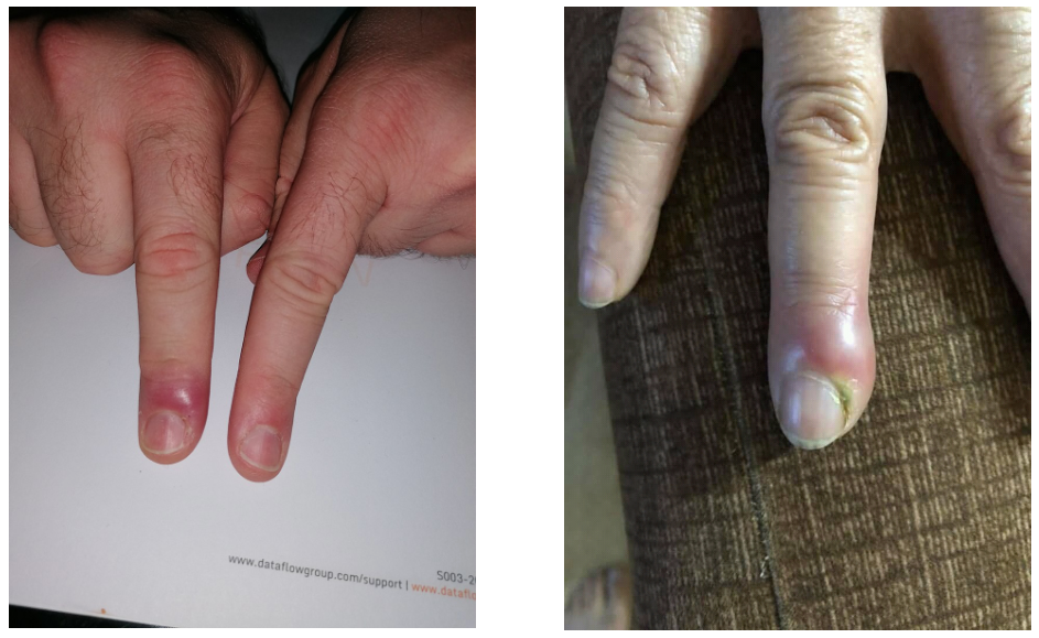
Left: simple paronychia and cellulitis. image from https://commons.wikimedia.org/wiki/File:Right_index_finger_paronychia_cellulitis2.jpg wikimedia commons, under CC attribution SA 4.0 international, accessed Oct 2023.
Right: complex paronychia with abscess. image from https://commons.wikimedia.org/wiki/File:Acute_paronychia.jpg wikimedia commons, under CC attribution SA 4.0 international, accessed oct 2023.

MANAGEMENT
Paronychias are further classified as simple or complex based on the absence or presence of an abscess, respectively. The most common organism implicated in complex paronychias is Staph aureus [6].
The management of simple paronychias involves warm saline soaks of the affected fingertip 3-4 times per day for 5-7 days [11]. Antibiotics are not indicated [7, 8]. These patients may be discharged with close outpatient follow-up, and should be instructed to return to the Emergency Department if they develop worsening and/or spreading pain and swelling, as this may be indicative of an infectious process.
Management of complex paronychias relies on source control via incision and drainage (I & D) [9]. Consider performing a digital block for analgesia prior to the procedure. Once adequate analgesia has been accomplished, an incision should be made at the area of greatest fluctuance, parallel to the nail. Patients should use warm saline soaks at home to encourage continued drainage. Antibiotics are not always indicated, but should be considered in severe cases or in patients with recurrent symptoms. A regimen of cefalexin plus TMP-SMX, doxycycline, or clindamycin is appropriate.
+ Felon
PRESENTATION
A felon is an infection of the pulp of the distal fingertip. Felons are the second most common hand infection after paronychias [13]. Patients typically present with tenderness and fluctuant swelling of the finger pad, distal to the distal interphalangeal joint [12].
The cause of the infection may be due to local trauma, overlying cellulitis, or spreading infection from a paronychia. However, up to 50% of patients have no identifiable source of infection preceding the felon [14]. Felons are typically polymicrobial in nature, with Streptococcus species and Staph aureus being the most common isolates.
MANAGEMENT
Prompt recognition and management is essential in preventing serious complications. If left untreated, felons can progress to osteomyelitis, tenosynovitis, septic arthritis, tendon rupture, and ischemia from compartment syndrome of the pulp [15, 16].
Before definitive management, X-rays of the affected finger should be considered, especially in cases where there is suspicion for a foreign body or clinical concern for associated fracture, osteomyelitis, or necrotizing soft tissue infection [17].
Similar to complex paronychias, treatment of most felons relies on incision and drainage. These patients will eventually require outpatient follow up with a hand surgery specialist, so consider getting the relevant consultant team involved early to facilitate this. Again, a digital nerve block is often an effective form of analgesia for this procedure. Once adequate analgesia has been accomplished, an incision should be made along the lateral aspect of the finger, avoiding the most palmar aspect of the fingertip. The incision should be made parallel to the nail, at least 0.5 cm from the nail plate, on the non-contact aspect of the digit (radial aspect of the thumb, or ulnar aspect of the fingers) in order to avoid the neurovascular bundles [18]. Blunt dissection should be performed with hemostats in order to separate the fibrous septa, allowing for decompression. Wound cultures should be obtained to guide future antibiotic therapy. Patients should be discharged from the Emergency Department with a course of cefalexin plus TMP-SMX, doxycycline, or clindamycin [19]. Outpatient follow up with a hand surgery specialist should be arranged prior to discharge.

Left: infection of the pulp space / felon. image from https://commons.wikimedia.org/wiki/File:Infection_of_the_pulp_space_of_the_thumb.jpg wikimedia commons, under CC attribution SA 3.0 unported, accessed oct 2023. added markings done by post author.
Right: suggested INCISION LOCATIONs FOR INCISION AND DRAINAGE OF FELON. original image.
References
[1] Tintinalli, JE, Stapczynski, JS, Ma, OJ, Yealy, DM, Meckler, GD, & Cline, D (2016). Arm, Forearm and Hand Lacerations. Tintinalli’s emergency medicine: A comprehensive study guide (Ninth edition.). New York: McGraw-Hill Education.
[2] Lemmon JA, Janis JE, Rohrich RJ. Soft-tissue injuries of the fingertip: methods of evaluation and treatment. An algorithmic approach. Plast Reconstr Surg. 2008 Sep;122(3):105e-117e. doi: 10.1097/PRS.0b013e3181823be0. PMID: 18766028.
[3] Leggit JC. Acute and Chronic Paronychia. Am Fam Physician. 2017 Jul 1;96(1):44-51. PMID: 28671378.
[4] Macneal P, Milroy C. Paronychia Drainage. In: StatPearls. Treasure Island (FL): StatPearls Publishing; June 29, 2022.
[5] Dulski A, Edwards CW. Paronychia. In: StatPearls. Treasure Island (FL): StatPearls Publishing; August 8, 2022.
[6] Fowler JR, Ilyas AM. Epidemiology of adult acute hand infections at an urban medical center. J Hand Surg Am. 2013;38(6):1189-1193. doi:10.1016/j.jhsa.2013.03.013
[7, 16, 18] Ong YS, Levin LS. Hand infections. Plast Reconstr Surg. 2009;124(4):225e-233e. doi:10.1097/PRS.0b013e3181b458c9
[8, 13] Barger J, Garg R, Wang F, Chen N. Fingertip Infections. Hand Clin. 2020;36(3):313-321. doi:10.1016/j.hcl.2020.03.004
[9, 11] Ritting AW, O'Malley MP, Rodner CM. Acute paronychia. J Hand Surg Am. 2012;37(5):1068-1070. doi:10.1016/j.jhsa.2011.11.021
[10] Ogunlusi JD, Oginni LM, Ogunlusi OO. DAREJD simple technique of draining acute paronychia. Tech Hand Up Extrem Surg. 2005;9(2):120-121. doi:10.1097/01.bth.0000163575.00615.69
[12] Ong YS, Levin LS. Hand infections. Plast Reconstr Surg. 2009;124(4):225e-233e. doi:10.1097/PRS.0b013e3181b458c9
[14] Nardi NM, McDonald EJ, Schaefer TJ. Felon. In: StatPearls. Treasure Island (FL): StatPearls Publishing; September 24, 2022.
[15] Watson PA, Jebson PJ. The natural history of the neglected felon. Iowa Orthop J. 1996;16:164-166.
[17] Nardi NM, McDonald EJ, Schaefer TJ. Felon. In: StatPearls. Treasure Island (FL): StatPearls Publishing; September 24, 2022.
[19] Tannan SC, Deal DN. Diagnosis and management of the acute felon: evidence-based review. J Hand Surg Am. 2012;37(12):2603-2604. doi:10.1016/j.jhsa.2012.08.002
Image references:
1. https://commons.wikimedia.org/wiki/File:507_Nails.jpg
2. https://commons.wikimedia.org/wiki/File:Right_index_finger_paronychia_cellulitis2.jpg
3. https://commons.wikimedia.org/wiki/File:Acute_paronychia.jpg
4. https://commons.wikimedia.org/wiki/File:Infection_of_the_pulp_space_of_the_thumb.jpg
post by Diana RODRIGUEZ, MD AND COLLEEN arnold, md
Dr. Rodriguez is a PGY-2 in Emergency Medicine at the University of Cincinnati
Dr. Arnold is a PGY-2 in Emergency Medicine at the University of Cincinnati and Mastering Minor Care Section Editor
editing by ALEXA SABEDRA, MD and ANITA GOEL, MD
Dr. Sabedra is an Assistant Professor at the University of Cincinnati and a graduate of the UC EM Class of 2019
Dr. Goel is an Assistant Professor at the University of Cincinnati and a graduate of the UC EM Class of 2018


