Article

Onopordum nervosum ssp. platylepis Flowers as a Promising Source of Antioxidant and Clotting Milk Agents: Behavior of Spontaneous and Cultivated Plants under Different Drying Methodologies

1
Research Laboratory Agrobiodiversity and Ecotoxicology (LR21AGR02), High Agronomic Institute of Chott Mariem B.P 47, University of Sousse, Sousse 4042, Tunisia
2
Department of Biology, College of Sciences, Imam Mohammad Ibn Saud Islamic University (IMSIU), Riyadh 11564, Saudi Arabia
*
Author to whom correspondence should be addressed.
Processes 2023, 11(10), 2962; https://doi.org/10.3390/pr11102962
Submission received: 12 September 2023 / Revised: 5 October 2023 / Accepted: 10 October 2023 / Published: 12 October 2023
(This article belongs to the Special Issue Advanced Drying Technologies in Food Processing)

Abstract

:
This study is a contribution to the valorization of the Onopordum nervosum ssp. platylepis flowers in the agri-food sector. It aims to evaluate the effect of different drying methodologies (room, microwave, convective, oven and freeze-drying) on the chemical composition, the microbiological quality of aqueous extracts and their biological activities, namely clotting milk and antioxidant activities for both spontaneous and cultivated plants. The results showed that the drying methodology has significantly affected the phenolic composition. The extract obtained from freeze-dried flowers showed the best amounts of total phenols (6.19 ± 0.50 mg GAE/g), flavonoids (2.35 ± 0.07 mg QE/g) and condensed tannins (0.038 ± 0.002 mg CE/g) followed by the microwave-dried flowers. However, microwave-dried flowers presented the lowest protein content with only 3.58 ± 0.62 mg BSAE/g. Finally, the biological activities were significantly dependent on the used drying process. The extract of freeze-dried flowers was the most potent to inhibit free radicals (diammonium 2,2′-azino-bis-(3-ethylbenzothiazoline-6-sulphonate) (ABTS), 1,1-diphenyl-2-picrylhydrazyl (DPPH)) and to reduce iron with the efficient concentrations EC50s of 0.48 ± 0.02 mg/mL, 0.16 ± 0.04 mg/mL and 1.52 ± 0.19 mg/mL, respectively. It also showed the highest clotting milk activity followed by room-dried and forced convective-dried flowers. As a consequence, freeze drying was the best method for preserving most chemical and biological properties of Onopordum nervosum ssp. platylepis flowers. However, in consideration of production costs and drying duration, the convective dryer is recommended as a more practical choice. All the obtained results do not reveal a significant difference between cultivated and spontaneous plants, which can suggest that the domestication of Onopordum nervosum ssp. platylepis can be useful for industrial applications.

1. Introduction

Asteraceae is the largest family of the flowering plants, and the most known and used thistle species are Cynara cardunculus, Cynara humilis, Cynara scolymus and Onopordum acanthium, which are used as sources of coagulant agent in the manufacture of raw ovine and/or caprine milk [1,2]. The crucial characteristic of thistles lies in their milk-clotting activity, which refers to the enzyme’s capacity to specifically break down κ-casein in milk. This enzymatic activity has been associated with aspartic proteinases. However, the application of plant proteases in cheese production is often hindered by their deactivation rates. This limitation can be attributed to various physicochemical phenomena, particularly the denaturation of the native protein molecule. Additionally, several factors, including the quality of the raw materials, catalyze certain undesirable alterations in the enzyme’s structure [3]. Furthermore, the thistle species have been used since ancient times as food and medicinal plant. Several species have different biological properties such as antidiabetic, choleretic, diuretic, cardiotonic, anti-hemorrhoidal, antioxidant, anti-inflammatory, cytotoxic and antimicrobial activities [2,4]. The different organs of thistle plants contain many bioactive compounds, particularly mono- and dicaf-feoylquinic acids, anthocyanins, and other flavonoids, such as apigenin and luteolin derivatives.
Onopordum L. is a genus of about 60 species of thistles belonging to the Asteraceae family, native to Europe (mainly the Mediterranean region: France, Spain, Italy, Greece), northern Africa (Tunisia, Algeria, Libya), the Canary Islands, the Caucasus and southwest and central Asia [4]. Several Onopordum are widely used in traditional medicine. Flowering branches of Onopordum acanthium are used as a diuretic and antipyretic and roots for diuretic, antipyretic, appetizing and abdominal pain. Onopordum tauricum seeds are used for the treatment of kidney disease in Turkey [4].
Onopordum nervosum ssp. platylepis is an endemic spicy of Tunisia [5]. A few studies have been conducted on this plant. The main important results reported that the chemical composition of seed oil included triglycerides, sterols, tocopherols and tocotrienols [5]. Subsequently, the investigation of clotting milk and antioxidant activities in Onopordum nervosum ssp. platylepis, as an Asteraceae plant, holds significant interest.
In recent decades, the number of industrial applications of the thistle species has been gradually increasing [2]. In addition, consumers, along with the food industry, are increasingly placing importance on the quality of food products. There is a growing demand for highly functional formulations, particularly those containing natural and nutraceutical ingredients [6]. This raises two significant considerations: The first pertains to plant cultivation, particularly in light of concerns related to climate change and the risk of losing numerous endemic plants, such as O. nervosum ssp. platylepis. The second revolves around the preservation of harvested plant material to prevent deterioration during storage. Drying is the ancient common processing method used to preserve plant materials [7,8]. However, the selection of drying method has a great influence on the quality, the chemical composition and the biological activities of dried plants [8,9,10]. Different plant drying methodologies have been studied such as room, hot air, freeze, vacuum, microwave drying, etc. [6,8,9,10]. For aromatic and sensitive plants, the natural drying method is used, but recently, with the advancements in technology, many new methods have been developed and used for industrial-scale drying processes [6]. Over time, several reports have proven the close relationship between the chemical composition of the species and the drying methodology [6,9,10]. To our knowledge there is no study reported on the drying effect on thistle flower’s quality, especially Onopordum nervosum ssp. platylepis. For that, all the findings of this study can present a new and interesting result that can enrich the knowledge about this plant.
In this context, the current study aims to evaluate the influence of five drying methodologies on the chemical composition, the antioxidant activity and the clotting milk activity of wild and cultivated O. nervosum ssp. platylepis flowers growing in central Tunisia to identify the best practical method for preserving the postharvest plant quality.

2. Material and Methods

2.1. Plant Material

Spontaneous and cultivated Onopordum nervosum ssp. platylepis flowers were obtained from the High Agronomic Institute of Chott Mariem (ISA-CM), Sousse, Tunisia plots. The localization coordinates were Latitude 35°55′01° N; 35°55′02° N and longitude 10°33′41″ E; 10°33′48″ E for spontaneous and cultivated plants, respectively. The plant material was identified by Prof. Rabiaa Haouala, a botanist from ISA-CM, University of Sousse.
The cultivated plants were grown on spaced lines (1.2 m) with spacing between the plants of 1.5 m. The plant materials were harvested during the flowering season (end of May 2021). At this stage of maturation, the flowers were completely open with a purple color, then petals are manually separated and subjected to different drying methods (Figure 1a–d).

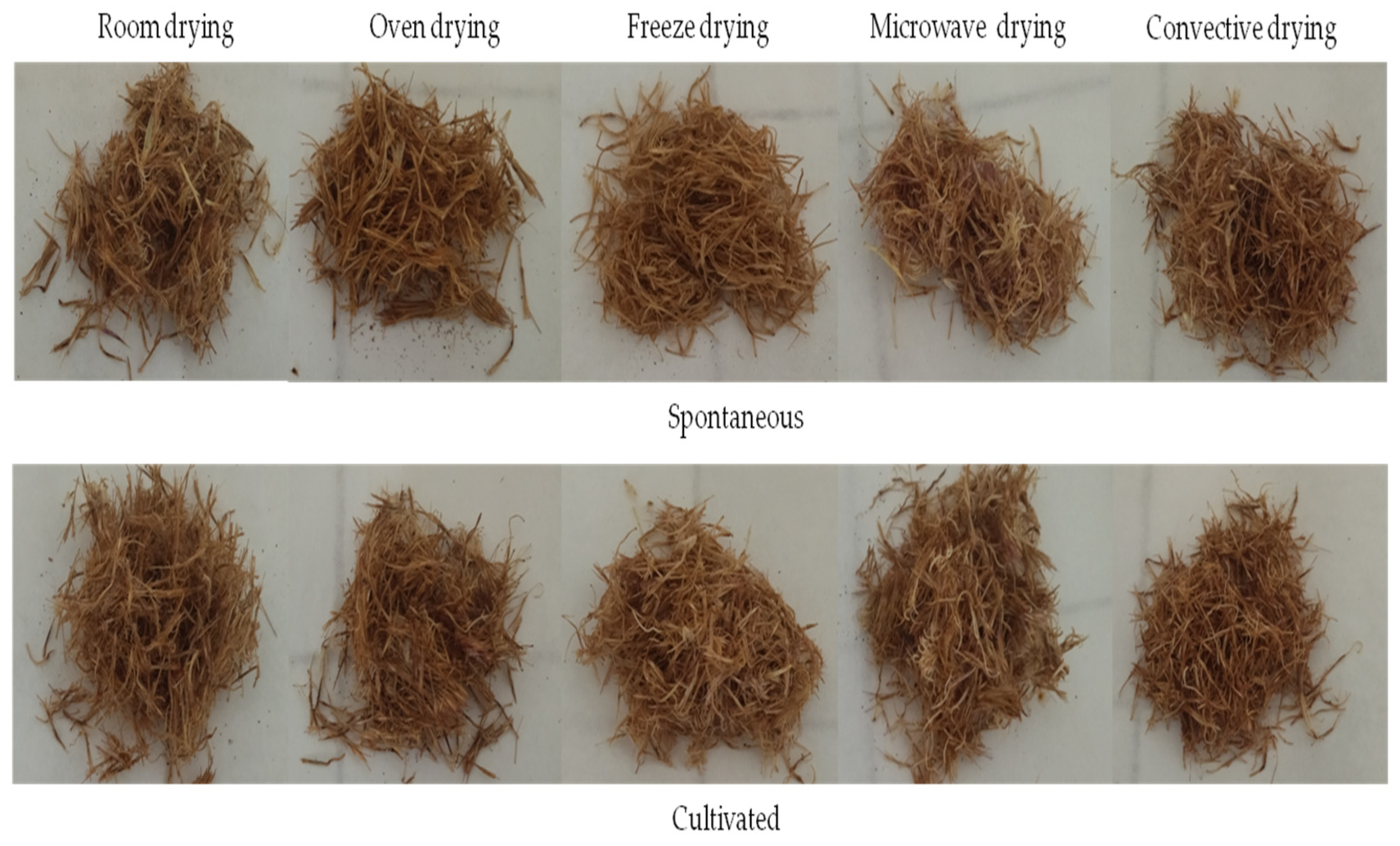
2.2. Drying Methodology

After the flowers were picked and the stem was removed, equal amounts of petal samples were treated via the following drying methods: room drying (RD) is achieved with natural air circulation. Plant materials are spread in thin layers in trays and mixed or turned frequently. The average temperature of the room is 22 ± 2 °C. The drying time was about 5 days. For oven drying (OD), samples were dried by circulating hot air at a temperature of 45 °C for 48 h in an electric thermostatic oven natural convection TCN 50 (Sozhou being medical device Co., Ltd., Kunshan, China). The freeze drying (FD) was performed at a temperature of −40 °C, and the vacuum was maintained at a pressure of 10−3 Torr for 36 h in a Telstar Lyoquest. The microwave (MD) treatment was performed at 300 W for 3 min, and finally, an electric forced convective dryer (CD) (BIOSEC dryer, Tauro Essiccatori, Camisano Vicentino, Italy) was used for flowers drying at 30 °C during 20 h.
The samples of each population were divided into three lots and collected (Figure 2) until reaching stable water content (10%). They were then ground into fine powders using a mill and preserved in spittoons, which were provided with the date of collection, name of the population and batch number.

2.3. Aqueous Extract Preparation

The extraction process follows the aqueous maceration method, involving prolonged contact of the plant material powder with a solvent to extract active ingredients. This procedure was conducted at room temperature.
Distilled water served as the solvent, with one gram of powder from each batch combined with 20 mL of distilled water. The resulting mixture was subjected to agitation on a magnetic stirrer at room temperature for a duration of 2 h. Subsequently, each batch underwent centrifugation twice at 4 °C and 8000× g rpm for 10 min. The supernatant was carefully collected and preserved at 4 °C for future use.

2.4. Chemical Characterization of O. nervosum ssp. platylepis Aqueous Extracts

2.4.1. Dry Matter

An empty capsule was weighed and its mass was noted. In this capsule, 1 mL of the extract was added, then was placed in the oven at 105 °C for 24 h.

2.4.2. Total Phenols Content

The adopted method was that of Singleton and Rossi, ref. [11] based on the quantification of the total concentration of hydroxyl groups that characterizes the polyphenols in the extract. A volume of 100 μL of each extract was added to 500 μL of Folin–Ciocalteu reagent diluted 10 times and 1000 μL of distilled water. This mixture was stirred at room temperature for 1 min. Then, 1500 μL of sodium carbonate (Na2CO3) 20% solution was added to this mixture. The whole solution was incubated for 2 h at room temperature to give the blue coloration, degraded according to the concentration of polyphenols in each solution. The absorbance of the mixture was measured using a UV-Vis spectrophotometer (ZUZI model 4201/50, ZUZI, Shenzhen, China) at a wavelength: λ = 760 nm. The total phenol (TP) content was calculated using calibration curve of gallic acid, and the results were expressed as mg gallic acid equivalent per g of extract (mg GAE/g).

2.4.3. Flavonoids Content

The determination of the flavonoid level was carried out according to the method described by Kim et al. [12] It is based on the formation of a stable complex between aluminum chloride AlCl3 and oxygen atoms located on carbons 4 and 5 of flavonoids to give a yellowish-red coloration. A volume of 1250 μL of distilled water and a volume of 75 μL of sodium nitrite NaNO2 at 5% were added to 250 μL of each extract. After 5 min, 150 μL of AlCl3 was added to the mixture. After 6 min, 500 μL of NaOH was added with 775 μL of distilled water to reach a total volume of 3000 μL. Without any incubation, the color of the mixtures immediately changed to yellow. The absorbance of the mixture was measured using a UV-Vis spectrophotometer (Zuzi model 4201/50, China) at a wavelength: λ = 510 nm against a blank. The total flavonoid (TF) content was determined using a calibration curve of quercetin, and the results were expressed as mg quercetin equivalent per g of extract (mg QE/g).

2.4.4. Condensed Tannins

The condensed tannins determination was carried out using the method of Tiitto [13] which is based on the reaction of vanillin with HCl. Vanillin reacts with the terminal flavonoid group of condensed tannins and forms a pinkish-red complex. Tannins, in fact, turn into anthocyanidols when they react with vanillin giving the pinkish-red coloration. A volume of 50 μL of each extract was added to a volume of 3000 μL of a 4% vanillin solution and then stirred. A volume of 1500 μL of concentrated HCl was added to this mixture. The latter is incubated in the dark for 15 min at room temperature. The absorbance of the mixture was measured using a UV-Vis spectrophotometer (Zuzi model 4201/50, China) at a wavelength of λ = 500 nm. The condensed tannins (CT) content was determined using a calibration curve of catechin, and the results were expressed as mg catechin equivalent per g of extract (mg CE/g).

2.4.5. Proteins Content

The proteins content of the plant aqueous extract directly affects its ability to clot milk. Extracts with higher protein content typically contain more proteases, resulting in more efficient milk coagulation [14].The proteins content in the O. nervosum platylepis extracts was determined according to the Bradford [15] method, based on the staining of the proteins of the solution using the Coomassie blue G250 dye. Once bound to proteins, its color turns from brown to blue. The intensity of the staining is proportional to the amount of protein present in the sample. The coloring reagent was prepared by dissolving 100 mg of bright Coomassie blue G250 in 50 mL of 95% ethanol. A volume of 130 mL of phosphoric acid (85%) was added, and the solution was supplemented to 1 L with distilled water and filtered on filter paper. The pH value of the dye reagent was adjusted by adding concentrated phosphoric acid to a value of 0.4. The reagent was stored at 4 °C, protected from light.
For protein determination, a volume of 200 µL was mixed with 2 mL of Bradford reagent. The contents of the tube were thoroughly mixed, and the absorbance was measured after 5 min using a UV-Vis spectrophotometer (ZUZI model 4201/50, China) at 595 nm using distilled water as blank.
The protein concentration was determined using a calibration curve of Bovine Serum Albumin (BSA) and was expressed as mg Bovine Serum Albumin equivalent/g of extract (mg BSAE/g).

2.5. Microbiological Characterization

To assess the microbiological quality of the extracts, the enumeration of total mesophilic aerobic flora (TMAF) was conducted using plate count agar (PCA) with an incubation period of 48 h at 30 °C [16].

2.6. Biological Activities

2.6.1. Clotting Milk Activity

Clotting milk activity was measured according to the method cited by Libouga [17]. The protocol consists of adding 1 mL of crude extract to 10 mL of prepared milk via spearing of 12 g of milk powder in 100 mL of distilled water, and then 0.01 M of CaCl2 was added with stirring. The tubes were placed in a water bath at 35 °C. The coagulation time corresponds to the time required for the appearance of the first flakes in a thin film of milk flowing on the wall of the tube.
The clotting milk activity was expressed either by the coagulant activity unit (C.A.U.) also called rennet unit (U.P) according to the method of Berridge [18].
The coagulant activity unit (CAU) is defined as the amount of enzyme per milliliter of enzymatic extract that causes the flocculation of 10mL of Berridge substrate in 100 s at 35 °C.
It was calculated according to the Formula (1):
C A U = 100 × V 10 × T × V
where the following are defined:
  • V: Volume of milk to coagulate (10 mL).
  • V’: Volume of the enzymatic solution (1 mL).
  • T: Flocculation time in seconds.

2.6.2. Antioxidant Activity

The antioxidant activity of O. nervosum ssp. platylepis was evaluated using radical scavenging tests of DPPH and ABTS+ radicals and the measure of the iron reducing power method (FRAP).

DPPH Assay

The adopted method in the DPPH test was that of Lee et al. [19]: one mL of the methanolic solution of DPPH (0.1 mM) was added to an equal volume of each aqueous solution of the extracts at different concentrations. The mixtures were left in the dark at room temperature for 30 min. The absorbance was measured using a UV-Vis spectrophotometer (Zuzi model 4201/50, China) against a blank at λ = 517 nm.
The inhibition percentage of DPPH radicals was given by the following Formula (2):
I P   % = 1 A s a m p l e A c o n t r o l × 100
Acontrol: absorbance of the control
Asample: absorbance of the sample
The values of the efficient concentration (EC50) representing the antioxidant concentration needed to reduce DPPH by 50% were calculated using logarithmic regression.

ABTS Assay

The ABTS radical scavenging capacity was evaluated using the method of Re et al. [20]. The radical cation ABTS•+ is generated by mixing a volume of 0.5 mL of a stock solution of ABTS at 7 mM and 0.5 mL of a solution of potassium persulfate K2S2O8 at 2.45 mM, protected from light at 4 °C for 12 to 16 h before use. The obtained solution was diluted with methanol to reach an absorbance of 0.7 to 734 nm. Then, 0.5 mL of the aqueous extract at different concentrations was mixed with 0.5 mL of the prepared solution. The mixture was left in the dark at room temperature for 10 min. The absorbance was measured using a UV-Vis spectrophotometer (Zuzi model 4201/50, China) against a blank prepared at λ = 734 nm.
The inhibition percentage of ABTS was given by the same method as described for DPPH test and the EC50 values were determined using logarithmic regression.

FRAP

The followed experimental protocol was that of Yildrim et al. [21]; indeed, 0.5 mL of the aqueous extract at different concentrations was mixed with 0.625 mL of phosphate buffer (0.2 M, pH = 6.6) and 0.625 mL of a solution of potassium ferricyanide K3Fe(CN)6 at 1%. The whole was incubated in a water bath at 50 °C for 30 min. Then, 0.625 mL of trichloroacetic acid (TCA) at 10% was added to stop the reaction. After 10 min, an aliquot of 1.250 mL was mixed with 1.250 mL of distilled water and 0.250 mL of an aqueous solution of FeCl3at 0.1%. The absorbance was then measured at 700 nm using a UV-Vis spectrophotometer (Zuzi model 4201/50, China).
The EC50 was determined graphically, it corresponds to the concentration that gives an absorbance of 0.5.

2.7. Statistical Analysis

All data were calculated by three replicates and expressed as mean ± standard error of mean. The SPSS version 20 statistical software package was employed for all statistical analysis. The ANOVA analysis of variance was performed and the Duncan test was used to determine significant differences between the different samples at the 5% level. Results were considered significant if the associated p value was less than 0.05.Correlation analysis was conducted to evaluate the relationship between the chemical composition and the biological activities. XLSTAT version 2023 software package was used to perform principle component analysis (PCA) and classification test (Dendrogram).

3. Results and Discussion

3.1. Effect of Domestication and Drying Methodology on Chemical Composition and Microbiological Properties of O. nervosum platylepis Aqueous Extract

3.1.1. Dry Matter of Aqueous Extract

The findings from the assessment of total solids in crude extracts of O. nervosum platylepis are displayed in Table 1, where values range from 1.30% to 2.0%. Statistical analysis reveals a notable disparity in solids linked to the flower drying method (p = 0.026). Nonetheless, no significant difference (p > 0.05) is observed concerning crop type (cultivated and spontaneous).The choice of drying method can impact the retention of bioactive compounds, including polyphenols, flavonoids, and proteins, which may contribute to the solid content of the extract. Specific drying methods must be selected to achieve desired solid content levels in plant extracts based on the research objectives, such as optimizing the concentration of bioactive compounds for various applications.

3.1.2. Phenolic Composition

According to Table 1, variability in total phenols, flavonoids and condensed tannins levels is observed in the studied plant material depending on drying method. The total phenol contents in O. nervosum ssp. platylepis flower extracts range from 2.956 to 5.54 mg GAE/g and from 3.01 to 6.19 mg GAE/g, flavonoids values are between 0.09 to 2.35 mg QE/g and 0.08 to 2.12 mg QE/g for cultivated and spontaneous samples, respectively. Small quantities of tannins were registered for both plant materials. No significant differences are recorded depending on the origin of the plant, which indicates that the chemical composition is not affected by the cultivation since it was conducted in similar conditions as spontaneous growth. According to these results, it is found that O. nervosum ssp. platylepis extracts are globally characterized by significant amounts of phenolic compounds in comparison with species of thistles. These results are comparable to the levels registered by Pandino et al. [22] for Cynara cardunculus var. Scolymus with a total phenol content of 4.43 mg GAE/g DM. However, they are less important than thosefound by Habibatni et al. [23], performed on O. acanthium, revealing higher values compared to our results where the total phenol content of the extract was 8.93 ± 1.33 mg GAE/g. It is also the case for the artichoke flower (Cynara scolymus L.) in research carried out by Mahmoudi et al. [23] indicating a total phenol content of 23.7 mg GAE/g). For condensed tannins, our results are consistent with the levels found by Mahmoudi et al. [24], who reported a CT value of 0.035 μg EC/g in C. scolymus L. flowers. This variability in results for thistle plant materials can be explained by the plant species and the extraction conditions which can significantly affect the obtained values.
The high significant variation (p = 0.000) of polyphenol contents depending on the drying method of the flowers indicated that the freeze and microwave drying are the best technologies for polyphenols extractability; however, the electric forced convective dryer and oven seem to be increasing the degradation of these substances. Those findings are in agreement with many studies; according to Lim and Murtijaya [25], the freeze and microwave drying of Phyllanthus amarus provided the same contents of total phenols (12.10 ± 0.56 mg GAE/g and 12.70 ± 1.17 mg GAE/g), respectively. The highest total phenolic content was also recorded for jasmine freeze-dried flowers [7]. Snoussi et al. [10] indicated that microwave heating of Myrtus communis leaves allows the highest values of phenolic compounds. However, the present results are in disagreement with those of Miao et al. [26] who found that freeze drying provides the lowest total flavonoids value in dried tea flowers. Broadly, freeze drying is usually more effective in preserving active components from degradation in plants, flowers and fruits than hot-air drying [7,27]. In fact, this method entails the creation of tiny ice crystals within the cell and their swift removal from the cell during the freezing process. The swift removal of these ice crystals enables the preservation of cell structures, thereby retaining phenolic compounds and vitamins. Additionally, complete elimination of moisture content facilitates the long-term preservation of plant samples. These attributes have made freeze drying a widely adopted pretreatment method for extracting plant materials during analysis [28].
Maghsoudlou et al. [29] indicated that the increase in total phenols content in dried plants can be explained by the fact that heat treatment promotes the release of phenolic compounds from the solid matrix. However, the lower amounts can be attributed to oxidation and thermal degradation of phenolic compounds with increased heat intensity and heat treatment duration [22,30].

3.1.3. Proteins Content

The analysis of the recorded results (Table 1) reveals a variability in proteins content in the plant subjected to different drying methods (p = 0.000). The most important proteins contents were registered in the extracts obtained from freeze-dried, room-dried and convective-dried flowers. The observed values for spontaneous flower extracts were 261.22, 84.53 and 79.02 mg BSAE/g, respectively. In contrast, the microwave-dried and oven-dried flowers have shown the lowest values with 3.58 and 28.99 mg BSAE/g, respectively. The results do not reveal a significant difference (p > 0.05) in terms of crop type (cultivated and spontaneous); this can be explained by the similarity between cultivation and natural conditions. In comparison with a study conducted on the species Onopordum tauricum, belonging to the same family, Mozzon et al. [31] revealed protein content levels comparable to our results, which show a value of about 132.61 mg BSAE/g. Based on our research, freeze drying is the most protective treatment for proteins; however, microwave drying is the most destructive for these substances. Those findings are in agreement with previous research which suggests that microwave heating has a more destructive effect than conventional heating [32].

3.1.4. Microbiological Properties of O. nervosum platylepis Aqueous Extract

The enumeration of TAMF results for microbiological analysis is presented in Table 1. The evaluation indicates that the microbiological quality of the studied extracts significantly depends on the drying methodology (p = 0.047). Notably, it was observed that O. nervosum platylepis extract obtained through microwave-dried flowers completely eliminated contamination. Conversely, extracts from room-dried flowers recorded the highest contamination levels (log CFU = 1.8). This difference can be attributed to the shorter drying time with microwaves (3 min) compared to the longer duration of room drying (5 days), which increases the risk of plant material contamination. Additionally, it has been documented that microwave radiation is efficient in eradicating bacteria [33].
On the other hand, the results did not indicate a significant difference (p > 0.05) between cultivated and spontaneous plants. The use of O. nervosum platylepis as a replacement for chemical additives should ensure an acceptable level of microbiological quality. In conclusion, based on the obtained results, it can be inferred that all the drying methods have provided hygienic O. nervosum platylepis extracts within the established standards.

3.2. Effect of Domestication and Drying Methodology on Biological Activity of O. nervosum platylepis Aqueous Extract

3.2.1. Clotting Milk Activity

The clotting milk activity is expressed in coagulation time and coagulant activity unit (CAU), and the results of the analysis are presented in Table 2.
According to the obtained results, the drying methodology significantly affects the coagulant activity of O. nervosum platylepis flower extracts (p = 0.000). The most potent activity was observed for freeze-dried flowers (CAU = 0.833)with the shorter clotting time (ClT) 120s, followed by the room-dried and forced convective-dried flowers with the CAU value of 0.556 and ClT of 180 s. The extract obtained from the oven-dried flowers showed a low coagulation activity; however, the microwave-dried samples were unable to coagulate the milk. The results were comparable for both cultivated and spontaneous plants.
Our results are lower than those obtained by Ben Amira et al. [3] for the flowers of C. cardunculus: a variation in the values of coagulant activity CAU was recorded ranging between 1.11 and 2.40 depending on repining stage. In addition, the study conducted by Mozzon et al. [31] has confirmed that O. tauricum flowers exhibited coagulation properties. These previous findings corroborate our results, supporting the notion that O. nervosum platylepis could serve as a potential source of clotting milk agents.

3.2.2. Antioxidant Activity

From the current point of view, in vitro antioxidant activity should be assessed using multiple methods to cover all aspects of antioxidant effectiveness; in the present study, three tests were performed to evaluate the antioxidant properties of O. nervosum platylepis flower extracts, and the results are presented in Figure 3 and Table 2.
The percentages of DPPH free radical inhibition as a function of crude extract concentrations of cultivated and spontaneous O. nervosum platylepis flowers are shown in Figure 3A(a,b).
The results showed that all extracts have radical scavenging activity depending on extract concentration; the higher extract concentrations led to the higher inhibition percentages. It has been observed that the used drying methodology significantly affect the radical scavenging activity of O. nervosum platylepis flowers (p = 0.000). The freeze-dried flower extracts showed the best DPPH radicals inhibition percentage for both spontaneous and cultivated plants.
The efficient concentration (EC50), which is inversely related to antioxidant activity, meaning that the smaller the EC50 value, the more potent the extract, is considered to have registered values ranging from 0.41 to 1.34 mg/mL, and a highly significant difference was recorded for the antioxidant capacity. Regarding the drying method, the extracts with the strongest DPPH scavenging activity were obtained from freeze-dried and microwave-dried flowers with the EC50values of 0.41 mg/mL and 0.67 mg/mL, respectively. However, the extracts obtained from dried flowers using the oven and the forced convective dryer showed the lowest DPPH inhibition, could inflict losses on antioxidant activity. The results do not reveal a significant difference (p > 0.05) in terms of crop type (cultivated and spontaneous) (Figure 3A(a,b)).
The reported results for the genus Onopordum indicate its radical scavenging ability; the study conducted by Habibatni [22] confirms the antioxidant properties of different Onopordum acanthium flower extracts with EC50 values of 134.4 μg/mL for the DPPH test.
The present results are in agreement with those of Cheng et al. [34], who indicated the significant effect of the drying method. The EC50 of native lignin of Brauns extracted from quince fruit subjected to different drying methodologies (microwave, sun and freeze drying) were about 0.14 mg/mL, 0.15 mg/mL and 0.18 mg/mL, respectively.
Total phenols, flavonoids and tannins are considered potentially antioxidant, and their presence indicates that the extracts have antioxidant activity, which varies depending on the drying method.
The obtained results for ABTS+ test (Figure 3B(a,b)) are similar to the DPPH test, and the crude extracts of freeze-dried and microwave-dried flowers showed the highest percentages of inhibition at the same concentrations of spontaneous and cultivated plants. Based on the obtained results (Table 2), the extracts of microwave-dried and freeze-dried flowers have the highest ABTS+ radical scavenging capacity (EC50 = 0.14 mg/mL and 0.16 mg/mL, respectively).
The evaluation of antioxidant activity showed that the studied extracts possessed antioxidant capacities, significantly depending on the drying methodology (p = 0.000). However, the results do not reveal a significant difference (p > 0.05) in terms of crop type (cultivated and spontaneous). Our results are in agreement with those obtained by Lim and Murtijaya [25], who found the best EC50(0.154 ± 0.012 mg/mL and 0.154 ± 0.065 mg/mL) for freeze-dried and microwave-dried Phyllanthus anarus, respectively.
Finally, for the FRAP test, the results of the variation in absorbance at λ = 700 nm as a function of the concentration of spontaneous and cultivated O. nervosum platylepis flower extracts are illustrated in Figure 3C(a,b). We observed that all the extracts exhibited the ability to reduce iron, albeit with varying capacities. As the concentration of extracts increased from 0.5 to 10 mg/mL, there was a corresponding rise in absorbance, from 0.29 to 1.47 and from 0.29 to 1.61 for the extracts of spontaneous and cultivated flowers, respectively. This can be attributed to an enhanced capability to convert Fe3+ to Fe2+, indicating improved antioxidant activity.
As can be seen in Table 2, the highest ferric reducing power is registered for the extracts obtained from freeze- and microwave-dried flowers with EC50 values of 1.52 mg/mL and 1.70 mg/mL, respectively, followed by the room-dried flower extract.
The statistical analysis revealed that there are no significant differences between the EC50 values of cultivated and spontaneous plant extracts (p > 0.05). These results are similar to the values found by El Hamdaoui et al. [35], who indicated no significant difference between the EC50s of essential oils obtained from cultivated and spontaneous lavender plants for the same tests. Nevertheless, our findings are in disagreement with those reported by Ceccanti et al. [6], who observed that the domestication of Sanguisorba minor Scop. had a profound impact on its phenolic composition and biological activity. The disparity in results can be attributed to the divergent agricultural practices employed across the various studies.

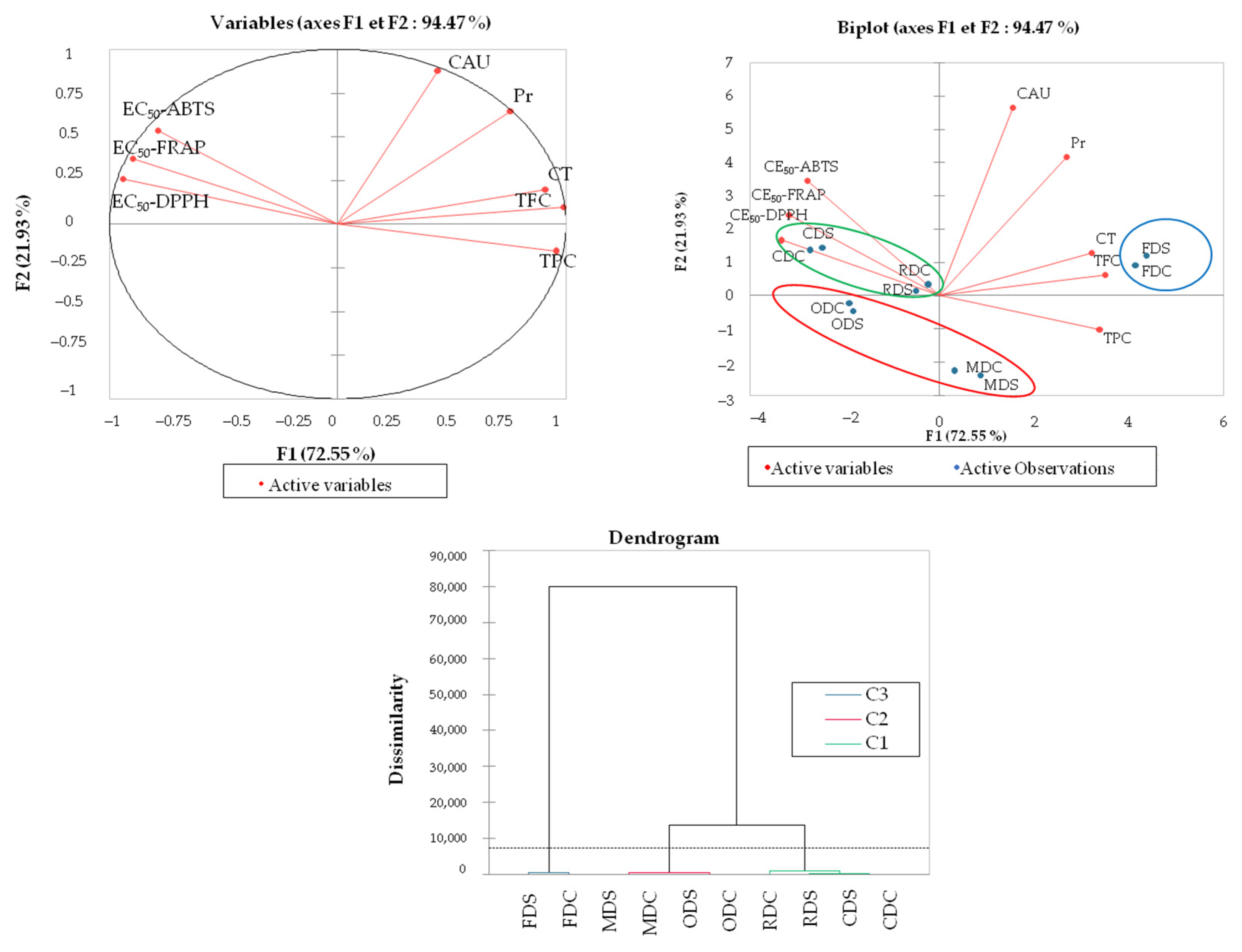
3.3. Correlation, PCA and Classification Analysis

Due to the differences in the chemical composition, both antioxidant and clotting milk activities showed significant differences. It is well known that different drying methods and drying temperatures can affect the chemical composition and biological activities of plants [26]. Correlation analysis (Table 3) was conducted between chemical composition (TP, TF, CT and PC) of O. nervosum platylepis aqueous extract and the biological activities (EC50 of DPPH, ABTS•+, FRAP, ClT and CAU). The results showed a negative correlation between TP, TF, CT and the EC50 of DPPH and ABTS radicals inhibition and FRAP with coefficient values varying between −0.540 and −0.894 with a high significant effect. Higher levels of TP, TF and CT in flower extracts were associated with higher antioxidant activity and lower EC50 values, suggesting that these substances may play an important role in free radical scavenging capacity and ferric reducing power. These results are in agreement with previous study of Lu et al. [9].
There was a negative correlation between protein content and antioxidant activity, suggesting that some proteins in O. nervosum platylepis aqueous extract can contribute to the antioxidant activity. Those findings are consistent with those obtained by Yu et al. [27], in which TF contributes significantly to antioxidant activity. The high concentration of flavonoids and phenols in G. jasminoides flower stamens and pistils results in high antioxidant capacity.
Negative and positive correlations were observed between protein content and the clotting milk activity represented by the ClT and the CAU parameters with the coefficient values of −0.733 and 0.921, respectively.
The PCA analysis (Figure 4) of the results for eight studied variables (Pr, TPC, TFC, CT, EC50 of DPPH, ABTS•+ and FRAP and CAU) showed that the correlation matrix has very strong coefficients, which indicates a dependent evolution of these variables. The axes 1 and 2 absorb 72.55 and 21.93% of the overall variation, respectively. The graphical representation of the variables in the plane formed by the axes 1 and 2 showed that the axis1 opposes the antioxidant activity to the chemical composition; however, the clotting milk activity is closely related to the protein content. These results are consistent with those issued previously by the correlation analysis. There is a tendency to isolate three groups of individuals regrouping the RD and the CD (class 1 green), OD and MD (class 2 red); however, the FD flowers represent a separated group (class 3 blue). We believe that the chemical and biological activities variables have a significant power in the separation of the O. nervosum platylepis extracts.
These results are also proven via the classification test (dendrogram representation), which indicated the presence of three groups.

4. Conclusions

The findings from this study lead to the conclusion that O. nervosum platylepis holds potential as a source of antioxidants and milk-clotting agents for cheese production, serving as a viable alternative to animal rennet. However, it is crucial to make a judicious choice of drying method to ensure the stability of the plant material.
On one hand, we observed significant effects of the drying method on both the chemical composition and biological activities of O. nervosum platylepis flowers. Notably, freeze drying emerged as the most protective method for preserving the plant material, yielding extracts with the highest efficacy in terms of antioxidant and milk-clotting activities.
On the other hand, our results did not reveal any significant differences based on the type of plant material (cultivated and spontaneous). This suggests the potential for domesticating O. nervosum platylepis, an endemic Tunisian plant facing a significant threat due to climate change. Domestication efforts could aid in its preservation and utilization.
In summary, this study underscores the potential of O. nervosum platylepis as a valuable resource for cheese production and the importance of selecting an appropriate drying method. Additionally, it highlights the opportunity for domestication to conserve this endangered Tunisian plant.

Author Contributions

I.E.: Conceptualization, Investigation, Methodology, Writing. N.D.: Methodology, Writing—review and editing. G.L.: Formal analysis, Methodology. R.K.: Methodology, review and editing. F.H.: Investigation, Methodology, writing—review and editing. A.M.A.: Writing—review and editing. H.A.A.: Writing—review and editing. B.D.A.: Supervision, Writing—review and editing. All authors have read and agreed to the published version of the manuscript.

Funding

This work was supported and funded by the Deanship of Scientific Research at Imam Mohammad Ibn Saud Islamic University (IMSIU) (grant number IMSIU-RG23096).

Data Availability Statement

Data are unavailable due to privacy.

Conflicts of Interest

The authors declare no conflict of interest.

References

  1. Brutti, C.B.; Pardo, M.F.; Caffini, N.O.; Natalucci, C.L. Onopordum acanthium L. (Asteraceae) flowers as coagulating agent for cheesemaking. LWT Food Sci. Technol. 2012, 45, 172–179. [Google Scholar] [CrossRef]
  2. Mandim, F.; Petropoulos, S.A.; Santos-Buelgab, C.; Ferreira, I.C.F.R.; Barros, L. Chemical composition of cardoon (Cynara cardunculus L. var. altilis) petioles as affected by plant growth stage. Food Res. Int. 2022, 156, 111330. [Google Scholar] [CrossRef] [PubMed]
  3. Ben Amira, A.; Besbes, S.; Attia, H.; Blecker, C. Milk-clotting properties of plant rennets and their enzymatic, rheological, and sensory role in cheese making: A review. Int. J. Food Prop. 2017, 20, 76–93. [Google Scholar] [CrossRef]
  4. Baştürk, A.; Peker, S. Antioxidant Capacity, Fatty Acid Profile and Volatile Components of the Onopordum Anatolicum and Onopordum Heteracanthum Species Seeds Grown in Van, Turkey. J. Inst. Sci. Technol. 2021, 11, 2810–2822. [Google Scholar] [CrossRef]
  5. Hachicha, S.F.; Barrek, S.; Skanji, T.; Ghrabi, Z.G.; Zarrouk, H. Composition chimique de l’huile des graines d’Onopordon nervosum subsp. platylepis Murb (Astéracées). J. Société Chim. Tunis. 2007, 2007, 23–28. [Google Scholar]
  6. Ceccanti, C.; Finimundy, T.C.; Heleno, S.A.; Pires, T.C.S.P.; Calhelha, R.C.; Guidi, L.; Ferreira, I.C.F.R.; Barros, L. Differences in the phenolic composition and nutraceutical properties of freeze dried and oven-dried wild and domesticated samples of Sanguisorba minor Scop. LWT Food Sci. Technol. 2021, 145, 111335. [Google Scholar] [CrossRef]
  7. Barman, M.; Soren, M.; Mishra, C.; Mitra, A. Dehydrated jasmine flowers obtained through natural convective solar drying retain scent volatiles and phenolics—A prospective for added-value utility. Ind. Crops Prod. 2022, 177, 114483. [Google Scholar] [CrossRef]
  8. Ziaa, M.P.; Alibas, I. Influence of the drying methods on color, vitamin C, anthocyanin, phenolic compounds, antioxidant activity, and in vitro bioaccessibility of blueberry fruits. Food Biosci. 2021, 42, 101179. [Google Scholar] [CrossRef]
  9. Lu, J.; Wang, Z.; Qin, L.; Shen, J.; He, Z.; Shao, Q.; Lin, D. Drying methods affect bioactive compound contents and antioxidant capacity of Bletilla striata (Thunb.). Reichb. f. flower. Ind. Crops Prod. 2021, 16, 113388. [Google Scholar] [CrossRef]
  10. Snoussi, A.; Essaidi, I.; Koubaier, H.B.H.; Zrelli, H.; Alsafari, I.; Živoslav, T.; Mihailovic, J.; Khan, M.; El Omri, A.; Veličković, T.Ć.; et al. Drying methodology effect on the phenolic content, antioxidant activity of Myrtus communis L. leaves ethanol extracts and soybean oil oxidative stability. BMC Chem. 2021, 15, 31. [Google Scholar] [CrossRef]
  11. Singleton, V.L.; Rossi, J.A. Colorimetry of Total Phenolics with Phosphomolybdic-Phosphotungstic Acid Reagent. Am. J. Enol. Vitic. 1965, 16, 144–158. [Google Scholar] [CrossRef]
  12. Kim, D.O.; Chun, O.K.; Kim, Y.J.; Moon, H.Y.; Lee, C.Y. Quantification of phenolics and their Antioxidant Capacity in Fresh Plums. J. Agric. Food Chem. 2003, 51, 6015–6509. [Google Scholar] [CrossRef] [PubMed]
  13. Tiitto, R.J. Phenolic constituents in the leaves of northem wiliows methods for the the analysis of certain phenolics. J. Agric. Food Chem. 1985, 33, 213–217. [Google Scholar] [CrossRef]
  14. Beya, N.; Debbebi, H.; Abidia, M.F.; Marzoukia, N.; Ben Salaha, A. The non-edible parts of fennel (Fœniculum vulgare) as a new milk-clottingprotease source. Ind. Crops Prod. 2018, 112, 181–187. [Google Scholar] [CrossRef]
  15. Bradford, M.M. A Rapid and sensitive method for the quantitation of microgram quantities of protein utilizing the principle of protein-dye binding. Anal. Biochem. 1976, 72, 248–254. [Google Scholar] [CrossRef]
  16. 1992,08-051:1-8; NAFNOR Norm of French Association of Standardisation. Microbiologie des Aliments. Dénombrement des Micro-Organismes par Comptage de Colonies Obtenues à 30 °C-Méthodes de Routines. AFNOR: Paris, France, 1992.
  17. Libouga, D.G.; Ngah, E.; Nono, Y.J.; Bitjoka, L. Clotting of cow (Bos taurus) and goat milk (Capra hircus) using calve and kid rennets. Afr. J. Biotechnol. 2008, 7, 3490–3496. [Google Scholar]
  18. Berridge, N.J. An improved method of observing the clotting of milk containing rennin. J. Dairy Res. 1952, 9, 328–329. [Google Scholar] [CrossRef]
  19. Lee, J.Y.; Hwang, W.I.; Lim, S.T. Antioxidant and anticancer activities of organic extracts from Platycodon grandiforum A. De Candolle roots. J. Ethnopharmacol 2004, 93, 409–415. [Google Scholar] [CrossRef]
  20. Re, R.; Pellegrini, N.; Proteggente, A.; Pannala, A.; Yang, M.; Rice-Evans, C. Antioxidant activity applying an improved ABTS radical cation decolourization assay. Free. Radic. Biol. Med. 1999, 26, 1231–1237. [Google Scholar] [CrossRef]
  21. Yildirim, A.; Mavi, A.; Kara, A.A. Determination of antioxidant and antimicrobial activities of Rumex crispus L. extracts. J. Agric. Food Chem. 2001, 49, 4083–4089. [Google Scholar] [CrossRef]
  22. Pandino, G.; Lombardo, S.; Mauromicale, G.; Williamson, G. Profile of polyphenols and phenolic acids in bracts and receptacles of globe artichoke (Cynara cardunculus var. scolymus) germplasm. J. Food Compos. Anal. 2011, 24, 148–153. [Google Scholar] [CrossRef]
  23. Habibatni, S.; Zohra, A.F.; Khalida, H.; Anwar, S.; Mansi, I.; Ali, N. Antioxydant in-vitro, inhibiteur de la xanthine oxydase et in-vivo Activité anti-inflammatoire, analgésique, antipyrétique d’Onopordum acanthium. J. Int. Phytomédecine 2017, 9, 92–100. [Google Scholar] [CrossRef]
  24. Mahmoudi, S.; Khali, M.; Mahmoudi, N. Etude de l’extraction des composés phénoliques de différentes parties de la fleur d’artichaut (Cynara scolymus L.). Nat. Technol. B Sci. Agron. Biol. 2013, 9, 35–40. [Google Scholar]
  25. Lim, Y.Y.; Murtijaya, J. Antioxidant properties of Phyllanthus amarus extracts as affected by different drying methods. LWT Food Sci. Technol. 2007, 40, 1664–1669. [Google Scholar] [CrossRef]
  26. Miao, J.; Liu, J.; Gao, X.; Lu, F.; Yang, X. Effects of different drying methods on chemical compositions, antioxidant activity and anti-α-glucosidase activity of Coreopsis tinctoria flower tea. Heliyon 2022, 8, e11784. [Google Scholar] [CrossRef] [PubMed]
  27. Yu, R.; Li, Y.; Si, D.; Yan, S.; Liu, J.; Si, J.; Zhang, X. Identification, quantitative and bioactivity analyses of aroma and alcohol-soluble components in flowers of Gardenia jasminoides and its variety during different drying processes. Food Chem. 2023, 420, 135846. [Google Scholar] [CrossRef] [PubMed]
  28. Garcìa, L.M.; Ceccanti, C.; Negro, C.; De Bellis, L.; Incrocci, L.; Pardossi, A.; Guidi, L. Effect of Drying Methods on Phenolic Compounds and Antioxidant Activity of Urtica dioica L. Leaves. Hortic. 2021, 7, 10. [Google Scholar] [CrossRef]
  29. Maghsoudlou, Y.; Ghajari, M.A.; Tavasoli, S. Effects of heat treatment on the phenolic compounds and antioxidant capacity of quince fruit and its tisane’s sensory properties. J. Food Sci. Technol. 2019, 56, 2365–2372. [Google Scholar] [CrossRef]
  30. Wojdyło, A.; Figiel, A.; Oszmiański, J. Effect of Drying Methods with the Application of Vacuum Microwaves on the Bioactive Compounds, Color, and Antioxidant Activity of Strawberry Fruits. J. Agric. Food Chem. 2009, 25, 1337–1343. [Google Scholar] [CrossRef] [PubMed]
  31. Mozzon, M.; Foligni, R.; Mannozzi, C.; Zamporlini, F.; Raffaelli, N.; Aquilanti, L. Clotting Properties of Onopordum tauricum (Willd.) Aqueous Extract in Milk of Different Species. Foods 2020, 9, 692. [Google Scholar] [CrossRef]
  32. Michalak, J.; Czarnowska-Kujawska, M.; Klepacka, J.; Gujska, E. Effect of Microwave Heating on the Acrylamide Formation in Foods. Molecules 2020, 25, 4140. [Google Scholar] [CrossRef] [PubMed]
  33. Mahmoud, M.E.; Shoaib, S.M.A.; Abdel Salam, M.; Elsayed, S.M. Efficient and fast removal of total and fecal coliform, BOD, COD and ammonia from raw water by microwave heating technique. Ground Water Sustain. Dev. 2022, 19, 100847. [Google Scholar] [CrossRef]
  34. Cheng, X.C.; Cui, X.Y.; Qin, Z.; Liu, H.M.; Wang, X.D.; Liu, Y.L. Effect of drying pretreatment methods on structural features and antioxidant activities of Brauns native lignin extracted from Chinese quince fruit. Process Biochem. 2021, 106, 70–77. [Google Scholar] [CrossRef]
  35. EL Hamdaoui, A.; Msanda, F.; Boubaker, H.; Leach, D.; Bombarda, I.; Vanloot, P.; Abbad, A.; Boudyach, H.; Achemchem, F. Essential oil composition, antioxidant and antibacterial activities of wild and cultivated Lavandula mairei Humbert. Biochem. Syst. Ecol. 2018, 76, 1–7. [Google Scholar] [CrossRef]
Figure 1. Onopordum nervosum ssp. platylepis: spontaneous plant (a), cultivated plant (b), flower (c) and used plant material for drying (d).
Processes 11 02962 g001
Figure 2. Onopordum nervosum ssp. platylepis dried flowers.
Processes 11 02962 g002
Figure 3. Antioxidant activity of O. nervosum ssp. platylepis dried flowers aqueous extract. Freeze drying (FD ), microwave drying (MD ×), room drying (RD ), oven drying (OD ) and convective drying (CD ); DPPH test (A), ABTS•+ test (B) and FRAP test (C); spontaneous (a) and cultivated plants (b).
Processes 11 02962 g003
Figure 4. Principle component analysis (PCA) and classification analysis of O. nervosum ssp. platylepis dried flowers (FDS: freeze dried; MD: microwave drying; RD: room drying; OD: oven drying and CD: convective drying; S: spontaneous; C: cultivated; EC: efficient concentration; CAU: clotting activity unit; Pr: proteins; CT: condensed tannins; TFC: total flavonoids content; TPC: total phenols content); Class 1 (green ellipse), Class 2 (red ellipse) and Class 3 (blue ellipse).
Processes 11 02962 g004
Table 1. Physicochemical composition and microbiological properties of different dried flower extracts from spontaneous and cultivated O. nervosum ssp. platylepis.
Drying MethodsRoom DryingOven DryingFreeze DryingMicrowave DryingConvective Drying
ParametersSFCFSFCFSFCFSFCFSFCF
DM (%)1.60 ± 0.04 b1.33 ± 0.03 b1.77 ± 0.06 a1.73 ± 0.04 a1.93 ± 0.06 a2.00 ± 0.02 a1.33 ± 0.06 b1.67 ± 0.06 b1.03 ± 0.04 c1.17 ± 0.03 c
TP (mg GAE/g)4.15 ± 0.50 b4.14 ± 0.77 b3.62 ± 0.21 bc3.75 ± 0.61 bc6.19 ± 0.50 a5.54 ± 0.67 a4.94 ± 0.15 ab4.20 ± 0.16 ab3.01 ± 0.32 c2.96 ± 0.22 c
TF (mg QE/g)0.45 ± 0.03 c0.51 ± 0.02 c0.18 ± 0.02 d0.19 ± 0.02 d2.35 ± 0.07 a2.12 ± 0.09 a0.88 ± 0.05 b0.99 ± 0.05 b0.09 ± 0.01 d0.08 ± 0.02 d
CT (mg CE/g)0.013 ± 0.002 bc0.013 ± 0.001 bc0.015 ± 0.003 bc0.017 ± 0.002 bc0.038 ± 0.002 a0.043 ± 0.001 a0.021 ± 0.003 b0.019 ± 0.001 b0.011 ± 0.00 c0.009 ± 0.001 c
PC (mg BSAE/g)84.53 ± 10.41 b99.84 ± 9.84 b29.85 ± 1.16 c28.99 ± 3.60 c263.05 ± 18.67 a261.23 ± 16.38 a3.58 ± 0.62 d4.15 ± 0.70 d79.02 ± 6.63 b78.87 ± 4.12 b
TAMF
(logCFU)
1.82 ± 0.20 a1.77 ± 0.30 a1.62 ± 0.16 b1.63 ± 0.10 b1.38 ± 0.12 c1.35 ± 0.14 cabs dabs d1.60 ± 0.22 b1.57 ± 0.25 b
SF: spontaneous flowers; CF: cultivated flowers; DM: dry matter; TP: total phenols; GAE: gallic acid equivalent); TF: total flavonoids; QE: quercetin equivalent; CT; condensed tannins; CE: catechin equivalent; PC: protein content; BSAE: bovine serum albumin equivalent, TAMF: total aerobic mesophilic flora; CFU: colony forming unit). a,b,c,d: different letters indicate significant difference between columns (p < 0.05).
Table 2. Clotting milk and antioxidant activities of different dried flower extracts from spontaneous and cultivated O. nervosum ssp. platylepis.
Drying MethodsRoom DryingOven DryingFreeze DryingMicrowave DryingConvective Drying
ParametersSFCFSFCFSFCFSFCFSFCF
ClT (s)180180490480120120NDND180190
CAU0.556 b0.556 b0.204 c0.208 c0.833 a0.833 a0 d0 d0.556 b0.526 b
EC50_DPPH (mg/mL)0.78 ± 0.08 b0.91 ± 0.04 b1.02 ± 0.02 a1.20 ± 0.02 a0.48 ± 0.02 c0.41 ± 0.03 c0.67 ± 0.09 b0.73 ± 0.04 b1.34 ± 0.08 a1.16 ± 0.01 a
EC50_ABTS (mg/mL)0.20 ± 0.02 b0.21 ± 0.01 b0.39 ± 0.03 a0.37 ± 0.03 a0.16 ± 0.04 b0.17 ± 0.01 b0.14 ± 0.01 b0.15 ± 0.02 b0.51 ± 0.01 a0.49 ± 0.02 a
EC50_FRAP 2.50 ± 0.03 d2.76 ± 0.12 d3.42 ± 0.06 b3.38 ± 0.09 b1.52 ± 0.19 c1.65 ± 0.06 c1.70 ± 0.07 c1.90 ± 0.05 c3.45 ± 0.03 a3.98 ± 0.09 a
SF: spontaneous flowers; CF: cultivated flowers; ND: not determined; ClT: clotting time; CAU: coagulant activity unit; EC: efficient concentration. a,b,c,d: different letters indicate significant difference between columns(p < 0.05).
Table 3. Pearson correlation between chemical composition and antioxidant and clotting milk activities of spontaneous and cultivated O. nervosum ssp. platylepis flower extracts.
EC50_DPPHEC50_ABTSEC50_FRAPClTCAU
TPCoefficient−0.894 **−0.769 **−0.864 **−0.3750.666 **
Sig0.0000.0000.0000.0710.000
TFCoefficient−0.876 **−0.750 **−0.848 **−0.560 **0.803 **
Sig0.0000.0000.0000.0040.000
CTCoefficient−0.780 **−0.540 **−0.689 **−0.3870.684 **
Sig0.0000.0020.0000.0620.000
PCCoefficient−0.530 **−0.275−0.432 *−0.733 **0.921 **
Sig0.0030.1410.0170.0000.000
TP: total phenols; TF: total flavonoids; CT: condensed tannins; PC: protein content; ClT: clotting time; CAU: coagulant activity unit; EC: efficient concentration. ** Correlation is significant at the 0.01 level. * Correlation is significant at the 0.05 level.
Disclaimer/Publisher’s Note: The statements, opinions and data contained in all publications are solely those of the individual author(s) and contributor(s) and not of MDPI and/or the editor(s). MDPI and/or the editor(s) disclaim responsibility for any injury to people or property resulting from any ideas, methods, instructions or products referred to in the content.

