Article

Large-Scale In Vitro Multiplication and Phytochemical Analysis of Himantoglossum affine (Boiss.) Schltr.: An Endangered Euro-Mediterranean Terrestrial Orchid

by
Mozhgan Fatahi
1,
Yavar Vafaee
1,2,*,
Nawroz Abdul-razzak Tahir
3 and
Jalal Khorshidi
1,2
1
Department of Horticultural Science and Engineering, Faculty of Agriculture, University of Kurdistan, Sanandaj 66177-15175, Iran
2
Research Center of Medicinal Plants Breeding and Development, University of Kurdistan, Sanandaj 66177-15175, Iran
3
Department of Horticulture, College of Agricultural Engineering Sciences, University of Sulaimani, Sulaimani 46001, Iraq
*
Author to whom correspondence should be addressed.
Diversity 2022, 14(12), 1137; https://doi.org/10.3390/d14121137
Submission received: 2 November 2022 / Revised: 29 November 2022 / Accepted: 2 December 2022 / Published: 18 December 2022
(This article belongs to the Special Issue Orchid Conservation and Associated Fungal Diversity)

Abstract

:
Himantoglossum affine is a threatened terrestrial orchid. We aimed to optimize asymbiotic seed germination and direct embryogenesis and to analyze the phytochemical profile and physico-biochemical analysis of leaf and tuber. The individual use of organic nitrogen compounds resulted in higher germination efficiencies, while the shortest times to germination were observed using coconut water plus casein hydrolysate. Plantlets grown on media supplemented with pineapple juice and peptone had the highest plantlet length and weight. For embryogenesis, the highest regeneration rate (44%) and embryo number/explant (10.12 ± 2.08) were observed in young protocorm-like body (PLB) explants with 0.5 mg/L naphthalene acetic acid (NAA) and 1 mg/L thidiazuron (TDZ). During the acclimatization process, the scattered vascular tubes converted to fully developed vascular tissues, ensuring maximum sap flux. Gas chromatography–mass spectrometry analysis identified 1,2,3-propanetriol, monoacetate, 4H-pyran-4-one, 2,3-dihydro-3,5-dihydroxy-6-methyl, and 2-butenedioic acid, 2-methyl-, (E)- as the most prevalent compounds. We reported higher contents of total phenolics and flavonoids and antioxidant activity compared to other terrestrial orchids. The glucomannan content (36.96%) was also higher than starch content (31.31%), comparable to those reported in other tuberous orchids. Based on the fragmentation of H. affine populations in the Middle East and Euro-Mediterranean countries due to over-harvesting, climate change, and/or human impact, our procedure offers a tool for the re-introduction of in vitro-raised plants to threatened areas.

1. Introduction

Among flowering plant families, Orchidaceae is the second most species-rich family (763 genera and 28,000 species), comprising different life forms such as epiphyte, lithophyte, aquatic, and terrestrial [1,2]. With 10% of the systematically verified angiosperm species, orchids are compatible with diverse niches, from tropical forests to high alpine regions [3]. The fascinating flower morphology, tiny and particular seed, specialized pollination system and reproduction cycle, as well as complicated symbiotic relationship with mycorrhizal fungi, have made orchids the species of interest for many comprehensive scientific studies. Between the various forms of orchids’ life, terrestrial orchids are usually grown in soil and generate round or palmate fleshy underground tubers [4].
The genus Himantoglossum, or lizard orchid, comprises 12 of the most charismatic Euro-Mediterranean terrestrial species. These species produce spectacular inflorescences with outstanding flowers distinguished by a unique, unusual elaborate labellum (lip), which is highly twisted and follow an exceptional flower ontogeny. The species within the Himantoglossum genus have attracted the interest of botanists, and their phylogeny is still under examination by evolutionary systematists. In this regard, H. affine (Boiss.) Schltr. is a Eurasian terrestrial orchid species with large and fleshy underground tubers and is widespread from Northern Europe to the Middle East, including the Iranian plateau and Anatolia. The main habitat of H. affine in Iran spans from poor grassland to oak forest, where they grow at an altitude range of 1400 to 2100 m in the western to central parts of the country [5]. The plants are robust, up to 80 cm tall, with large and ovate-oblong tubers, erect and broad leaves, and a stout and long inflorescence. The flowering period extends from the end of May to June [5].
The herbal use of underground tubers of terrestrial orchids for the cure of various illnesses and disorders has a long history [6]; however, dried tubers are currently exploited for the production of Salep to prepare special ice creams and beverages with pleasant rheological features in Iran and Turkey [7]. This is why Salep is still utilized in Iranian herbal folk and medicine, where practitioners in herbal pharmacies prescribe Salep as a nutritional supplement for malnourished children and also as a stimulant, expectorant for breathing problems and for painful joints and bone strengthening. Moreover, H. affine Salep powder is utilized as a starter to prepare yogurt with unique properties [8,9]. An annual volume of 5–6 million tubers of terrestrial orchids is collected from Iran’s main diversity hotspots and exported to neighboring countries such as Turkey, Pakistan, and India. In this regard, tubers of H. affine constitute one of the main parts of Iranian collected and exported Salep, along with other terrestrial orchids [8]. H. affine is listed under Appendix II of the Convention on International Trade in Endangered Species of Wild Fauna and Flora (CITES), and its populations are at a greater risk of depletion compared to other terrestrial orchid species with smaller underground tubers, as local collectors prefer to collect big tubers [10,11]. The vulnerability of the Iranian H. affine populations has been confirmed by lower intra-population genetic diversity and gene flow [11,12,13,14]. In addition to the effect of overexploitation, global climate change is one of the main reasons for the broad extent of fragmentation observed in H. affine populations on a global scale [15,16]. We decided to investigate H. affine as domestication and commercial Salep production using species with big tubers could be more economical and could help alleviate exploitation pressure on species with small tubers.
Providing the presence of particular mycorrhizal fungi, which supply required nutrients for seeds and young plantlets, only a small percentage of seeds in terrestrial species can germinate in nature [17,18]. Individual orchid capsules can produce about 0.2–2 million seeds [19], and asymbiotic germination techniques can be employed for large-scale propagation of terrestrial orchids by the generation of a high number of in vitro raised plantlets over a short time period [20]. Despite over-collection, Iranian terrestrial orchid species have not previously been domesticated and are exclusively gathered from nature driving intense collection pressures, resulting in the local distinction of several species [4,11]. In this connection, little effort has been made for in vitro symbiotic or asymbiotic seed germination and micropropagation of terrestrial orchid species grown in Iran, in particular, H. affine. In this connection, asymbiotic seed germination and embryogenesis of Epipactis veratrifolia collected from Iran were successfully optimized [21]. It was shown in Orchis simia that protocorms developed on media containing pineapple juice (PJ) in combination with Aminoven (AV) or casein hydrolysate (CH) or peptone (Pep) were bigger in size and, therefore, had higher weight (4.5-fold higher) compared to protocorms grown on other media [22]. Finding suitable medium ingredients and their optimized concentrations could not only improve the efficiency and success of orchid in vitro cultures, but also might introduce low-cost and economically suitable formulations. However, some mineral salt-rich substrates are commonly used for tropical epiphytic orchids; their high concentration of inorganic nitrate and ammonium is toxic for most terrestrial orchids [23]. In this connection, Malmgren [24] developed a basal medium containing simple organic compounds, including PJ, coconut water (CW), boiled potato, and other similar ingredients, but each terrestrial species needs a unique combination of organic constituents in which finding the best formulation requires more study and attention.
The phytochemical profile and bioactive constituents with given pharmacological and therapeutic activities and their biosynthetic pathways in epiphytic orchids’ organs are well-known and well-documented [25,26]. However, only limited information is available on the phytochemistry of terrestrial orchid species used in the food industry and traditional medicine, and their physiological or therapeutic activities are not yet understood [6]. Salep powder prepared from dry tubers of terrestrial orchids contains glucomannan (7.7–54.6%), starch (5.44–38.7%), and protein (3.11–4.95%). Glucomannan is a carbohydrate composing polymers of 1,4-β-D-mannose and 1,4-β-D-glucose units responsible for special rheological attributes of traditional ice creams and beverages consumed in the Middle East and Mediterranean countries [27]. As an important carbohydrate, glucomannan controls appetite by delaying gastric depletion time and providing a feeling of satiety, introduced as a potential candidate in slimming diets [28].
Based on the aforementioned background, the present research study was performed on H. affine to (i) optimize asymbiotic seed germination using modified Malmgren medium, (ii) to analyze in vitro direct regeneration potential of old and young protocorm-like bodies (PLBs), and crown explants in terms of somatic embryogenesis, and (iii) to investigate phytochemical profile, glucomannan, starch, and protein content, and antioxidant activity of leaf and tuber ethanolic and methanolic. In addition, we performed seed viability via fluorescein diacetate (FDA) and tetrazolium chloride (TTZ) bioassays, and we also compared tissue histology patterns of root, stem, protocorm, and leaf of native and in vitro-raised plants.

2. Materials and Methods

2.1. Plant Material and Explant Preparation for In Vitro Study

Representative wild H. affine plants were collected from Javanrood in Kermanshah province in Western Iran (latitude 46°26′, longitude 34°53′, altitude 1644 m) during June 2019 and transferred to the research greenhouse at the University of Kurdistan until the experiments. The taxonomic identity of the studied species was confirmed at the Herbarium of the University of Kurdistan, where a voucher specimen has been lodged (HA-0398). Before capsule dehiscence, the seed samples were collected from a single plant. The seed samples were then stored in small paper envelopes at 4 °C to eliminate any potential dormancy until seed scanning electron microscopy (SEM), seed viability, and asymbiotic germination analyses. Seeds were surface-sterilized for 2 min in 1% NaOCl solution following three times 90 s rinsing in sterile double-distilled water. An aseptic inoculating loop was used for sowing the seed, and the disinfected seed spread out on the medium surface by a sterile glass spreader. An average of 200 seeds per petri dish were sown with five Petri dishes as replicates. For embryogenesis experiments, the organs of aseptically in vitro plantlets (raised on same medium), including crown, young protocorm, and old protocorm dissected to corresponding sizes to prepare required explants.

2.2. Media Preparation and Treatments

We used modified Malmgren [24] as basal medium for asymbiotic seed germination, protocorm formation, and plantlet development comprising 7 g/L agar (Merck, Darmstadt, Germany), 10 g/L sucrose, 1 g/L activated charcoal, 90 mg/L (Ca)3PO4, 184 mg/L MgSO4·7H2O, 90 mg/L KH2PO. We studied the individual employment of PJ and CW as the main organic constituents in combination with AV (Fresenius Kasi, Vienna, Austria), Pep (Merck, Darmstadt, Germany), and CH (Duchefa, Haarlem, The Netherlands) as the organic nitrogenous sources. For asymbiotic seed germination experiments, treatments included PJ + AV, PJ + Pep, PJ + CH, PJ + AV + Pep, PJ + AV + CH, PJ + CH + Pep, PJ + AV, CW + Pep, CW + CH, CW + AV + Pep, CW + AV + CH, CW + CH + Pep, where the used constituents were as follows: 20 mL/L PJ; 100 mL/L CW; 4 mL/L AV; 2 mL/L CH; 1 g/L Pep. Different parameters, including germination rate; time to germination; plantlet length and weight; shoot length and diameter; protocorm length, width, and weight; tuber length, width, and weight, were evaluated. For the somatic embryogenesis experiment, the crown, young PLB, and old PLB explants of H. affine were sown on the Murashige and Skoog [29] basal medium supplemented with naphthalene acetic acid (NAA) (0, 0.5, 1 mg/L) together with thidiazuron (TDZ) (0, 0.5, 1, 2 mg/L) and benzyl aminopurine (BAP) (0, 0.5, 1, 2 mg/L), respectively.

2.3. Extraction and Gas Chromatography–Mass Spectrometry (GC–MS) Analysis

The ethanol and methanol solvents were used to prepare extracts from H. affine tubers and leaves. For drying tubers, the conventional method of the local people was employed. First, the tubers were washed with cold tap water and then boiled in water for 2 min and washed again with cold water. The tuber samples were drained of water, loaded on the 40 × 80 cm trays, and dried in the shade under the open field until complete hardening. The leaf materials were also dried in the same conditions. The dried tuber and leaf samples were finely ground to powder by an A11B electric pulverizer (IKA, Staufen, Germany). The resulting tuber and leaf powders (10 g each) were extracted in 20 mL methanol and ethanol solvents (Merck, Darmstadt, Germany) for 12 h using a Soxhlet apparatus and evaporated at 37 °C to produce crude ethanol and methanol powders. The obtained extract powders were then dissolved in diethyl ether, and after solvent evaporation under a chemical hood, they were finally dissolved in analytical-grade methanol and maintained in amber bottles at 4 °C in a refrigerator until GC–MS analysis. Extraction was conducted in triplicate, where each replication comprised the mixed materials of five plants. Phytochemicals present in the ethanolic and methanolic extracts of leaf and tuber were characterized and quantified using GC–MS by a Thermoquest-Finnigan Trace GC–MS instrument (Thermo Finnigan, San Jose, CA, USA) equipped with a DB-17MS column (30 × 0.32 mm ID and film thickness 0.25 mm) (Agilent Technologies, Santa Clara, USA). The GC–MS conditions used by Mozafari et al. [30] were employed: column temperature, 40–290 °C at 5 °C; injector and detector temperatures 250 and 290 °C; volume injection, 0.1 µL; split ratio, 1:50; carrier gas, helium with flow rate 1.2 mL/min; ionization potential, 70 eV; ionization current, 150 µA; mass range, 35–465 mUI.

2.4. Determination of Total Phenolic Content (TP), Total Flavonoids Content (TF), and Total Antioxidant Activity (TAA)

TP content was determined according to the Folin–Ciocalteu method [31]. The absorbance was determined at 725 nm, and the gallic acid (50–500 ppm) standard curve was employed for the final determination of TF, which was expressed as mg of gallic acid equivalent per 100 g fresh weight (FW) leaf or tuber sample. TF was measured based on the protocol previously described by Toor and Savage [32]. The absorbance was recorded at 510 nm, TF content was standardized against quercetin, and data were presented as mg of quercetin equivalents per 100 g FW leaf or tuber sample. Total antioxidant activity (TAA) of extracts was determined spectrophotometrically based on 2,2-diphenyl-1-picrylhydrazyl (DPPH) method, as developed by Brand-Williams et al. [33]. To measure the reduction of the DPPH radicals, absorbance was determined at a fixed wavelength of 517 nm, and antiradical activity was then estimated as inhibition percentage.

2.5. Tuber Bio-Physiochemical Analyses

Ash and moisture of the tuber samples were determined using gravimetric methods at 500 °C and 105 °C based on the standard methods of the Association of Official Analytical Chemists (AOAC) [34]. Total soluble proteins were extracted by grinding tuber samples in liquid nitrogen and resuspending 200 mg tuber tissue in 2 mL protein extraction buffer. The homogenates prepared based on the Bradford [35] method were centrifuged at 10,000× g for 10 min at 4 °C, and protein concentration was consequently estimated according to Bradford assay. The glucomannan content of dried tubers was measured based on the Professional Standard of the People’s Republic of China for Konjac flour [36]. In brief, 200 mg tuber powder was resuspended in 50 mL formic acid–NaOH buffer (0.1 M), and was then electro-magnetically agitated at 30 °C overnight. The samples were then diluted with the same buffer to a volume of 100 mL and centrifuged at 4000 rpm for 20 min. The resulting supernatant containing glucomannan was heated and digested with H2SO4 (3 M) for 90 min and was then re-cooled. As the final step, 2.5 mL NaOH (6 M) was added to the solution, and the resulting mixture was spectrophotometrically analyzed at 550 nm. The starch content was measured in dried tuber powder using the KI method, as described by McCready et al. [37]. To determine the viscosity of Salep samples, in the first step, one unit of powder was mixed with ten units of cold distilled water to obtain a 1% solution. The resulting mixture was then thoroughly mixed with hot distilled water, bringing it to 100 units. The suspension was added to a Vibro AND–SV-10 viscometer (A&D Ltd., Tokyo, Japan) based on the tuning-fork vibration procedure by detecting the running electrical current needed for the resonance of two sensor plates. The vibration frequency was 30 Hz, and the temperature was 25 °C.

2.6. SEM and Seed Viability Analyses

An Olympus BX51 stereo-microscope (Olympus Optical Co., Ltd., Tokyo, Japan) was employed to take the light microscopic images. SEM experiment was performed using FESEM Mira 3 scanning electron microscope (Tescan, Breno, Czech Republic) at 30 kV voltage to reveal seed testa micro-structure details. Fluorescein diacetate (FDA) assay based on the Pritchard [38] protocol and TTZ assay according to Van Waes and Debergh [39] method were performed to determine H. affine seed viability.

2.7. Histochemical Investigation

The anatomical study was conducted based on the protocols and terminologies provided by Aybeke [40] and Aybeke [41]. Here, 1–2 cm pieces of protocorm, leaf, stem, and root of in vitro-raised plantlets and fully acclimatized plants were fixed by incubation in Formalin–Aceto–Alcohol (FAA) solution (63% (v/v) ethyl alcohol, 5% (v/v) glacial acetic acid, and 1.85% (v/v) formalin) for 48 h. The transversal and frontal sections were directly prepared by a razor blade as hand-free sections. Carmine and Fast green were used for the staining of tissues. The leucoplasts containing glucomannan were stained with Lugol’s solution (0.5 g I2, 1 g KI, and 150 mL distilled water) for 10 s. The well-stained tissues were photographed using an Olympus DP72 camera fitted on an Olympus BX51 microscope.

2.8. Data Analyses

For seed germination study, a split-plot experimental design was used in which the main organic constituent (PJ and CW) and organic nitrogen source (AV, Pep, and CH) were considered as whole plot and sub-plot treatment factors, respectively. A factorial experiment based on a completely randomized design with two factors was employed for the embryogenesis study. The first factor was auxin (NAA) and the second factor was cytokinin (TDZ and BAP). Data were analyzed using analysis of variance (ANOVA) and mean values compared by a least significant difference (LSD) test at 5% significance level using SAS software v. 9.1 (SAS Institute Inc, Cary, NC, USA).

3. Results

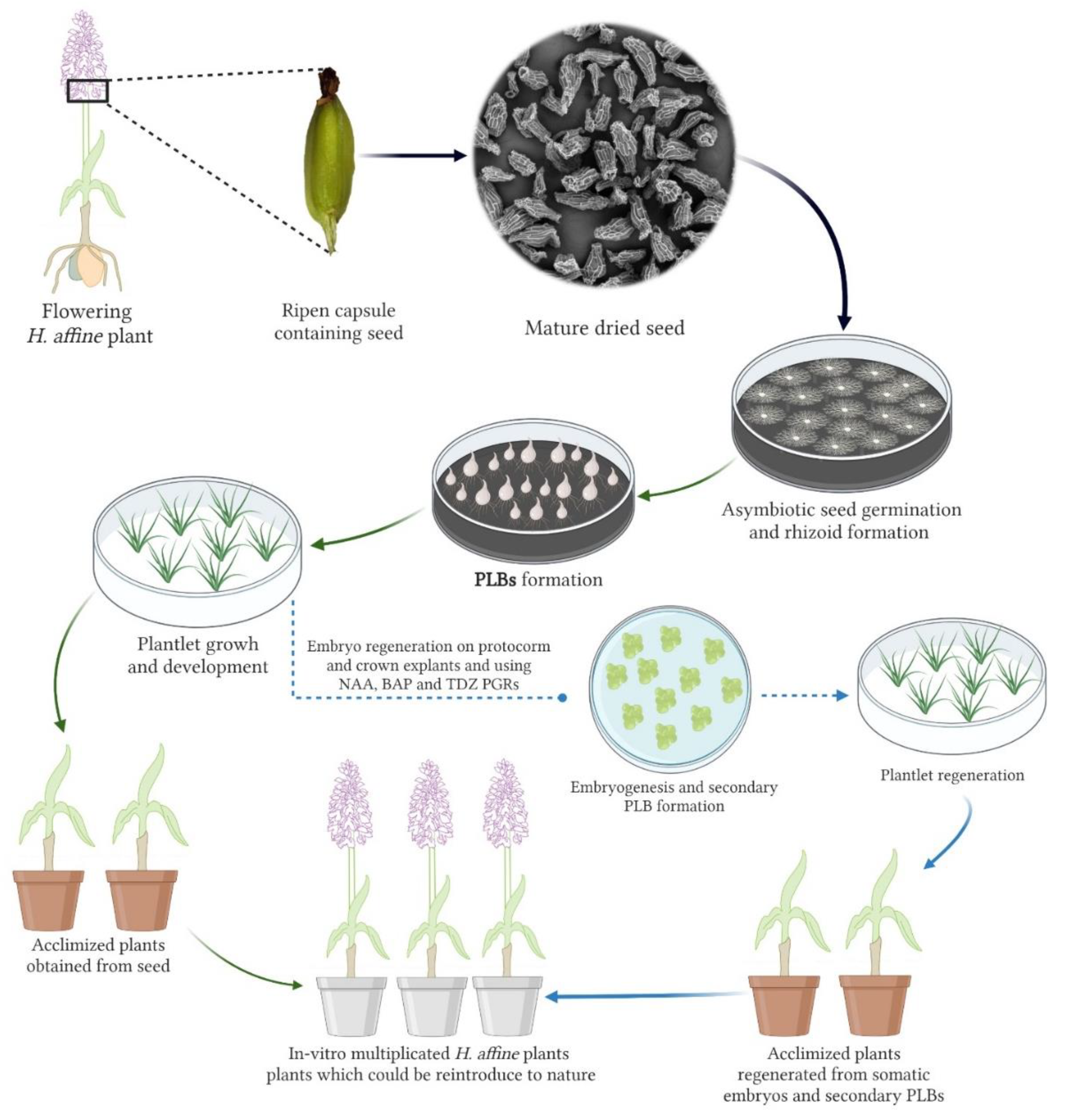
3.1. Asymbiotic Seed Germination

Prior to the seed germination study, an SEM analysis was carried out to show seed microstructure and testa micro-morphology details. As shown in Figure 1E–G, H. affine has an overall flask-shaped seed. A mean seed length of 538.89 ± 12.52 µm, seed width of 208.33 ± 12.55 µm, and cell number of 70.65 ± 2.87 were recorded for the studied seed. The anticlinal walls were prominent, and seed cells were big in the middle and small in the chalazal and polar ends. Based on the light microscopic imaging (Figure 1H), the seed testa color was brown. On the other hand, seed viability analysis by TTZ and FDA assays confirmed the viability of embryos in H. affine seed (Figure 1I,J) with an average rate of 95.33 ± 1.69% and 91.66 ± 2.05% viability for FDA and TTZ, respectively.
Figure 2 shows the stages of seed germination, protocorm development, and plantlet growth. Both the type of organic constituent as well as organic nitrogen source significantly impacted the asymbiotic seed germination and in vitro plantlet growth attributes (Table 1). In this regard, the highest germination rates were obtained with PJ in combination with AV (99.46 ± 0.38%), Pep (98.77 ± 0.37%), and CH (99.00 ± 0.47%), while implementation of CW resulted in lower germination rates. Generally, the individual use of organic nitrogen compounds resulted in higher asymbiotic germination efficiencies. On the other hand, the shortest time to germination was observed using CW, particularly in combination with CH (16.6 ± 0.4 days) and AV + CH (17 ± 0.31 days), whereas seed sown on media supplemented with PJ in combination with AV and Pep required more time to germinate. Regarding the protocorm growth, the media supplemented with CW in combination with Pep and CH resulted in higher protocorm dimensions in comparison to other combinations, and at the end of the experiment, the highest (0.094 ± 0.01 g) and lowest (0.036 ± 0.00 g) protocorm weight were obtained with CW + AV and PJ + CH, respectively. The attributes of in vitro-raised plantlets were also significantly impacted by organic ingredients, where plantlets grown on media supplemented with PJ in combination with Pep represented the highest plantlet (4.2 ± 0.04 cm), shoot length (1.96 ± 0.042 cm), and plantlet weight (0.58 ± 0.002 g). The highest number of roots was observed with CW + AV (5.2 ± 0.20) followed by CW + Pep (3.2 ± 0.020) and CW + CH (3 ± 0.031) treatments (Table 1). The first sign of asymbiotic seed germination was embryo swelling followed by testa rupturing (Figure 2A,D,E), the formation of protocorm and rhizoids (Figure 2B,C), activation of shoot and root meristem, and subsequently plantlet growth (Figure 2F) and finally micro-tuber development (Figure 2G). The in vitro-raised plantlets were successfully acclimatized (Figure 2H) and transferred to a research field, where they restarted the growth in the next growing season (Figure 2I).

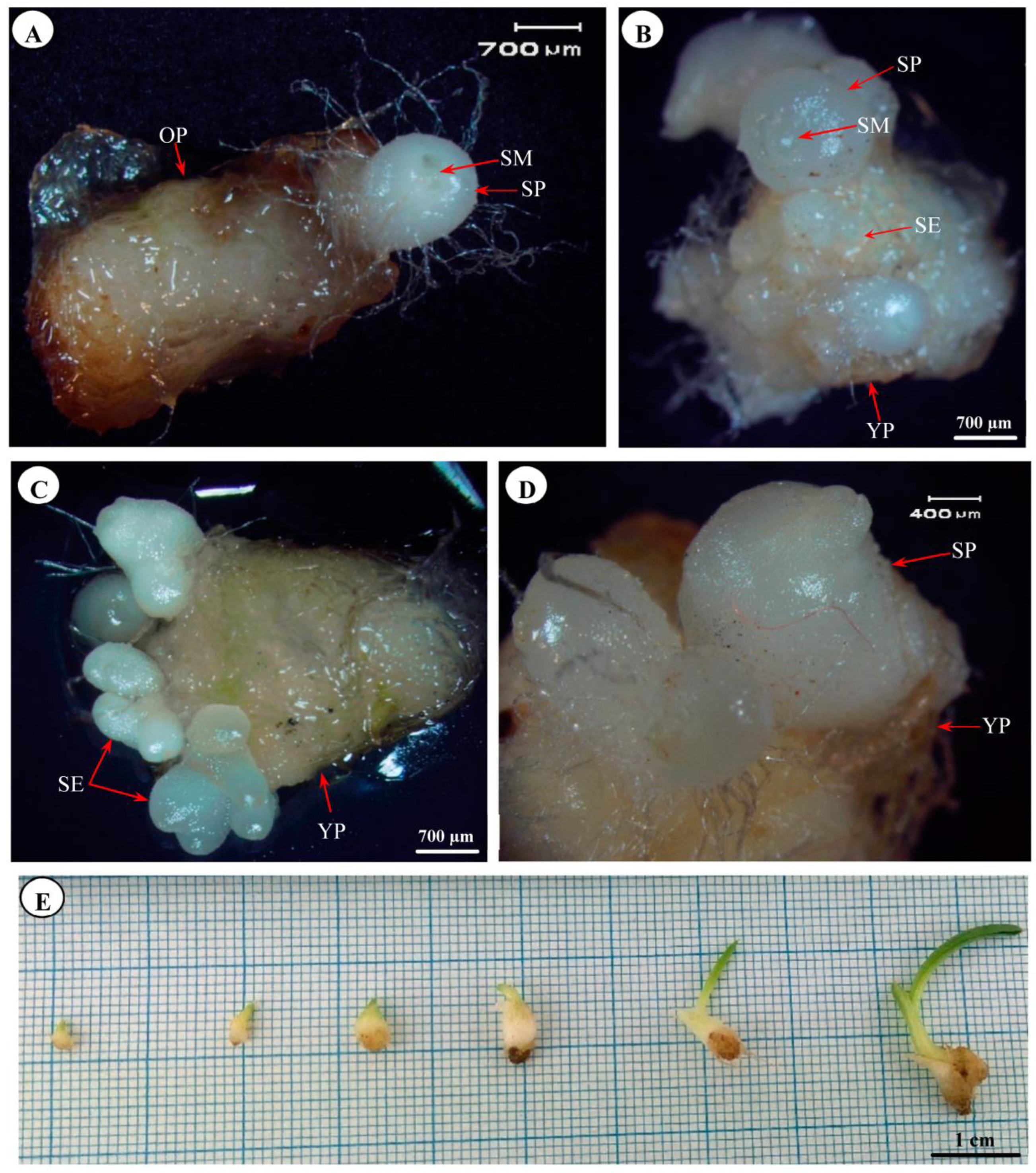
3.2. Somatic Embryogenesis and Secondary Protocorm Formation

The explants derived from in vitro-raised plantlets represented significantly different rates of somatic embryogenesis and number of embryos per explant in response to the used plant growth regulator (PGR) combinations (Table 2). However, there was no embryogenesis, and therefore no produced embryo with individual application of auxin or cytokinin. The highest regeneration rate (44%), as well as the highest number of embryos per explant (10.12 ± 2.08), were observed in young PLB explants on MS medium supplemented with 0.5 mg/L NAA and 1 mg/L TDZ. Overall, the young PLB explants represented higher embryogenesis potential and more somatic embryos compared to old PLB and crown explants, while old PLBs were more efficient explants than the crown explant. Moreover, TDZ in combination with NAA, was shown to be more effective than BAP in terms of somatic embryogenesis. Furthermore, the embryogenesis rate and frequency were dose-dependent, in which medium concentrations of NAA, TDZ, and BAP proved to be more efficient, while overall low and high concentrations resulted in lower regeneration rates. The different stages of somatic embryo formation and development and their turning to secondary PLBs are shown in Figure 3A–D. After four weeks of explant regeneration, the produced embryos matured and converted to secondary PLBs resembling the original PLBs resulting from seed germination. The secondary PLBs subcultured again on the new Malmgren medium and successfully grown to healthy and ready to acclimatization plantlets (Figure 3E).

3.3. Histology of In Vitro-Raised and Acclimatized Plantlets

The in vitro formation of protocorms in H. affine coincided with the presence of high-density rhizoids (Figure 4A), which seem to be necessary for the maximum absorption of nutrients from the surrounding medium. However, after the polarized growth of PLBs and thus the formation of root and shoot primordia, primary roots continue the nutrient uptake instead of rhizoids. During the acclimatization process of in vitro-raised plantlets, the scattered vascular tubes converted to fully developed tissues, which are probably involved in ensuring maximum sap flux (Figure 4C). Orchids are among typical monocotyledon species, and this was obvious from the histochemical pattern of studied H. affine plant parts. Furthermore, the developing layers of leaves had only a few vascular bundles (Figure 4D), which were converted to more organized structures in acclimatized plants (Figure 4E). In the transverse section of the protocorm, there was a central stele-like structure with a few small xylem and phloem tubes (Figure 4F). In addition, some parenchyma cells of protocorm had amyloplast grains containing glucomannan and starch. Figure 4G represents the histology of the stem in well-established H. affine plants in which vascular bundles within the cortex comprising sieve tubes and xylem vessels are distinct. The histology of small tubers produced under in vitro conditions showed a high density of plastids containing glucomannan and starch within storage parenchyma cells (Figure 4H). The number of these organelles was surprisingly decreased in fully developed tubers of acclimatized plants (Figure 4I). Finally, there was a substantial difference between the in vitro-raised plantlets and field-established plants regarding epidermal and stomatal cells. In this connection, the acclimatized plants had a higher frequency of stomata and bigger guard cells (Figure 4J) in comparison to in vitro and non-acclimatized plantlets (Figure 4K).

3.4. Phytochemical Analysis

GC–MS analysis of methanolic and ethanolic extracts prepared from tuber and leaf powder revealed a broad range of phytochemicals. The compounds higher than 0.3% are shown in Table 3. In this respect, 1,2,3-propanetriol,monoacetate was the most prevalent phytochemical present in the methanolic extract of tuber (47.63 ± 0.82%) and leaf (42.65 ± 1.8%), while 4H-pyran-4-one, 2,3-dihydro-3,5-dihydroxy-6-methyl, and 2-butenedioic acid, 2-methyl-, (E)- were found at the highest percentages in tuber (37.53 ± 1.12%) and leaf (19.40 ± 2.95%) ethanolic extract, respectively. Carbohydrates and their derivatives, including glycerin, 6-acetyl-beta-d-mannose, D-melezitose, ribitol, furaneol, and 1,3-Dioxolane were also present in high ratios in all four studied extracts. On the other hand, a high level of phenolic compounds was interestingly observed in both tuber and leaf material extracted by methanol and ethanol solvents. In this regard, 26.75 ± 1.23% of tuber methanolic extract belonged to 1-methoxy-4-(1-propenyl)-, while phenol, 4-(ethoxymethyl)- was the primary phenolic derivative in tuber ethanolic (8.26 ± 0.92%) and leaf methanolic (6.96 ± 0.91%) and ethanolic (19.01 ± 1.55%) extracts. 2-Methyl-1-cyclohexenyl) formaldehyde semicarbazone (4.55 ± 0.34 in tuber methanolic extract), alpha-pinene (14.30 ± 1.08 in tuber ethanolic extract), and 1,3-Diazacyclooctane-2-thione (13.08 ± 2.11% in leaf ethanolic extract) were other prevalent phytochemical constituents present in H. affine plant parts.

3.5. Physico-Biochemical Evaluation

As shown in Table 4, high contents of TP and TF and high rates of antioxidant activity were observed for leaf and tuber samples extracted by methanol and ethanol solvents. The highest TP (0.497 ± 0.012 mg/100 g FW) and TF (0.396 ± 0.011 mg/100 g FW) content, and antioxidant activity (0.766 ± 0.010%) were recorded for the methanolic extract of tuber, while the lowest values were observed in leaf ethanolic extracts. In general, methanolic extract had higher TP and TF content, and antioxidant activity than ethanolic extracts. Moreover, samples with higher TP and TF content represented higher antioxidant activity. Table 5 represents the physico-biochemical properties of Salep powder obtained from dried tubers of H. affine. The recorded glucomannan content in our study (36.96%) was higher than starch content (31.31%), and a viscosity of 17.96 ± 0.85 cP was obtained resulting from both carbohydrates.

4. Discussion

The observed seed microstructure qualitative and quantitative data were comparable with those reported by Vafaee et al. [11], Gholami et al. [12], and Şenel et al. [42]. As the orchid seed microstructure of terrestrial orchids is species-specific, the knowledge of their morphometry can be helpful when the origin of the seed used for asymbiotic in vitro germination is unknown and/or seed collection has been conducted after the flowering period. By knowing the exact species and sub-species of a target seed sample, we can confidentially provide optimum media and environmental conditions from the seed asymbiotic germination to PLB formation, plantlet growth, tuber formation, and even flowering. This is important as terrestrial orchid species are solely identified based on the floral characteristics, which sometimes are missing.
In the current study, the employed TTZ and FDA bioassays revealed high viability rates for H. affine seed sample. As preliminary tests to determine the initial viability of seed bulks, TTZ and FDA have also been utilized in other epiphytic and terrestrial orchid species, including Cyrtochilum aemulun [43], Dendrobium lasianthera [44], Cypripedium spp. [45], and Cephalanthera falcata [46]. The knowledge of the initial seed viability could help the interpretation of symbiotic and asymbiotic seed germination results, and it also aims at the long-term screening of seed viability in tropical and temperate orchid species for germplasm renewing and maintenance [47].
It is generally accepted that germination and PLB formation are more challenging and complex in terrestrial orchids than in epiphytic species because of different factors such as physical and physiological barriers and dependency on the mycorrhiza fungi [23]. The last factor is needed to provide essential nutrients, including macro and micro-elements, hydrocarbons, and amino acids, and also to adjust osmotic potential [48,49]. For the successful establishment of symbiotic cultures, mycorrhizal isolate preparation and the development of symbionts are required, while asymbiotic germination could alternatively provide an in vitro propagation platform for terrestrial orchids without symbiosis. Nevertheless, developing an optimized medium for asymbiotic germination of a single terrestrial species or even sub-species is still an important and understudied issue [50,51]. The strength of constituents and, in particular, the concentration of NO3 and NH4 used in conventional media for in vitro germination of epiphytic orchids is too high and poisonous for most terrestrial orchids [52,53,54]. In this regard, Malmgren basal medium [24] comprises organic constituents, including coconut milk, PJ, and amino acid mixture, which is suitable, and eliminates dependency on mycorrhizal fungi by supplying required minerals, vitamins, and phytohormones in optimum concentrations and combinations [55,56]. The observed seed germination (the seed developed to PLB) rates obtained in the present research were higher than those reported in H. robertianum (24%) [57], H. adriaticum (5.1%) [56], and H. jankae (77%) [58]. Our findings regarding the greater efficiency of PJ for the improvement of asymbiotic seed germination and also the final growth performance of in vitro-raised plantlets were in line with the results obtained in H. jankae [58]. Moreover, Ophrys species have represented better plantlet growth using PJ-enriched Malmgren media [59]. This enhancing impact of PJ might be attributed to the elimination of explant-exudated phenolic compounds and the presence of ready-to-uptake minerals and vitamins [59]. On the other hand, the faster germination of H. affine seed occurred with media containing CW. CW contains growth regulators, including auxin, cytokines, gibberellins, and abscisic acid. The auxin in CW is IAA (the main auxin in plants) [60]. Cytokines in CW are involved in cell division and thus accelerate growth. They are mainly used to amplify orchid-like protochromes in the plant industry [60]. It has been stated that CW can supply the required energy of the cells more quickly, and some of its components, such as cytokinin, trigger cell division [60], resulting in faster germination and PLB formation [58]. In the present study, the single implementation of organic nitrogen compounds was generally more efficient than their combinational use in enhancing seed germination and plantlet growth attributes. The simultaneous use of nitrogenous organic constituents may negatively affect water potential and thus disrupt water and nutrient uptake by the embryo and PLBs [23,61]. Nevertheless, both individual and combinational applications of CH, AV, and Pep eliminated dependency on inorganic nitrogen forms or mycorrhizal fungi, where CH and AV were more effective in seed time to germination and plantlet growth, respectively. These enhancing impacts have also been observed in other terrestrial orchids, including Orchis militaris [51], H. jankae [58], and Ophrys spp. [59]. CH comprises 18 amino acids, various vitamins, different forms of phosphates, and several microelements, explaining why it is known as a germination and growth-inducing factor in orchid tissue culture media [62]. It is believed that ready amino acid mixture (in available commercial forms like AV or Vamine) could be metabolized more effectively than inorganic counterparts by orchid PLBs developed under in vitro conditions as they may redirect some pathways of nitrogen assimilation [23,24].
The orchids grown in temperate regions, in particular terrestrial orchids, have been considered highly recalcitrant species with weak responses of direct and indirect regeneration, in particular, somatic embryogenesis [63]. This could be one of the reasons for the low embryogenesis rate and embryo number per explant obtained in the present study compared to those reported in tropical species. Nevertheless, there are several reports on the somatic embryogenesis of tropical orchids, including Spathoglottis plicata [64], Paphiopedilum niveum [65], Tolumnia [66], and Anoectochilus elatu [67]. It has been reported that both explant type and/or auxin and cytokinin combination can affect embryogenesis in all kinds of orchids. Here, we found the combination of TDZ and BAP, along with young PLB explants, as the most effective treatments in terms of embryogenesis rate and generated embryo number. In this regard, and in accord with our results, Moradi et al. [21] reported higher embryogenesis ratios and embryo number per explant in Epipactis veratrifolia using BAP and protocorm explant. The difference between the regeneration and embryogenesis potential of orchids’ explants might be related to their different status of indigenous hormones [68]. Both somatic embryogenesis and embryo maturity (somatic or zygotic) of terrestrial orchid species need cytokinin as a crucial factor [67], and it has been observed in S. plicata [64] that the presence of exogenous auxins is only required at the beginning of somatic embryogenesis induction; however, Mahendran and Narmatha Bai [69] reported high embryo number in Malaxis densiflora only with 2,4-D auxin. They stated that auxins activate embryogenesis by inducing the expression of stress-related genes responsible for molecular reprogramming of the somatic cells to start embryo induction and generation. Nevertheless, in our study, there was no or low rate of somatic embryogenesis induction in the crown and young or old PLB explants with the individual use of NAA, TDZ, or BAP. This could imply the simultaneous role of auxins and cytokinins toward efficient embryogenesis to generate a high number of somatic embryos [70]. Because of the high genetic diversity intrinsic in the Orchidaceae family arising from sexual reproduction [71], the secondary PLBs resulting from somatic embryos can also be utilized in orchid breeding and domestication programs as a propagule tool for clonal and vegetative maintenance of bred material.
A broad spectrum of phytochemicals with different natures, structures, and molecular weights was identified in leaf and tuber’s methanolic and ethanolic extracts of H. affine, among which there were some biologically important and active constituents. One of the most prevalent constituents quantified in both ethanolic and methanolic extracts of leaf and tuber was 1,2,3-propanetriol monoacetate, which has also been identified in male floral extracts of Lomandra leucocephala ssp. robusta [72] and ethanolic extract of Vanilla [73]. Studies show that 1,2,3-propanetriol monoacetate has not only strong antifungal activity but also poses broad antibacterial and insecticidal properties [74]. Benzene, 1-methoxy-4-(1-propenyl) has also been found in high percentages in rosemary essential oil [75] and Carum roxburghianum extract [76] with profound antifungal activity (against Aspergillus niger) [75], and antibacterial activity [77]. As a well-known component, glycerin is extensively used in the cosmetic, fragrance, pharmaceutical, and polymer industries [78]. On the other hand, 4H-Pyran-4-one, 2,3-dihydro-3,5-dihydroxy-6-methyl—found in Plukenetia conophora seed [79], Clerodendrum viscosum [80], and Castanopsis fissa [81]—is a flavonoid fraction with antifungal and protective effects. This particular sugar residue enables scavenging superoxide radicals mediated by saponins through the formation of hydroperoxide intermediates which, therefore, inhibit structural damage induced by free radicals [79]. Several other phytochemicals identified in our study have also been reported in extracts obtained from various organs of orchid species, including Pleione maculata (benzenamine, 4-methoxy-2-methyl-) [82], Orchis maculata (2-Butenedioic acid, 2-methyl) [83], Pleione maculate [82] (phenol, 4-(ethoxymethyl)), and Vanda cristata [84] (6-acetyl-beta-d-mannose). Other than mycorrhizal fungi, there are also some endophytic bacterial species associated with underground orchid tubers; thus, the exact origin of the identified phytochemical constituents should be traced more carefully. For example, 6-acetyl-beta-d-mannose has been identified in bacterial extract isolated from Vanda cristata orchid [84], while in our study, it was present in ethanolic extract of the tuber.
H. affine is among the terrestrial orchid species whose leaf- and tuber-derived materials are used in Kurdish and Persian folk herbal medicines. This application may be partly explained by the observed high TP and TF contents and TAA. The reported TP and TF contents in the present study are higher than those quantified in other terrestrial orchids, including H. robertianum [85] and Anacamptis pyramidalis [86]. Overall, H. affine tuber sample had more TP and TF than leaf material, which is in agreement with findings in Dactylorhiza maculate, in which the aerial part had more phenolic content [87]. This can be due to the effect of species, growing environment, and/or drying method. On the other hand, there were higher TP and TF contents in Dendrobium crepidatum extracts prepared by methanol compared to other solvents [88], which is also in line with the present findings. There was a similar condition for TAA in Dactylorhiza romana, where extract prepared by methanol represented more TAA than ethanol, and authors related this effect to the difference in polarity of solvents. Moreover, we found more TAA in samples with higher TC and TF contents, which probably shows the role of phenolic and flavonoid compounds in the scavenging of free radicals [25]. The glucomannan content is an indicator of Salep powder quality, which also determines the rheological properties of Salep-derived products, such as beverages and ice creams, in cooperation with other carbohydrates [27]. The recorded glucomannan content in our study (36.96%) was higher than starch content (31.31%), where both attributes were comparable to those reported in H. affine (36.64% glucomannan and 35% starch) [89]. On the contrary, glucomannan content was higher than values reported in terrestrial Turkish species [28]. However, this controversy could be due to the techniques used to quantify glucomannan. Overall, probably due to the large size of tubers and medium to high content of glucomannan, which results in good quality Salep powder, H. affine is among the most overexploited species in the Middle East.

5. Conclusions

Tuberous orchid species are at the front line of extinction, with many threatened taxa compared to other orchid life forms; thus, our optimized asymbiotic seed germination and secondary PLB regeneration procedures could assist in planning conservational strategies. Taking into consideration the fragmentation of H. affine populations in Iran, Turkey, and Euro-Mediterranean countries due to tuber over-harvesting, climate change, and/or human impact, our procedure could be used as a tool for large-scale propagation of in vitro-raised plants for re-introduction to threatened areas (Figure 5). Furthermore, the phytochemical and histochemical results provide preliminary data to perform more comprehensive research on terrestrial orchids as neglected species. Last but not least, due to the outstanding traits and attractive and long-living flowers and inflorescence, the present research provides an impulse for the exploitation of H. affine not only as a plant in herbal medicine and food industries but also as an ornamental garden, potted, or cut flower.

Author Contributions

Y.V., conceptualization, writing—original draft preparation, supervision; M.F., methodology; J.K. and N.A.-r.T., writing—review and editing. All authors have read and agreed to the published version of the manuscript.

Funding

This research received no external funding.

Institutional Review Board Statement

Not applicable.

Data Availability Statement

Not applicable.

Acknowledgments

We thank Svante Malmgren, Ali Akbar Mozafari, Abdolbaset Ghorbani, Sirvan Babaei, and Kolsom Ahmadzade for their kind help. We also thank Biorender.com for providing the tool to draw Figure 5.

Conflicts of Interest

The authors declare no conflict of interest.

References

  1. Shrestha, M.; Dyer, A.; Dorin, A.; Ren, Z.X.; Burd, M. Rewardlessness in orchids: How frequent and how rewardless? Plant Biol. 2020, 22, 555–561. [Google Scholar] [CrossRef] [PubMed]
  2. Shefferson, R.P.; Jacquemyn, H.; Kull, T.; Hutchings, M.J. The demography of terrestrial orchids: Life history, population dynamics and conservation. Bot. J. Linn. Soc. 2020, 192, 315–332. [Google Scholar] [CrossRef]
  3. Gaskett, A.C.; Gallagher, R.V. Orchid diversity: Spatial and climatic patterns from herbarium records. Ecol. Evol. 2018, 8, 11235–11245. [Google Scholar] [CrossRef] [PubMed] [Green Version]
  4. de Boer, H.J.; Ghorbani, A.; Manzanilla, V.; Raclariu, A.C.; Kreziou, A.; Ounjai, S.; Osathanunkul, M.; Gravendeel, B. DNA metabarcoding of orchid-derived products reveals widespread illegal orchid trade. Proc. Biol. Sci. 2017, 284, 20171182. [Google Scholar] [CrossRef] [PubMed]
  5. Renz, J. Flora Iranica. Part 126: Orchidaceae; Naturhistorisches Museum: Vienna, Austria, 1978. [Google Scholar]
  6. Teoh, E.S. Introduction. In Medicinal Orchids of Asia; Springer International Publishing: Cham, Switzerland, 2016; pp. 3–17. [Google Scholar]
  7. Farhoosh, R.; Riazi, A. A compositional study on two current types of salep in Iran and their rheological properties as a function of concentration and temperature. Food Hydrocoll. 2007, 21, 660–666. [Google Scholar] [CrossRef]
  8. Ghorbani, A.; Gravendeel, B.; Zare, S.; De Boer, H.J. Illegal wild collection and international trade of CITES-listed terrestrial orchid tubers in Iran. Traffic Bull. 2014, 26, 52–58. [Google Scholar]
  9. Ghorbani, A.; Gravendeel, B.; Naghibi, F.; de Boer, H. Wild orchid tuber collection in Iran: A wake-up call for conservation. Biodivers. Conserv. 2014, 23, 2749–2760. [Google Scholar] [CrossRef]
  10. Ghorbani, A.; Gravendeel, B.; Selliah, S.; Zarre, S.; de Boer, H. DNA barcoding of tuberous Orchidoideae: A resource for identification of orchids used in Salep. Mol. Ecol. Resour. 2017, 17, 342–352. [Google Scholar] [CrossRef] [Green Version]
  11. Vafaee, Y.; Mohammadi, G.; Nazari, F.; Fatahi, M.; Kaki, A.; Gholami, S.; Ghorbani, A.; Khadivi, A. Phenotypic characterization and seed-micromorphology diversity of the threatened terrestrial orchids: Implications for conservation. S. Afr. J. Bot. 2021, 137, 386–398. [Google Scholar] [CrossRef]
  12. Gholami, S.; Vafaee, Y.; Nazari, F.; Ghorbani, A. Exploring genetic variations in threatened medicinal orchids using start codon targeted (SCoT) polymorphism and marker-association with seed morphometric traits. Physiol. Mol. Biol. Plants 2021, 27, 769–785. [Google Scholar] [CrossRef]
  13. Kaki, A.; Vafaee, Y.; Khadivi, A. Genetic variation of Anacamptis coriophora, Dactylorhiza umbrosa, Himantoglossum affine, Orchis mascula, and Ophrys schulzei in the western parts of Iran. Ind. Crops Prod. 2020, 156, 112854. [Google Scholar] [CrossRef]
  14. Gholami, S.; Vafaee, Y.; Nazari, F.; Ghorbani, A. Molecular characterization of endangered Iranian terrestrial orchids using ISSR markers and association with floral and tuber-related phenotypic traits. Physiol. Mol. Biol. Plants 2021, 27, 53–68. [Google Scholar] [CrossRef] [PubMed]
  15. Bateman, R.M.; Molnar, V.A.; Sramko, G. In situ morphometric survey elucidates the evolutionary systematics of the Eurasian Himantoglossum clade (Orchidaceae: Orchidinae). PeerJ 2017, 5, e2893. [Google Scholar] [CrossRef] [PubMed] [Green Version]
  16. Sramko, G.; Attila, M.V.; Hawkins, J.A.; Bateman, R.M. Molecular phylogeny and evolutionary history of the Eurasiatic orchid genus Himantoglossum s.l. (Orchidaceae). Ann. Bot. 2014, 114, 1609–1626. [Google Scholar] [CrossRef]
  17. Phillips, R.D.; Reiter, N.; Peakall, R. Orchid conservation: From theory to practice. Ann. Bot. 2020, 126, 345–362. [Google Scholar] [CrossRef]
  18. Zhao, D.K.; Selosse, M.A.; Wu, L.; Luo, Y.; Shao, S.C.; Ruan, Y.L. Orchid reintroduction based on seed germination-promoting mycorrhizal fungi derived from protocorms or seedlings. Front. Plant Sci. 2021, 12, 701152. [Google Scholar] [CrossRef] [PubMed]
  19. Barthlott, W.; Große-Veldmann, B.; Korotkova, N. Orchid seed diversity: A scanning electron microscopy survey. Englera 2014, 32, 1–245. [Google Scholar]
  20. Batty, A.L.; Dixon, K.W.; Brundrett, M.; Sivasithamparam, K. Constraints to symbiotic germination of terrestrial orchid seed in a mediterranean bushland. New Phytol. 2001, 152, 511–520. [Google Scholar] [CrossRef]
  21. Moradi, S.; Dianati Daylami, S.; Arab, M.; Vahdati, K. Direct somatic embryogenesis in Epipactis veratrifolia, a temperate terrestrial orchid. J. Hortic. Sci. Biotechnol. 2016, 92, 88–97. [Google Scholar] [CrossRef]
  22. Fatahi, M.; Vafaee, Y.; Nazari, F.; Tahir, N.A.-r. In vitro asymbiotic seed germination, protocorm formation, and plantlet development of Orchis simia Lam.: A threatened terrestrial orchid species. S. Afr. J. Bot. 2022, 151, 156–165. [Google Scholar] [CrossRef]
  23. Rasmussen, H.N.; Dixon, K.W.; Jersakova, J.; Tesitelova, T. Germination and seedling establishment in orchids: A complex of requirements. Ann. Bot. 2015, 116, 391–402. [Google Scholar] [CrossRef] [PubMed] [Green Version]
  24. Malmgren, S. Orchid propagation: Theory and practice. In Proceedings of the North American Native Orchids: Propagation and Production, Germantown, MD, USA, 16–17 March 1996; pp. 63–71. [Google Scholar]
  25. Gutiérrez, R.M.P. Orchids: A review of uses in traditional medicine, its phytochemistry and pharmacology. J. Med. Plants. Res. 2010, 4, 592–638. [Google Scholar]
  26. Wang, Z.; Zhao, M.; Cui, H.; Li, J.; Wang, M. Transcriptomic landscape of medicinal Dendrobium reveals genes associated with the biosynthesis of bioactive components. Front. Plant Sci. 2020, 11, 391. [Google Scholar] [CrossRef] [PubMed]
  27. Ece Tamer, C.; Karaman, B.; Utku Copur, O. A Traditional Turkish Beverage: Salep. Food Rev. Int. 2006, 22, 43–50. [Google Scholar] [CrossRef]
  28. Acemi, A.; Cobanoglu, O.; Turker-Kaya, S. FTIR-based comparative analysis of glucomannan contents in some tuberous orchids, and effects of pre-processing on glucomannan measurement. J. Sci. Food Agric. 2019, 99, 3681–3686. [Google Scholar] [CrossRef]
  29. Murashige, T.; Skoog, F. A revised medium for rapid growth and bio assays with tobacco tissue cultures. Physiol. Plant. 1962, 15, 473–497. [Google Scholar] [CrossRef]
  30. Mozafari, A.A.; Vafaee, Y.; Shahyad, M. Phytochemical composition and in vitro antioxidant potential of Cynodon dactylon leaf and rhizome extracts as affected by drying methods and temperatures. J. Food Sci. Technol. 2018, 55, 2220–2229. [Google Scholar] [CrossRef]
  31. Singleton, V.L.; Orthofer, R.; Lamuela-Raventós, R.M. [14] Analysis of total phenols and other oxidation substrates and antioxidants by means of folin-ciocalteu reagent. Methods Enzymol. 1999, 299, 152–178. [Google Scholar]
  32. Toor, R.K.; Savage, G.P. Antioxidant activity in different fractions of tomatoes. Food Res. Int. 2005, 38, 487–494. [Google Scholar] [CrossRef]
  33. Brand-Williams, W.; Cuvelier, M.-E.; Berset, C. Use of a free radical method to evaluate antioxidant activity. LWT-Food Sci. Technol. 1995, 28, 25–30. [Google Scholar] [CrossRef]
  34. AOAC. Official Methods of Analysis of the Association of Official Analytical Chemists, 14th ed.; Association of Official Analytical Chemists: Washington, DC, USA, 2019. [Google Scholar]
  35. Bradford, M.M. A rapid and sensitive method for the quantification of microgram quantities of protein utilizing the principle of protein-dye binding. Anal. Biochem. 1976, 72, 248–254. [Google Scholar] [CrossRef] [PubMed]
  36. Liu, P.; Zhang, S.; Zhu, G.; Chen, Y.; Ouyang, H.; Han, M. Professional Standard of the People’ Republic of China for Konjac Flour; Ministry of Agriculture of the People’s Republic of China: Beijing, China, 2002. [Google Scholar]
  37. McCready, R.; Guggolz, J.; Silviera, V.; Owens, H. Determination of starch and amylose in vegetables. Anal. Chem. 1950, 22, 1156–1158. [Google Scholar] [CrossRef]
  38. Pritchard, H. Determination of orchid seed viability using fluorescein diacetate. Plant Cell Environ. 1985, 8, 727–730. [Google Scholar]
  39. Van Waes, J.; Debergh, P. Adaptation of the tetrazolium method for testing the seed viability, and scanning electron microscopy study of some Western European orchids. Physiol. Plant. 1986, 66, 435–442. [Google Scholar] [CrossRef]
  40. Aybeke, M. Morphological and histochemical investigations on Himantoglossum robertianum (Loisel.) P. Delforge (Orchidaceae) seeds. Plant Syst. Evol. 2013, 300, 91–97. [Google Scholar] [CrossRef]
  41. Aybeke, M. Comparative anatomy of selected rhizomatous and tuberous taxa of subfamilies Orchidoideae and Epidendroideae (Orchidaceae) as an aid to identification. Plant Syst. Evol. 2012, 298, 1643–1658. [Google Scholar] [CrossRef]
  42. Şenel, G.; Şeker, Ş.S.; Akbulut, M.K.; Akçin, Ö.E. An integrative anatomical, morphological, micromorphological and molecular approach to Turkish epidendroid and orchidoid species (Orchidaceae). Nord. J. Bot. 2018, 36, e01700. [Google Scholar] [CrossRef]
  43. Salazar Mercado, S.A.; Caleno, J.D.Q.; Suarez, J.P.R. Optimization of the tetrazolium test in three species of orchids of the Andean forest. Aust. J. Crop Sci. 2020, 14, 822–829. [Google Scholar] [CrossRef]
  44. Utami, E.S.W.; Hariyanto, S.; Manuhara, Y.S.W. In vitro propagation of the endangered medicinal orchid, Dendrobium lasianthera J.J.Sm through mature seed culture. Asian Pac. J. Trop. Biomed. 2017, 7, 406–410. [Google Scholar] [CrossRef]
  45. Vujanovic, V.; St-Arnaud, M.; Barabé, D.; Thibeault, G. Viability testing of orchid seed and the promotion of colouration and germination. Ann. Bot. 2000, 86, 79–86. [Google Scholar] [CrossRef] [Green Version]
  46. Yamazaki, J.; Miyoshi, K. In vitro asymbiotic germination of immature seed and formation of protocorm by Cephalanthera falcata (Orchidaceae). Ann. Bot. 2006, 98, 1197–1206. [Google Scholar] [CrossRef] [PubMed]
  47. Seaton, P.T.; Hosomi, S.T.; Custódio, C.C.; Marks, T.R.; Machado-Neto, N.B.; Pritchard, H.W. Orchid seed and pollen: A toolkit for long-term storage, viability assessment and conservation. In Orchid Propagation—From Laboratories to Greenhouses; Yeung, L.Y., Chee-Tak, E., Eds.; Humana Press: New York, NY, USA, 2018; pp. 71–98. [Google Scholar]
  48. Gao, Y.; Zhao, Z.; Li, J.; Liu, N.; Jacquemyn, H.; Guo, S.; Xing, X. Do fungal associates of co-occurring orchids promote seed germination of the widespread orchid species Gymnadenia conopsea? Mycorrhiza 2020, 30, 221–228. [Google Scholar] [CrossRef] [PubMed]
  49. Perotto, S.; Rodda, M.; Benetti, A.; Sillo, F.; Ercole, E.; Rodda, M.; Girlanda, M.; Murat, C.; Balestrini, R. Gene expression in mycorrhizal orchid protocorms suggests a friendly plant-fungus relationship. Planta 2014, 239, 1337–1349. [Google Scholar] [CrossRef] [PubMed]
  50. Quiroz, K.; Saavedra, J.; Vogel, H.; Verdugo, G.; Caligari, P.D.S.; Garcia-Gonzales, R. In vitro asymbiotic germination for micropropagation of the recalcitrant terrestrial orchid Chloraea crispa (Orchidaceae). Appl. Plant Sci 2017, 5, apps1600142. [Google Scholar] [CrossRef] [PubMed]
  51. Nabieva, A.Y. Asymbiotic seed germination and in vitro seedling development of Orchis militaris, an endangered orchid in Siberia. J. Genet. Eng. Biotechnol. 2021, 19, 122. [Google Scholar] [CrossRef]
  52. Swarts, N.D.; Dixon, K.W. Conservation Methods for Terrestrial Orchids; Ross Publishing: Richmond, VA, USA, 2017; p. 240. [Google Scholar]
  53. Figura, T.; Tylova, E.; Jersakova, J.; Vohnik, M.; Ponert, J. Fungal symbionts may modulate nitrate inhibitory effect on orchid seed germination. Mycorrhiza 2021, 31, 231–241. [Google Scholar] [CrossRef]
  54. Figura, T.; Weiser, M.; Ponert, J. Orchid seed sensitivity to nitrate reflects habitat preferences and soil nitrate content. Plant Biol. 2020, 22, 21–29. [Google Scholar] [CrossRef]
  55. Stewart, S.L.; Kane, M.E. Asymbiotic seed germination and in vitro seedling development of Habenaria macroceratitis (Orchidaceae), a rare Florida terrestrial orchid. Plant Cell Tissue Organ Cult. 2006, 86, 147–158. [Google Scholar] [CrossRef]
  56. Del Vecchio, S.; Pierce, S.; Fantinato, E.; Buffa, G. Increasing the germination percentage of a declining native orchid (Himantoglossum adriaticum) by pollen transfer and outbreeding between populations. Plant Biol. 2019, 21, 935–941. [Google Scholar] [CrossRef]
  57. Calevo, J.; Copetta, A.; Marchioni, I.; Bazzicalupo, M.; Pianta, M.; Shirmohammadi, N.; Cornara, L.; Giovannini, A. The use of a new culture medium and organic supplement to improve in vitro early stage development of five orchid species. Plant Biosyst. Int. J. Deal. All Asp. Plant Biol. 2022, 156, 143–151. [Google Scholar] [CrossRef]
  58. Dulić, J.; Ljubojević, M.; Ognjanov, V.; Barać, G.; Dulić, T. In vitro germination and seedling development of two European orchid species, Himantoglossum jankae Somlyay, Kreutz & Óvári and Spiranthes spiralis (L.) Chevall. In Vitro Cell. Dev. Biol. Plant 2019, 55, 380–391. [Google Scholar] [CrossRef]
  59. Kitsaki, C.K.; Zygouraki, S.; Ziobora, M.; Kintzios, S. In vitro germination, protocorm formation and plantlet development of mature versus immature seeds from several Ophrys species (Orchidaceae). Plant Cell Rep. 2004, 23, 284–290. [Google Scholar] [CrossRef] [PubMed]
  60. Yong, J.W.; Ge, L.; Ng, Y.F.; Tan, S.N. The chemical composition and biological properties of coconut (Cocos nucifera L.) water. Molecules 2009, 14, 5144–5164. [Google Scholar] [CrossRef] [PubMed] [Green Version]
  61. Utami, E.S.W.; Hariyanto, S. Organic compounds: Contents and their role in improving seed germination and protocorm development in orchids. Int. J. Agron. 2020, 2020, 2795108. [Google Scholar] [CrossRef]
  62. Kunakhonnuruk, B.; Inthima, P.; Kongbangkerd, A. In vitro propagation of Epipactis flava Seidenf., an endangered rheophytic orchid: A first study on factors affecting asymbiotic seed germination, seedling development and greenhouse acclimatization. Plant Cell Tissue Organ Cult. 2018, 135, 419–432. [Google Scholar] [CrossRef]
  63. Jørgensen, B.I. Hardy orchids: Symbiotic in vitro propagation and cultivation. Environ. Eff. Control Plant Tissue Cult. 1994, 393, 165–172. [Google Scholar] [CrossRef]
  64. Manokari, M.; Priyadharshini, S.; Shekhawat, M.S. Direct somatic embryogenesis using leaf explants and short term storage of synseeds in Spathoglottis plicata Blume. Plant Cell Tissue Organ Cult. 2021, 145, 321–331. [Google Scholar] [CrossRef]
  65. Soonthornkalump, S.; Nakkanong, K.; Meesawat, U. In vitro cloning via direct somatic embryogenesis and genetic stability assessment of Paphiopedilum niveum (Rchb.f.) Stein: The endangered Venus’s slipper orchid. In Vitro Cell. Dev. Biol. Plant 2019, 55, 265–276. [Google Scholar] [CrossRef]
  66. Shen, H.J.; Chen, J.T.; Chung, H.H.; Chang, W.C. Plant regeneration via direct somatic embryogenesis from leaf explants of Tolumnia Louise Elmore ‘Elsa’. Bot. Stud. 2018, 59, 4. [Google Scholar] [CrossRef] [Green Version]
  67. Sherif, N.A.; Franklin Benjamin, J.H.; Senthil Kumar, T.; Rao, M.V. Somatic embryogenesis, acclimatization and genetic homogeneity assessment of regenerated plantlets of Anoectochilus elatus Lindl., an endangered terrestrial jewel orchid. Plant Cell Tissue Organ Cult. 2017, 132, 303–316. [Google Scholar] [CrossRef]
  68. Parthibhan, S.; Rao, M.V.; Teixeira da Silva, J.; Senthil Kumar, T. Somatic embryogenesis from stem thin cell layers of Dendrobium aqueum. Biol. Plant. 2018, 62, 439–450. [Google Scholar] [CrossRef]
  69. Mahendran, G.; Narmatha Bai, V. Direct somatic embryogenesis of Malaxis densiflora (A. Rich.) Kuntze. J. Genet. Eng. Biotechnol. 2016, 14, 77–81. [Google Scholar] [CrossRef] [PubMed] [Green Version]
  70. Chugh, S.; Guha, S.; Rao, I.U. Micropropagation of orchids: A review on the potential of different explants. Sci. Hortic. 2009, 122, 507–520. [Google Scholar] [CrossRef]
  71. Cardoso, J.C.; Zanello, C.A.; Chen, J.T. An overview of orchid protocorm-like bodies: Mass propagation, biotechnology, molecular aspects, and breeding. Int. J. Mol. Sci. 2020, 21, 985. [Google Scholar] [CrossRef] [PubMed] [Green Version]
  72. Draper, J.T.; Conran, J.G.; Crouch, N.; Weinstein, P.; Simpson, B.S. Sexual dimorphism in the dioecious monocot Lomandra leucocephala ssp. robusta and its potential ecosystem and conservation significance. Aust. J. Bot. 2020, 68, 275–287. [Google Scholar] [CrossRef]
  73. McCormick, D. Characterisation of Vanilla Extracts Based on Sensory Properties and Chemical Composition: A Thesis Presented in Partial Fulfilment of the Requirements for the Degree of Doctor of Philosophy in Food Technology at Massey University, New Zealand. Ph.D. Thesis, Massey University, Palmerston North, New Zealand, 2018. [Google Scholar]
  74. Casuga, F.P.; Castillo, A.L.; Corpuz, M.J.-A.T. GC–MS analysis of bioactive compounds present in different extracts of an endemic plant Broussonetia luzonica (Blanco)(Moraceae) leaves. Asian Pac. J. Trop. Biomed. 2016, 6, 957–961. [Google Scholar] [CrossRef] [Green Version]
  75. Salih, T.F.M.; Mohammed, L.M.; Qader, K.O. Chemical Analysis and Growth Inhibitory Effect of Rosemary Plant on Aspergillus niger. Kurd. J. Appl. Res. 2017, 2, 151–154. [Google Scholar] [CrossRef] [Green Version]
  76. Chowdhury, J.U.; Bhuiyan, M.N.I.; Begum, J. Constituents of leaf and fruits essential oil of Carum roxburghianum Benth. J. Sci. Res. 2009, 1, 160–163. [Google Scholar] [CrossRef]
  77. Wang, Q.; Ou, Z.; Lei, H.; Zeng, X.; Ying, Y.; Bai, W. Antimicrobial activities of a new formula of spice water extracts against foodborne bacteria. J. Food Process. Preserv. 2012, 36, 374–381. [Google Scholar] [CrossRef]
  78. Jungermann, E.; Sonntag, N.O. Glycerine: A Key Cosmetic Ingredient; CRC Press: Boca Raton, FL, USA, 1991; Volume 11. [Google Scholar]
  79. Olaniyan, O.T.; Kunle-Alabi, O.T.; Raji, Y. Protective effects of methanol extract of Plukenetia conophora seeds and 4H-Pyran-4-One 2,3-Dihydro-3,5-Dihydroxy-6-Methyl on the reproductive function of male Wistar rats treated with cadmium chloride. JBRA Assist. Reprod. 2018, 22, 289–300. [Google Scholar] [CrossRef]
  80. Ghosh, G.; Panda, P.; Rath, M.; Pal, A.; Sharma, T.; Das, D. GC-MS analysis of bioactive compounds in the methanol extract of Clerodendrum viscosum leaves. Pharmacogn. Res. 2015, 7, 110–113. [Google Scholar] [CrossRef] [Green Version]
  81. Peng, W.-X.; Wu, S.-B.; Yang, L.; Li, K.-F.; Peng, W.-L.; Hong, S.-Y. Determination of Chemical Components of Benzene/Ethanol Extractives of Castanopsis Fissa Wood by GC/MS. In Proceedings of the 2008 2nd International Conference on Bioinformatics and Biomedical Engineering, Shanghai, China, 16–18 May 2008; pp. 3165–3167. [Google Scholar]
  82. Sympli, H.D. Estimation of drug-likeness properties of GC-MS separated bioactive compounds in rare medicinal Pleione maculata using molecular docking technique and SwissADME in silico tools. Netw. Model. Anal. Health Inf. Bioinform. 2021, 10, 14. [Google Scholar] [CrossRef] [PubMed]
  83. Khadartsev, A.; Sukhikh, G.T.; Volochaeva, M.V.; Platonov, V.V.; Dunaeva, I.V. Chromato-mass spectrometry of Orchis maculata ethanol extract (Orchidaceae), e-edition. J. New Med. Technol. 2019, 13, 102–121. [Google Scholar]
  84. Shah, S.; Chand, K.; Rekadwad, B.; Shouche, Y.S.; Sharma, J.; Pant, B. A prospectus of plant growth promoting endophytic bacterium from orchid (Vanda cristata). BMC Biotechnol. 2021, 21, 16. [Google Scholar] [CrossRef] [PubMed]
  85. Bazzicalupo, M.; Burlando, B.; Denaro, M.; Barreca, D.; Trombetta, D.; Smeriglio, A.; Cornara, L. Polyphenol Characterization and Skin-Preserving Properties of Hydroalcoholic Flower Extract from Himantoglossum robertianum (Orchidaceae). Plants 2019, 8, 502. [Google Scholar] [CrossRef] [PubMed] [Green Version]
  86. Hurkan, K.; Yuksel, M.B.; Hurkan, Y.K.; Demir, N. Determination of total phenolic and flavonoid contents, antioxidant and antimicrobial activities of some important salep orchids. Eskişehir Teknik Üniversitesi Bilim ve Teknoloji Dergisi-C Yaşam Bilimleri ve Biyoteknoloji 2019, 8, 191–202. [Google Scholar] [CrossRef]
  87. Dalar, A.; Guo, Y.; Esim, N.; Bengu, A.S.; Konczak, I. Health attributes of an endemic orchid from Eastern Anatolia, Dactylorhiza chuhensis Renz&Taub.—In vitro investigations. J. Herb. Med. 2015, 5, 77–85. [Google Scholar] [CrossRef] [Green Version]
  88. Bhattacharyya, P.; Kumaria, S.; Job, N.; Tandon, P. En-masse production of elite clones of Dendrobium crepidatum: A threatened, medicinal orchid used in Traditional Chinese Medicine (TCM). J. Appl. Res. Med. Aromat. Plants 2016, 3, 168–176. [Google Scholar] [CrossRef]
  89. Şen, M.A.; Palabiyik, I.; Kurultay, Ş. Composition, viscosity and solubility of saleps from twenty different orchid (Orchidaceae) species. J. Food Meas. Charact. 2018, 12, 1334–1339. [Google Scholar] [CrossRef]
Figure 1. (A,B) Inflorescence and flower morphology; (C) underground tuber morphology; (DF) scanning electron microscopy (SEM) micrograph of seed testa micromorphology; (G) light microscopy of seed color and structure; (H) immature white seed within the capsule; (I,J) fluorescein diacetate (FDA) and tetrazolium chloride (TTZ) analyses of seed viability, respectively (red and white arrowheads show the viable and dead or deeding seeds, respectively); (K) seed with softened testa after disinfection with sodium hypochlorite.
Diversity 14 01137 g001
Figure 2. Seed germination and plantlet growth of H. affine (Boiss.) Schltr. (A) Embryo enlargement and protocorm formation 16 days after seed sown. (B,C) Protocorm development and rhizoid formation 24–30 days after seed sown. (D,E) SEM micrograph of testa rupturing and asymbiotic seed germination. (F) Fully developed plantlets ready for acclimatization 3–4 months after seed sown. (G) The stages of in vitro protocorm development, rooting, and plantlet growth. (H) In vitro-raised plantlets successfully acclimatized and transferred to plugs. (I) H. affine plant established under field condition in the next spring. SS, swelling seed; RT, rupturing testa; DP, developing protocorm-like bodies (PLBs); RZ, rhizoids; LP, leaf primordium; IT, in vitro formed tubers.
Diversity 14 01137 g002
Figure 3. Embryogenesis and secondary protocorm-like body (PLB) formation in H. affine (Boiss.) Schltr. (A) The formation of secondary PLB with developed rhizoids on old protocorm as explant; (AD) different stage of embryo development on PLB explants and turning to secondary PLB. (E) Different stage of plantlet development from secondary PLBs. OP, old PLB explant; SP, secondary PLB; SM, shoot meristem; YP, young PLB explant.
Diversity 14 01137 g003
Figure 4. Histology of in vitro-raised plantlet and acclimatized and established plants of H. affine. (A) High-density rhizoids on PLB generated during asymbiotic seed germination. (B) Transverse section of root in in vitro raised plantlets showing central stele containing procambium developing vascular system. (C) Root histology in field-established plants showing differentiated xylem and phloem vascular elements. (D) Histochemical view of shoot in seedling developed under in vitro condition. (E) The central vascular bundle in leaf of acclimatized plant. (F,G) Cross-sections of stem in in vitro raised plantlets and field-acclimatized plants, respectively. (H) Anatomy of tuber initially formed under in vitro condition, which shows amyloplast containing starch and glucomannan. (I) The anatomy of tuber in well-established plants in the field. (J,K) The epidermis and stomata of leaf’s lower surface in in vitro-raised plantlets and field-acclimatized plants, respectively. AG, amyloplast grains; DA, developing endodermis; DL, developing leaves; GC, guard cells; LE, leaf epidermis; LV, leaf vein; PV, phloem vessels; RP, ray parenchyma; SB, single vascular bundle; VB, vascular bundle; XV, xylem vessels.
Diversity 14 01137 g004
Figure 5. Flow diagram showing the strategy for in vitro large-scale micropropagation of H. affine (Boiss.) Schltr. via asymbiotic seed germination and somatic embryogenesis. Created with BioRender.com.
Diversity 14 01137 g005
Table 1. The effect of pineapple juice (PJ) and coconut water (CW) as the main organic constituent and Aminoven (AV), peptone (Pep), and casein hydrolysate (CH) as the organic nitrogenous sources on the seed germination and plantlet growth attributes of endangered terrestrial orchid species H. affine (Boiss.) Schltr.
TraitPJCW
AVPepCHAV + PepAV + CHPep + CHAVPepCHAV + PepAV + CHPep + CH
Germination percent (%)99.46 ± 0.38 a98.77 ± 0.37 a99.00 ± 0.47 a95.38 ± 1.51 b64.85 ± 2.01 e75.99 ± 1.43 cd61.28 ± 1.17 f58.2 ± 0.76 g50.06 ± 1.44 h77.64 ± 0.73 c75.46 ± 0.53 cd66.62 ± 0.49 e
Time to germination (day)51.20 ± 1.46 i56.00 ± 1.64 h34.20 ± 0.20 g32.80 ± 0.91 f31.00 ± 0.77 f27.60 ± 0.40 e19.60 ± 0.50 c22.60 ± 0.50 d16.60 ± 0.40 a18.00 ± 0.00 b17.00 ± 0.31 a38.00 ± 0.83 i
Protocorm length (cm)0.56 ± 0.04 b0.30 ± 0.02 b0.26 ± 0.02 c0.58 ± 0.06 b0.25 ± 0.03 d0.38 ± 0.03 d0.64 ± 0.04 a0.64 ± 0.05 a0.48 ± 0.06 c0.32 ± 0.03 e0.34 ± 0.02 e0.42 ± 0.04 d
Protocorm width (cm)0.28 ± 0.040 b0.20 ± 0.05 cd0.20 ± 0.03 c0.26 ± 0.05 b0.17 ± 0.02 d0.20 ± 0.00 c0.32 ± 0.05 a0.27 ± 0.03 b0.24 ± 0.02 c0.20 ± 0.04 cd0.19 ± 0.02 d0.26 ± 0.08 bc
Protocorm weight (g)0.07 ± 0.00 b0.04 ± 0.00 d0.03 ± 0.00 ef0.08 ± 0.00 ab0.03 ± 0.00 ef0.04 ± 0.00 de0.09 ± 0.01 a0.08 ± 0.00 ab0.66 ± 0.00 c0.03 ± 0.00 e0.04 ± 0.00 de0.06 ± 0.00 c
Plantlet length (cm)3.58 ± 0.03 d4.20 ± 0.04 a2.58 ± 0.03 d3.10 ± 0.05 b3.06 ± 0.09 b2.44 ± 0.01 e2.52 ± 0.03 d2.54 ± 0.02 d2.72 ± 0.03 c2.50 ± 0.04 d2.16 ± 0.02 g2.24 ± 0.02 f
Plantlet weight (g)0.38 ± 0.00 f0.58 ± 0.00 a0.25 ± 0.00 e0.34 ± 0.00 cd0.30 ± 0.003 c0.22 ± 0.00 g0.26 ± 0.00 ef0.27 ± 0.00 d0.26 ± 0.00 b0.22 ± 0.00 g0.02 ± 0.00 i0.23 ± 0.00 h
Shoot length (cm)1.76 ± 0.02 d1.96 ± 0.04 a1.22 ± 0.03 e1.86 ± 0.03 b1.62 ± 0.02 b1.09 ± 0.02 e1.20 ± 0.03 c1.36 ± 0.02 f1.64 ± 0.02 f1.33 ± 0.01 cd0.87 ± 0.01 g1.04 ± 0.05 g
Shoot diameter (cm)0.19 ± 0.10 b0.20 ± 0.00 b0.19 ± 0.00 b0.19 ± 0.00 b0.09 ± 0.00 e0.10 ± 0.00 e0.23 ± 0.02 a0.20 ± 0.00 b0.20 ± 0.01 b0.10 ± 0.00 e0.12 ± 0.00 d0.15 ± 0.00 c
Root number (cm)1.60 ± 0.024 d1.60 ± 0.024 d1.00 ± 0.01 g1.40 ± 0.02 e1.20 ± 0.02 f1.20 ± 0.02 f5.20 ± 0.20 a3.20 ± 0.02 b3.00 ± 0.03 c0.40 ± 0.02 i0.80 ± 0.03 h0.40 ± 0.02 i
Different letters in each row indicate significance by LSD tests at p < 0.05. Values represent the means ± standard error.
Table 2. The effect of plant growth regulators (PGRs): naphthalene acetic acid (NAA), thidiazuron (TDZ), and benzyl aminopurine (BAP) on the regeneration and number of embryos per explant in the endangered terrestrial orchid species H. affine (Boiss.) Schltr. using different explants (crown, young PLB, and old PLB).
PGRs (mg/L)Regeneration (%)The Number of Embryos per Explants
CrownYoung PLBOld PLBCrownYoung PLBOld PLB
NAATDZ
000.00 ± 0.00 e0.00 ± 0.00 e0.00 ± 0.00 e0.00 ± 0.00 e0.00 ± 0.00 e0.00 ± 0.00 e
00.50.00 ± 0.00 e0.00 ± 0.00 e0.00 ± 0.00 e0.00 ± 0.00 e0.00 ± 0.00 e0.00 ± 0.00 e
010.00 ± 0.00 e4.00 ± 8.00 d4.00 ± 8.00 d0.00 ± 0.00 e5.00 ± 0.00 c0.00 ± 0.00 e
020.00 ± 0.00 e4.00 ± 8.00 d8.00 ± 9.79 cd0.00 ± 0.00 e3.50 ± 0.50 ef3.25 ± 0.47 f
0.500.00 ± 0.00 e0.00 ± 0.00 e0.00 ± 0.00 e0.00 ± 0.00 e0.00 ± 0.00 e0.00 ± 0.00 e
0.50.58.00 ± 9.79 cd12.00 ± 9.79 cd8.00 ± 9.79 cd4.50 ± 0.50 de8.50 ± 0.50 a3.50 ± 0.50 ef
0.5132.00 ± 8.00 b44.00 ± 0.00 a32.00 ± 8.00 b8.33 ± 1.20 ab10.12 ± 2.08 a4.87 ± 1.05 cf
0.5212.00 ± 9.79 cd20.00 ± 8.00 bc16.00 ± 0.00 c4.66 ± 1.24 cd7.20 ± 0.74 b5.75 ± 0.43 c
108.00 ± 9.79 cd4.00 ± 8.00 d0.00 ± 0.00 e3.85 ± 0.50 ef4.00 ± 0.00 e0.00 ± 0.00 e
10.512.00 ± 9.79 cd24.00 ± 9.79 b4.00 ± 8.00 d6.33 ± 0.47 bc6.20 ± 0.89 bc4.00 ± 0.00 e
1112.00 ± 9.79 cd28.00 ± 9.79 b24.00 ± 9.79 b5.60 ± 0.94 b–d8.42 ± 0.72 ab8.33 ± 0.94 ab
120.00 ± 0.00 e12.00 ± 9.79 cd20.00 ± 8.00 bc0.00 ± 0.00 e4.33 ± 0.47 de2.50 ± 0.50 h
NAABAP
00.50.00 ± 0.00 e0.00 ± 0.00 e0.00 ± 0.00 e0.00 ± 0.00 e0.00 ± 0.00 e0.00 ± 0.00 e
010.00 ± 0.00 e0.00 ± 0.00 e0.00 ± 0.00 e0.00 ± 0.00 e0.00 ± 0.00 e0.00 ± 0.00 e
024.00 ± 8.00 d4.00 ± 8.00 d8.00 ± 9.79 cd2.00 ± 0.00 i2.00 ± 0.00 i2.00 ± 0.00 i
0.50.512.00 ± 9.79 cd8.00 ± 9.79 cd4.00 ± 8.00 d3.00 ± 0.00 h4.66 ± 1.24 c-f2.50 ± 0.50 f-i
0.518.00 ± 9.79 cd24.00 ± 0.00 b4.00 ± 8.00 d2.50 ± 0.50 h6.50 ± 0.50 bc4.00 ± 0.00 e
0.5212.00 ± 9.79 cd16.00 ± 0.00 c8.00 ± 9.79 cd2.33 ± 0.47 hi8.00 ± 0.00 ab4.25 ± 0.43 de
10.524.00 ± 8.00 b8.00 ± 9.79 cd4.00 ± 8.00 c4.6 ± 1.01 cd4.80 ± 0.74 cd3.00 ± 0.00 h
118.00 ± 9.79 cd12.00 ± 9.79 cd16.00 ± 8.00 cd2.00 ± 1.00 i3.25 ± 0.43 f1.75 ± 0.43 ij
124.00 ± 8.00 d12.00 ± 9.79 cd20.00 ± 8.00 bc2.00 ± 0.00 i2.00 ± 0.00 i1.60 ± 0.48 ij
PLB, protocorm-like body. Different letters indicate significance by LSD tests at p < 0.05. Values represent the means ± standard error.
Table 3. The phytochemical components of methanolic and ethanolic extracts of tuber and leaf in H. affine (Boiss.) Schltr. identified by gas chromatography–mass spectrometry (GC–MS) analysis.
Methanolic Extract
TuberLeaf
CompoundFormulaRT%MWCompoundFormulaRT%MW
1,2,3-propanetriol, monoacetateC10H12O17.42047.63 ± 0.821341,2,3-propanetriol, monoacetateC10H12O17.12842.65 ± 1.84134
Benzene, 1-methoxy-4-(1-propenyl)-C5H10O417.96426.75 ± 1.231484H-Pyran-4-one, 2,3-dihydro-3,5-dihydroxy-6-methylC6H8O414.67511.32 ± 1.86136
GlycerinC3H8O328.81916.42 ± 0.66926-Acetyl-.beta.-d-mannoseC8H14O720.11610.45 ± 1.80222
(2-Methyl-1-cyclohexenyl) formaldehyde semicarbazoneC9H15N3O20.1664.55 ± 0.34137alpha-D-glucopyranosyl-(1->3)C18H32O1620.91410.16 ± 1.02504
Bicyclo[2.2.1]heptan-2-one, 1,3,3-trimethyl-C10H16O12.2341.10 ± 0.50152Phenol, 4-(ethoxymethyl)-C9H12O220.2656.96 ± 0.91152
2,5-Methylene-d,l-rhamnitolC7H14O514.3510.73 ± 0.28178RibitolC5H12O515.7202.59 ± 0.32152
Ethyl(dimethyl)allyloxysilaneC7H16OSi9.5120.71 ± 0.121442-Propanone, 1,1-diethoxy-C7H14O315.3992.43 ± 0.49146
SorbitolC6H14O620.9580.46 ± 0.18182Hexadecanoic acid, 3-hydroxy-, methyl esterC17H34O315.8092.34 ± 0.27286
TrimethylsilylmethanolC4H12OSi9.7650.43 ± 0.20104Hexadecanoic acid, methyl esterC17H34O238.2491.89 ± 0.36270
Phenol, 3-methyl-C7H8O1612.8320.33 ± 0.11108TrimethylsilylmethanolC4H12OSi10.1991.41 ± 0.27104
Isosorbide DinitrateC6H8N2O811.6260.31 ± 0.16236L-LyxoseC5H10O59.7981.37 ± 0.44150
Propionic acid, 2-mercapto-, allyl esterC6H10O2S10.1760.26 ± 0.92146D-Glucose, 6-O-.alpha.-D-galactopyranosyl-C12H22O119.3511.09 ± 0.90342
d-Mannitol, 1-decylsulfonyl- Undecanoic acid, 3-hydroxy-, methyl esterC12H24O318.9280.95 ± 0.28216
N-Methyl-N-(toluene-4-sulfonylmethyl)-acetamideC11H15NO3S13.9940.81 ± 0.55241
beta.-D-Glucopyranose, 4-O-.beta.-D-galactopyranosyl-C12H22O1124.6260.56 ± 0.21170
1,2,3,4,5-CyclopentanepentolC5H10O58.2550.54 ± 0.16155
Decanoic acid, 3-hydroxy-, methyl esterC11H22O319.1100.45 ± 0.17202
Ethanolic Extract
TuberLeaf
Compound CompoundFormulaRT%MW
4H-Pyran-4-one, 2,3-dihydro-3,5-dihydroxy-6-methylC6H8O418.02837.53 ± 1.121442-Butenedioic acid, 2-methyl-, (E)-C5H6O412.81819.40 ± 2.95130
alpha.-PineneC10H169.67214.30 ± 1.08136Phenol, 4-(ethoxymethyl)-C9H12O219.81519.01 ± 1.55152
2,4-Octadienoic acid, 7-hydroxy-6-methyl-C9H14O322.73113.93 ± 1.101701,3-Diazacyclooctane-2-thioneC6H12N2S13.07313.08 ± 2.11144
2-Furancarboxaldehyde, 5-(hydroxymethyl)-C6H6O322.13411.70 ± 0.88121Nonanediamide, N,N′-di-benzoyloxy-C23H26N2O614.6346.60 ± 0.49426
Phenol, 4-(ethoxymethyl)-C9H12O224.5588.26 ± 0.921525-Methyl-1-phenylhex-5-en-1-oneC13H16O15.2336.48 ± 1.41188
2,4-Dihydroxy-2,5-dimethyl-3(2H)-furan-3-oneC6H8O411.6523.06 ± 0.281441,3-DioxolaneC3H6O28.7906.09 ± 1.5074
2-n-PropylthianeC8H16S18.4981.50 ± 0.411441,3-Dioxolane, 2-(dichloromethyl)-C4H6Cl2O28.3955.37 ± 1.17156
IsopinocarveolC10H16O16.8171.20 ± 0.551526-Acetyl-.beta.-d-mannoseC8H14O717.2924.72 ± 0.67222
d-Gala-l-ido-octonic amideC8H17NO813.0440.92 ± 0.37255Benzenemethanol, 2-methoxy-.alpha.-2-propenyl-C11H14O217.9542.91 ± 0.89178
D-Glucose, 6-O-.alpha.-D-galactopyranosylC12H22O1111.8710.91 ± 0.473421,2,3,4-CyclohexanetetrolC6H12O49.4072.53 ± 1.01148
2,2-Dimethyl-3-vinyl-bicyclo[2.2.1]heptaneC11H1815.7460.86 ± 0.101502H-Indeno[1,2-b]furan-2-one, 3,3a,4,5,6,7,8,8b-octahydro-8,8-dimethylC13H18O222.5692.52 ± 0.52206
Bicyclo[3.1.1]hept-3-en-2-ol, 4,6,6-trimethyl-C10H16O17.1040.83 ± 0.16152Bicyclo[3.2.1]oct-6-ene-6,8-dimethanol, 1,7-dimethyl-4-isopropyl-, bis(3,5-dinitrobenzoate)C29H30N4O1215.6061.99 ± 0.38626
N-Methyl-N-(toluene-4-sulfonylmethyl)-acetamideC11H15NO3S13.9940.81 ± 0.16241Geranyl isovalerateC15H26O222.8621.97 ± 0.44238
l-Gala-l-ido-octonic lactoneC8H14O813.1580.70 ± 0.09238Dithiocarbamate, S-methyl-,N-(2-methyl-3-oxobutyl)-C7H13NOS216.0401.66 ± 0.32191
OcimeneC10H1612.5020.53 ± 0.11136D-MelezitoseC18H32O1611.2001.58 ± 0.47504
Cyclohexane, 1,4-dimethoxy-2-methyl-, stereoisomerC9H18O217.1830.44 ± 0.08158Cyclohexanone, 5-(1-hydroxy-2-propenyl)-2,2-dimethyl-,C11H18O217.4521.50 ± 0.28182
Isobornyl acetateC12H20O221.1440.32 ± 0.091962-Nitrohept-2-en-1-olC7H13NO39.7391.03 ± 0.27159
1,5,5-Trimethyl-6-methylene-cyclohexeneC10H1615.1680.30 ± 0.04136E-9-Methyl-8-tridecen-2-ol, acetateC16H30O217.6860.67 ± 0.30254
RT, retention time determined on DB-17MS capillary column. Components are listed in order of percentage for each organ. MW, molecular weight. Values represent the means ± standard error.
Table 4. The content of total phenolics (TP), total flavonoids (TF), and total antioxidant activity (TAA) in endangered terrestrial orchid species H. affine (Boiss.) Schltr. as affected by organ and solvent type.
OrganSolventTP (mg/100 g FW)TF (mg/100 g FW)TAA (%)
LeafMethanol0.40 ± 0.01 c0.31 ± 0.01 c0.63 ± 0.02 bcd
Ethanol0.38 ± 0.01 d0.28 ± 0.01 d0.61 ± 0.02 d
TuberMethanol0.49 ± 0.01 a0.39 ± 0.01 a0.76 ± 0.01 a
Ethanol0.44 ± 0.01 b0.33 ± 0.01 b0.64 ± 0.01 bc
Different letters indicate significance by LSD tests at p < 0.05. FW, fresh weight. Values represent the means ± standard error.
Table 5. Biochemical properties of Salep powder obtained from endangered terrestrial orchid species H. affine (Boiss.) Schltr.
TraitValue
Moisture (%)11.55 ± 0.70
Ash (g/100g)1.76 ± 0.05
Protein (g/100g)3.95 ± 0.04
Starch (%)31.31 ± 0.60
Glucomannan (%)36.96 ± 1.22
Viscosity (cP)17.96 ± 0.85
Values represent the means ± standard error.
Publisher’s Note: MDPI stays neutral with regard to jurisdictional claims in published maps and institutional affiliations.

